Case Reports
. 2021 Sep 9;16(11):3499-3503.
doi: 10.1016/j.radcr.2021.08.038. eCollection 2021 Nov.

Early rebleeding of a foramen magnum dural arteriovenous fistula: A case report and review of the literature

Affiliations
Case Reports

Early rebleeding of a foramen magnum dural arteriovenous fistula: A case report and review of the literature

Takanari Okamoto et al. Radiol Case Rep. .

Abstract

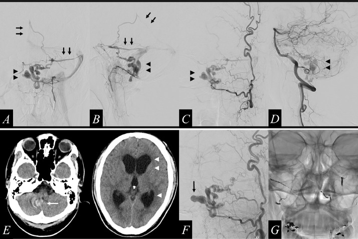
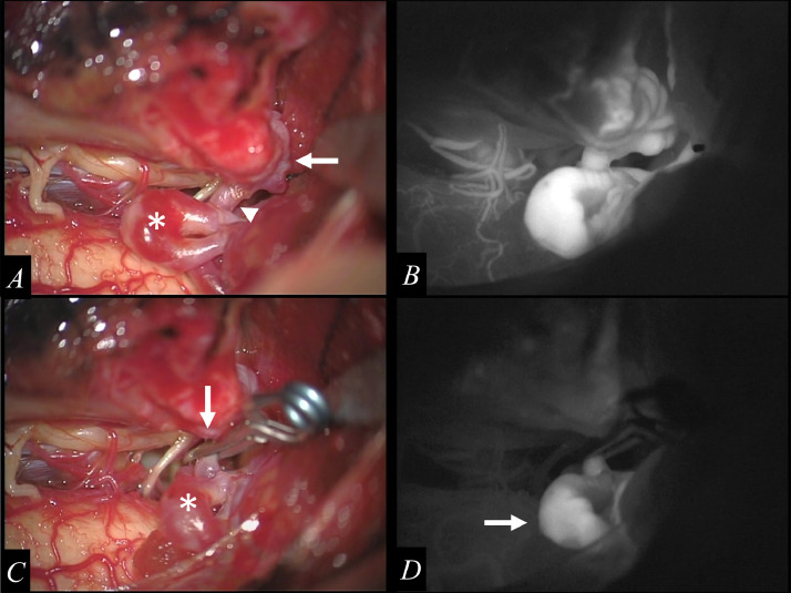
Foramen magnum dural arteriovenous fistula (FM-DAVF) is a subset of craniocervical junction arteriovenous fistulas. We report a rare case of FM-DAVF with early rebleeding and review the literature. A 50-year-old man experienced 3 episodes of intracranial bleeding from a vessel malformation in the acute stage. We identified an FM-DAVF, supplied by multiple feeding arteries (eg, left ascending pharyngeal artery) that drained into the straight sinus and left superior petrosal sinus. The draining vein had venous varices. We performed transarterial feeder embolization and surgical disconnection of the DAVF. Early rebleeding of FM-DAVF is rare. High-risk patients require risk assessment and appropriate treatment as soon as possible in the acute stage.

Keywords: AVM, arteriovenous malformation; Ascending pharyngeal artery; CT, computed tomography; Craniocervical junction arteriovenous fistula; DAVF, dural arteriovenous fistula; DSA, digital subtraction angiography; Early rebleeding; Foramen magnum dural arteriovenous fistula; GKS, gamma knife surgery; ICG, indocyanine green; MRI, magnetic resonance imaging; PICA, posterior inferior cerebellar artery; SAH, subarachnoid hemorrhage.


Figures

Fig 1 –
Fig. 1
Initial computed tomography (CT) displays normal findings (A). The same-day magnetic resonance imaging (MRI) shows a subarachnoid hemorrhage in the bilateral occipital lobe (B, arrows). The next-day CT reveals bleeding, primarily in the third ventricle (C, arrows). 12 days after the initial bleeding event, CT reveals a third intraventricular bleeding event in the fourth ventricle and the lateral ventricle with intracranial ventriculomegaly (D, arrows)
Fig 2 –
Fig. 2
Digital subtraction angiography (DSA) shows a dural arteriovenous fistula (DAVF), which is fed by the jugular branch and hypoglossal branch of the left ascending pharyngeal artery (A and B), the jugular branch of the left occipital artery (C), and the posterior meningeal artery from the left vertebral artery (D) that drains into the straight sinus and the left superior petrosal sinus (A, B; arrows) through the vein of the lateral recess of the fourth ventricle, which is dilated and tortuous with large varices (A-D, arrowheads). 28 days after the initial bleeding event, the CT image shows a radical increase in the size of the varix (E, white arrow) and severe ventriculomegaly (E, white arrowheads). DSA imaging shows a radical increase in the size of the varix (F, black arrow), compared to its previous size. We performed transarterial coil embolization on the main feeders (G)
Fig 3 –
Fig. 3
The vascular mass suspected of being a “shunted pouch” is confirmed inside of the dura of the left lateral margin of the foramen magnum (A, arrows), which is connected to vein of the lateral recess of the fourth ventricle through a single drainer (A, arrowheads). The inferior portion of the varix is identified (A, asterisk). Intraoperative indocyanine green (ICG) angiography shows retrograde flow to the varix through the single drainer in the early arterial phase (B). The single drainer is obliterated as close as possible to the shunted pouch by using an 11-mm straight clip (C, arrow). The surface of the varix is darker and the variceal wall tension is reduced (C, asterisk). ICG angiography shows stagnation of blood flow in the varix (D, arrow) after the occlusion of the draining vein.

References

    1. Kim H, Lee Y-S, Kang H-J, Lee M-S, Suh S-J, Lee J-H. A rare case of subarachnoid hemorrhage caused by ruptured venous varix due to dural arteriovenous fistula at the foramen magnum fed solely by the ascending pharyngeal artery. J Cerebrovasc Endovasc Neurosurg. 2018;20:120–126. doi: 10.7461/jcen.2018.20.2.120. - DOI - PMC - PubMed
    1. Jellema K, Tijssen CC, van Gijn J. Spinal dural arteriovenous fistulas: a congestive myelopathy that initially mimics a peripheral nerve disorder. Brain. 2006;129:3150–3164. doi: 10.1093/brain/awl220. Pt 12. - DOI - PubMed
    1. Motebejane MS, Choi IS. Foramen magnum dural arteriovenous fistulas: clinical presentations and treatment outcomes, a case-series of 12 patients. Oper Neurosurg (Hagerstown) 2018;15:262–269. doi: 10.1093/ons/opx229. - DOI - PubMed
    1. Nakamura M, Miyazaki T, Shinozaki N, Izumi M, Itabashi T. Clinical characteristics of craniocervical junction arteriovenous fistulas. No Shinkei Geka. 2017;45:879–888. doi: 10.11477/mf.1436203611. Japanese. - DOI - PubMed
    1. Kinouchi H, Mizoi K, Takahashi A, Nagamine Y, Koshu K, Yoshimoto T. Dural arteriovenous shunts at the craniocervical junction. J Neurosurg. 1998;89:755–761. doi: 10.3171/jns.1998.89.5.0755. - DOI - PubMed

Publication types

LinkOut - more resources