. 2021 Oct;20(10):e13467.
doi: 10.1111/acel.13467. Epub 2021 Sep 23.

Late-in-life treadmill training rejuvenates autophagy, protein aggregate clearance, and function in mouse hearts

Affiliations

Late-in-life treadmill training rejuvenates autophagy, protein aggregate clearance, and function in mouse hearts

Jae Min Cho et al. Aging Cell. 2021 Oct.

Abstract

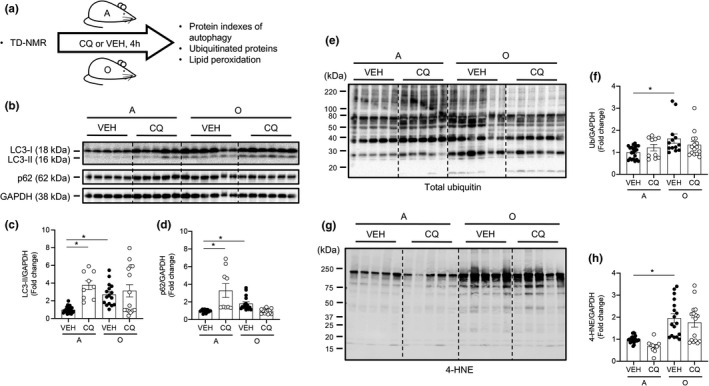
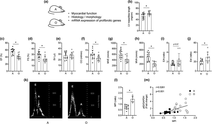
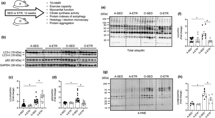
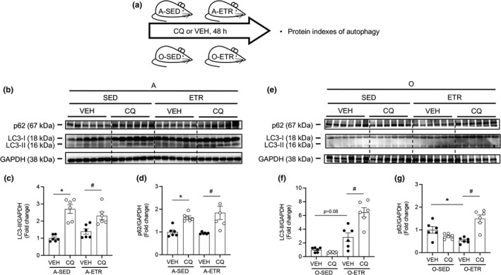
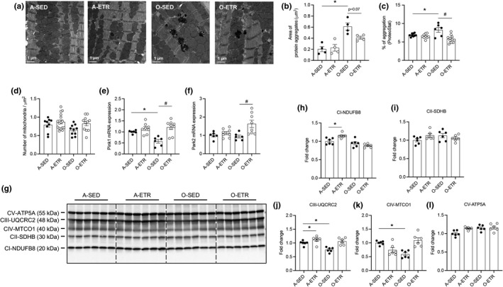
Protein quality control mechanisms decline during the process of cardiac aging. This enables the accumulation of protein aggregates and damaged organelles that contribute to age-associated cardiac dysfunction. Macroautophagy is the process by which post-mitotic cells such as cardiomyocytes clear defective proteins and organelles. We hypothesized that late-in-life exercise training improves autophagy, protein aggregate clearance, and function that is otherwise dysregulated in hearts from old vs. adult mice. As expected, 24-month-old male C57BL/6J mice (old) exhibited repressed autophagosome formation and protein aggregate accumulation in the heart, systolic and diastolic dysfunction, and reduced exercise capacity vs. 8-month-old (adult) mice (all p < 0.05). To investigate the influence of late-in-life exercise training, additional cohorts of 21-month-old mice did (old-ETR) or did not (old-SED) complete a 3-month progressive resistance treadmill running program. Body composition, exercise capacity, and soleus muscle citrate synthase activity improved in old-ETR vs. old-SED mice at 24 months (all p < 0.05). Importantly, protein expression of autophagy markers indicate trafficking of the autophagosome to the lysosome increased, protein aggregate clearance improved, and overall function was enhanced (all p < 0.05) in hearts from old-ETR vs. old-SED mice. These data provide the first evidence that a physiological intervention initiated late-in-life improves autophagic flux, protein aggregate clearance, and contractile performance in mouse hearts.

Keywords: aging; cardiac function; exercise; protein aggregates.


Conflict of interest statement

None of the authors has any conflicts of interest to disclose.

Figures

FIGURE 1
FIGURE 1
Hearts from old mice display impaired autophagic flux, accumulation of ubiquitinated proteins, and oxidative stress. (a) Body composition was assessed using TD‐NMR in adult (A; 8‐mo) and older (O; 24‐mo) mice. Vehicle (VEH; phosphate‐buffered saline, PBS) or chloroquine (CQ; 75 mg/kg lean muscle mass) was administered to A and O mice and tissues were obtained 4 h later. Representative images (b, e, g) and mean data ± standard error are shown for LC3‐II (c), p62 (d), poly‐ubiquitin (total ubiquitin; f), and 4‐hydroxy‐2‐nonenal (4‐HNE; h). LC3‐II and p62 were greater in hearts from O vs. A mice treated with VEH. In A mice, LC3‐II and p62 increased further in CQ vs. VEH‐treated cohorts, whereas these endpoints were similar in O mice treated with CQ. These findings demonstrate that autophagic flux is robust in A but not O mice. Total ubiquitin and 4‐HNE were elevated in hearts from O vs. A mice, but CQ did not alter responses in either group. For panels (c, d), n = 9–24, *< 0.05 vs. A‐VEH mice. For (f) and (h), n = 10–21, *< 0.05 vs. A‐VEH. For panels (c, d, f, h), data are expressed as fold change relative to values obtained from A‐VEH mice
FIGURE 2
FIGURE 2
Cardiac function is impaired in older vs. adult mice. (a) Transthoracic echocardiography was performed on adult (A; 8‐mo) and older (O; 24‐mo) mice. Mean data ± standard error are shown (b‐j, l). Left ventricular (LV) mass normalized to tibial length was greater in O vs. A animals (b). Ejection fraction (EF, %; c), fractional shortening (FS, %; d), cardiac output (CO, ml/min; f), passive diastolic filling (MVE, mm/s; g), active diastolic filling (MVA, mm/s; h), and an estimate of end‐diastolic filling pressure (E/e’, ratio; j) were impaired in O vs. A mice, whereas stroke volume (SV, μl; e) and the E/A ratio (i) were not different between groups. These data, together with our observation that the myocardial performance index (MPI, l) is greater in hearts from O vs. A mice, indicate that systolic and diastolic dysfunction exists in O vs. A mice. (k) Representative images of blood flow velocity obtained during the assessment of MVE and MVA from both groups. (m) The correlation between protein expression of p62/GAPDH and MPI was strong in hearts from A and O mice. For (b–j, l), n = 9–13; for (m), n = 16–17. For (b–j, l), *< 0.05 vs. A
FIGURE 3
FIGURE 3
Late‐in‐life exercise training improves steady‐state autophagy, clearance of ubiquitinated proteins, and oxidative stress in mouse hearts. (a) Adult (A, 5 mo) and older (O, 21 mo) mice did (ETR) or did not (SED) complete 12 weeks of treadmill running. At least 24 h following the last exercise bout, A (8 mo) and O (24 mo) hearts were excised and prepared for immunoblotting. Representative images (b, e, g) and mean data ± standard error are shown for LC3‐II (c), p62 (d), total ubiquitin (f), and 4‐HNE (h). LC3‐II (c) and p62 (d) were greater in hearts from O‐SED vs. A‐SED mice. While no differences existed between A‐SED and A‐ETR mice, LC3‐II increased (c) and p62 decreased (d) in hearts from O‐ETR vs. O‐SED mice. Total ubiquitin (f) and 4‐HNE (h) were elevated in hearts from O‐SED vs. A‐SED mice. Although no differences existed between A‐SED and A‐ETR mice, total ubiquitin (f) and 4‐HNE (h) decreased in hearts from O‐ETR vs. O‐SED mice. For panels (c, d), n = 9–24, *< 0.05 vs. A‐SED mice. For (f) and (h), n = 6–15, *< 0.05 vs. A‐SED. For panels (c, d, f, h), data are expressed as fold change relative to values obtained from A‐SED mice
FIGURE 4
FIGURE 4
Late‐in‐life exercise training improves steady‐state autophagy and autophagic flux in mouse hearts. (a) Adult (A, 5 mo) and older (O, 21 mo) mice did (ETR) or did not (SED) complete 12 wk of treadmill running. Mice were treated with VEH or CQ 48, 24, and 4 h prior to tissue collection. Representative images (b, e) and mean data ± standard error are shown for LC3‐II (c, f) and p62 (d, g). In A hearts treated with VEH (b–d), ETR did not influence steady‐state LC3‐II or p62. Robust CQ‐evoked increases in LC3‐II and p62 observed in A hearts were similar regardless of exercise training (b–d). In O hearts treated with VEH, (e–g), LC3‐II trended upwards (= 0.08) and p62 decreased in response to ETR. These data indicate that late‐in‐life exercise training improves steady‐state autophagy. While CQ did not influence LC3‐II or p62 in hearts from O‐SED mice (e–g), significant accumulation of LC3‐II (f) and p62 (g) was observed in hearts from O‐ETR mice. These findings indicate late‐in‐life exercise training improves autophagic flux in mouse hearts. For (b–g), n = 6. For (c, d), *< 0.05 vs. A‐SED‐VEH; #< 0.05 vs. A‐ETR‐VEH. For (f, g), *< 0.05 vs. O‐SED‐VEH; #< 0.05 vs. O‐ETR‐VEH. For (c, d, f, g), data are expressed as fold change relative to values obtained from A‐SED‐VEH mice or O‐SED‐VEH mice
FIGURE 5
FIGURE 5
Late‐in‐life exercise training improves cardiac function in mice. Older (O, 21 mo) mice did (ETR) or did not (SED) complete 12 wk of treadmill running. At least 24 h following the last exercise bout, transthoracic echocardiography was completed on O‐SED and O‐ETR mice (24 mo). Mean data ± standard error are shown (a–i, k). Left‐ventricular mass normalized to tibia length was not different between groups (a). Ejection fraction (EF,%; b), fractional shortening (FS, %; c), stroke volume (SV, μl; d), and cardiac output (μl/min, e) were greater in O‐ETR vs. O‐SED mice, whereas passive diastolic filling (MVE, mm/s, f), active diastolic filling (MVA, mm/s, g), the MVA/MVE ratio (E/A, h), and an estimate of diastolic filling pressure (E/e’, ratio; i) were not different between groups. (j) Representative images of blood flow velocity obtained during the assessment of MVE and MVA from both groups. The myocardial performance index (MPI, k) was less in O‐ETR vs. O‐SED mice, indicating that function improved in trained mice. (l) The correlation between protein expression of p62/GAPDH and MPI was strong in hearts from O‐SED and O‐ETR mice. For panels (a–i) and (k), n = 18–33, *< 0.05 vs. O‐SED. For (l), n = 11. Data are expressed as mean ± SEM
FIGURE 6
FIGURE 6
Late‐in‐life exercise training attenuates protein aggregate accrual, and improves indexes of mitophagy and mitochondrial quality in mouse hearts. Adult (A, 5 mo) and older (O, 21 mo) mice did (ETR) or did not (SED) complete 12 wk of treadmill running. At least 24 h following the last exercise bout, hearts were obtained from A‐SED, A‐ETR (8 mo), and O‐SED, O‐ETR (24 mo) mice. Protein aggregates (a–c) and mitochondrial number (d; electron microscopy, EM), PTEN‐induced kinase 1 (Pink1; e) and Parkinson protein 2 E3 ubiquitin protein ligase isoform 3 (Park2, f; qPCR), and Complex I‐V protein expression (g–l; immunoblotting) were assessed. (a) Representative EM images (2700×) from each group. Aging‐associated protein aggregation (a, b) was attenuated (= 0.07) by late‐in‐life exercise training. Alternative procedures (ProteoStat assay kit; c) substantiate results shown in (b). Mitochondrial number did not differ among groups (d). The age‐associated reduction in Pink1 mRNA was restored by late‐in‐life exercise training (e), whereas training improved Park2 mRNA in older mice (f). The age‐associated reduction in UQCRC2 (j, complex III) and MTCO1 (k, complex IV) was restored by exercise training. Aging did not impact NDUFB8 (h; complex I), SDHB (i, complex II), or ATP5A (l, complex V). For (b), n = 4 mice per group, n = 8–16 fields of view, data are expressed as area of protein aggregation (µm2); (c), n = 6–11, data are expressed as % of protein aggregation; (d), n = 9–16, data expressed as number of mitochondria per area (µm2); (e, f), n = 6–8 mice; (g–l), n = 6 mice; (b–k), *< 0.05 vs. A‐SED; #< 0.05 vs. O‐SED

Similar articles

Cited by

References

    1. Benjamin, E. J. , Muntner, P. , Alonso, A. , Bittencourt, M. S. , Callaway, C. W. , Carson, A. P. , Chamberlain, A. M. , Chang, A. R. , Cheng, S. , Das, S. R. , Delling, F. N. , Djousse, L. , Elkind, M. S. V. , Ferguson, J. F. , Fornage, M. , Jordan, L. C. , Khan, S. S. , Kissela, B. M. , Knutson, K. L. , … Virani, S. S. (2019). Heart disease and stroke statistics‐2019 update: A report from the American Heart Association. Circulation, 139(10), e56–e528. 10.1161/CIR.0000000000000659 - DOI - PubMed
    1. Bharath, L. P. , Cho, J. M. , Park, S.‐K. , Ruan, T. , Li, Y. , Mueller, R. , Bean, T. , Reese, V. , Richardson, R. S. , Cai, J. , Sargsyan, A. , Pires, K. , Anandh Babu, P. V. , Boudina, S. , Graham, T. E. , Symons, J. D. (2017). Endothelial Cell Autophagy maintains shear stress‐induced nitric oxide generation via glycolysis‐dependent purinergic signaling to endothelial nitric oxide synthase. Arteriosclerosis, Thrombosis, and Vascular Biology, 37(9), 1646–1656. 10.1161/ATVBAHA.117.309510 - DOI - PMC - PubMed
    1. Bharath, L. P. , Ruan, T. , Li, Y. , Ravindran, A. , Wan, X. , Nhan, J. K. , Walker, M. L. , Deeter, L. , Goodrich, R. , Johnson, E. , Munday, D. , Mueller, R. , Kunz, D. , Jones, D. , Reese, V. , Summers, S. A. , Babu, P. V. A. , Holland, W. L. , Zhang, Q.‐J. , … Symons, J. D. (2015). Ceramide‐initiated protein phosphatase 2A activation contributes to arterial dysfunction in vivo. Diabetes, 64(11), 3914–3926. 10.2337/db15-0244 - DOI - PMC - PubMed
    1. Bhuiyan, M. S. , Pattison, J. S. , Osinska, H. , James, J. , Gulick, J. , McLendon, P. M. , Hill, J. A. , Sadoshima, J. , Robbins, J. (2013). Enhanced autophagy ameliorates cardiac proteinopathy. Journal of Clinical Investigation, 123(12), 5284–5297. 10.1172/JCI70877 - DOI - PMC - PubMed
    1. Boyle, A. J. , Shih, H. , Hwang, J. , Ye, J. , Lee, B. , Zhang, Y. , Kwon, D. , Jun, K. , Zheng, D. , Sievers, R. , Angeli, F. , Yeghiazarians, Y. , Lee, R. (2011). Cardiomyopathy of aging in the mammalian heart is characterized by myocardial hypertrophy, fibrosis and a predisposition towards cardiomyocyte apoptosis and autophagy. Experimental Gerontology, 46(7), 549–559. S0531‐5565(11)00060‐X [pii]; 10.1016/j.exger.2011.02.010 - DOI - PMC - PubMed

Publication types

Substances