. 2021 Sep 23;14(1):234.
doi: 10.1186/s12920-021-01084-w.

Whole genome sequencing in the diagnosis of primary ciliary dyskinesia

Affiliations

Whole genome sequencing in the diagnosis of primary ciliary dyskinesia

Gabrielle Wheway et al. BMC Med Genomics. .

Abstract

Background: It is estimated that 1-13% of cases of bronchiectasis in adults globally are attributable to primary ciliary dyskinesia (PCD) but many adult patients with bronchiectasis have not been investigated for PCD. PCD is a disorder caused by mutations in genes required for motile cilium structure or function, resulting in impaired mucociliary clearance. Symptoms appear in infancy but diagnosis is often late or missed, often due to the lack of a "gold standard" diagnostic tool and non-specific symptoms. Mutations in > 50 genes account for around 70% of cases, with additional genes, and non-coding, synonymous, missense changes or structural variants (SVs) in known genes presumed to account for the missing heritability.

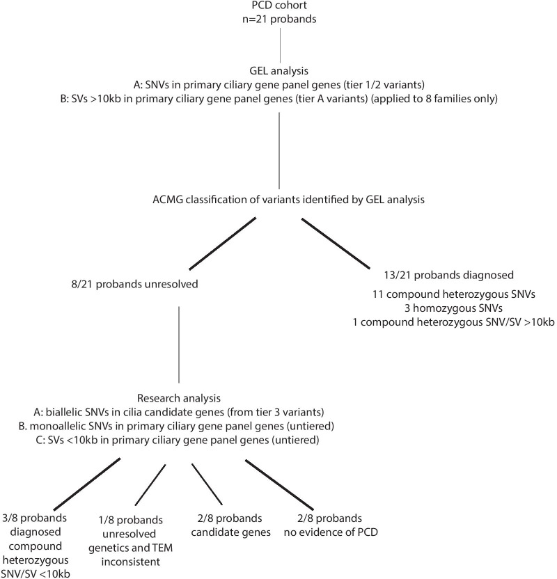
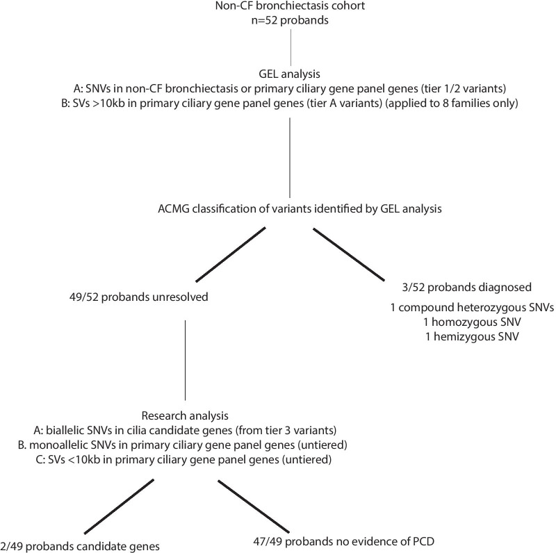
Methods: UK patients with no identified genetic confirmation for the cause of their PCD or bronchiectasis were eligible for whole genome sequencing (WGS) in the Genomics England Ltd 100,000 Genomes Project. 21 PCD probands and 52 non-cystic fibrosis (CF) bronchiectasis probands were recruited in Wessex Genome Medicine Centre (GMC). We carried out analysis of single nucleotide variants (SNVs) and SVs in all families recruited in Wessex GMC.

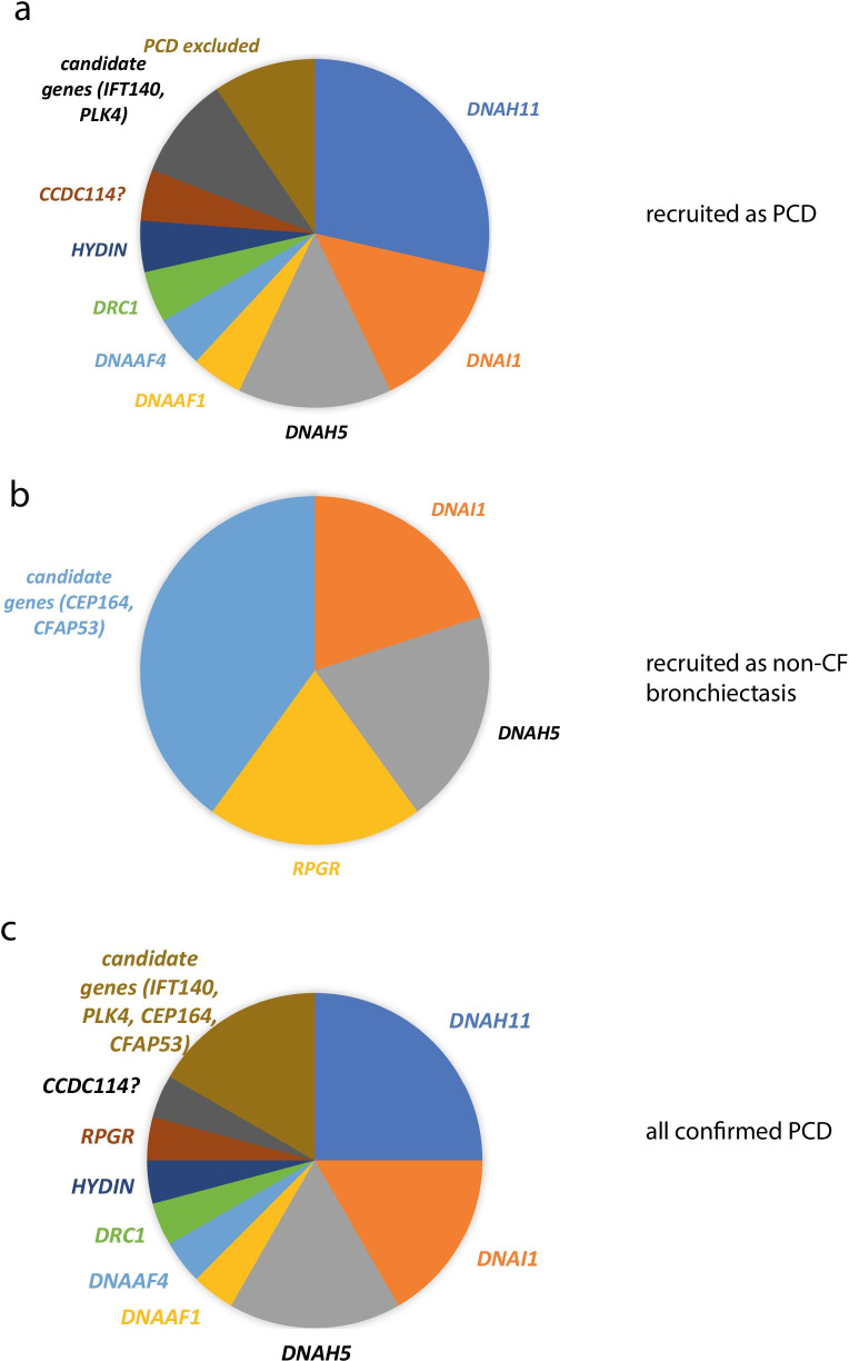
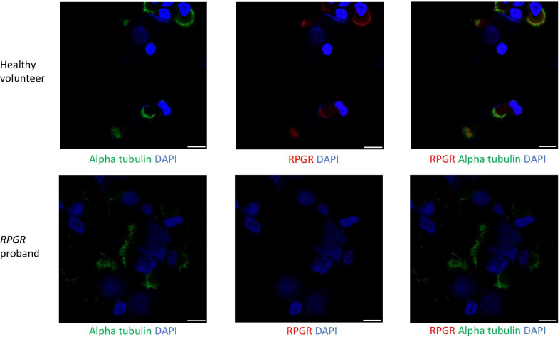
Results: 16/21 probands in the PCD cohort received confirmed (n = 9), probable (n = 4) or possible (n = 3) diagnosis from WGS, although 13/16 of these could have been picked up by current standard of care gene panel testing. In the other cases, SVs were identified which were missed by panel testing. We identified variants in novel PCD candidate genes (IFT140 and PLK4) in 2 probands in the PCD cohort. 3/52 probands in the non-CF bronchiectasis cohort received a confirmed (n = 2) or possible (n = 1) diagnosis of PCD. We identified variants in novel PCD candidate genes (CFAP53 and CEP164) in 2 further probands in the non-CF bronchiectasis cohort.

Conclusions: Genetic testing is an important component of diagnosing PCD, especially in cases of atypical disease history. WGS is effective in cases where prior gene panel testing has found no variants or only heterozygous variants. In these cases it may detect SVs and is a powerful tool for novel gene discovery.

Keywords: Diagnosis; Gene discovery; Non-CF bronchiectasis; Primary ciliary dyskinesia; Whole genome sequencing.


Conflict of interest statement

JSL reports grants, personal fees and non-financial support from Circassia. GW is employed by Illumina Inc. Other authors declare no competing interests.

Figures

Fig. 1
Fig. 1
Genomics England Ltd (Genomics England Ltd) variant filtering and tiering strategy for single nucleotide variants (SNVs) or small insertions or deletions. Tier 1 and tier 2 variants were returned to recruiting Genome Medicine Centres (GMC) for variant review and classification following American College of Medical Genetics guidelines. Tier 3 variants were available for review in the research environment. Primary ciliary disorders PanelApp gene panel can be found in Table 1, non-CF bronchiectasis PanelApp gene panel can be found in Table 2
Fig. 2
Fig. 2
Structural variants in known PCD genes identified in unsolved patients in the Wessex PCD cohort. IGV screenshot showing heterozygous ~ 3.5 kb SV in DNAAF4 deleting exon 7 in case 8 in Table 3. This was found in trans with a stop gain variant in DNAAF4, providing a confirmed diagnosis for this patient. a IGV screenshot showing heterozygous ~ 9.5 kb SV in DNAH11 deleting exon 20 and 21 in case 9 in Table 3. This results in an in-frame deletion of amino acids 1256–1337 containing a coiled-coil domain which may take part in protein–protein interactions. Multiple alignment of DNAH11 protein sequence across multiple species shows that this region of the protein is well conserved and likely to be functionally important, so that the deletion would be deleterious to protein function. The ACMG classification was likely pathogenic. This was found in trans with a pathogenic stop gain variant in DNAH11, providing a confirmed diagnosis for this patient. b IGV screenshot showing heterozygous ~ 9.8 kb SV in HYDIN deleting exon 18 in case 13, affected sibling and mother but not father. This results in an in-frame deletion of amino acids 793–843 of the 5121 amino acid protein. Multiple alignment of HYDIN protein sequence across multiple species shows that this region of the protein is conserved and likely to be functionally important, but the ACMG classification was variant of uncertain significance because we couldn’t show that the deleted interval was critical to protein function. This was found in trans with a stop gain variant in HYDIN (also found in affected sibling and father), providing a probable diagnosis for this patient and their affected sibling. c Immunofluorescence confocal image of SPEF2 (red), ciliary axoneme marker alpha tubulin (green) and DAPI, overlaid with phase contrast image of single multiciliated cells from the nasal epithelium of health volunteer (left) and PCD case 13, who has a 9.8 k deletion in HYDIN in trans with a stop gain variant in HYDIN. HYDIN is required for trafficking of SPEF2 into the cilia, and so absence of SPEF2 in the cilia of this patient is supportive of the genetic findings in this patient
Fig. 3
Fig. 3
Flowchart showing clinical and research analyses carried out and all results for Wessex PCD cohort
Fig. 4
Fig. 4
Causative genes in PCD cases recruited as PCD or recruited as non-CF bronchiectasis. a Pie chart chowing proportion of patients recruited into PCD disease category with causative variants in different known and candidate PCD genes. b Pie chart chowing proportion of patients recruited into non-CF disease category with causative variants in different known and candidate PCD genes. c Pie chart chowing proportion of patients recruited into PCD and non-CF disease categories with causative variants in different known and candidate PCD genes
Fig. 5
Fig. 5
Flowchart showing clinical and research analyses carried out and all results for Wessex non-CF bronchiectasis cohort
Fig. 6
Fig. 6
Immunofluorescence confocal images of multiciliated nasal epithelial cells from non-CF bronchiectasis patient with mutation in RPGR. Immunflourescence confocal images of RPGR (red), ciliary axoneme marker alpha tubulin (green) and DAPI in multiciliated cells from the nasal epithelium of health volunteer (left) and non-CF bronchiectasis case C, who has a predicted damaging hemizygous p.H201R missense mutation in RPGR. Absence of RPGR in the cilia of this patient is supportive of the genetic findings in this patient. Scale bar = 10 µm

References

    1. O’Callaghan C, Chetcuti P, Moya E. High prevalence of primary ciliary dyskinesia in a British Asian population. Arch Dis Child. 2010;95(1):51. doi: 10.1136/adc.2009.158493. - DOI - PubMed
    1. Kuehni CE, Frischer T, Strippoli MP, Maurer E, Bush A, Nielsen KG, et al. Factors influencing age at diagnosis of primary ciliary dyskinesia in European children. Eur Respir J. 2010;36(6):1248–1258. doi: 10.1183/09031936.00001010. - DOI - PubMed
    1. Behan L, Dunn Galvin A, Rubbo B, Masefield S, Copeland F, Manion M, et al. Diagnosing primary ciliary dyskinesia: an international patient perspective. Eur Respir J. 2016;48(4):1096–1107. doi: 10.1183/13993003.02018-2015. - DOI - PMC - PubMed
    1. Lucas JS, Barbato A, Collins SA, Goutaki M, Behan L, Caudri D, et al. European Respiratory Society guidelines for the diagnosis of primary ciliary dyskinesia. Eur Respir J. 2017;49(1). - PMC - PubMed
    1. Shapiro AJ, Davis SD, Polineni D, Manion M, Rosenfeld M, Dell SD, et al. Diagnosis of primary ciliary dyskinesia an official american thoracic society clinical practice guideline. Am J Respir Crit Care Med. 2018;197(12):e24–e39. doi: 10.1164/rccm.201805-0819ST. - DOI - PMC - PubMed

LinkOut - more resources