Review
. 2022 Feb;23(2):107-124.
doi: 10.1038/s41580-021-00413-2. Epub 2021 Sep 23.

The mechanisms of integral membrane protein biogenesis

Affiliations
Review

The mechanisms of integral membrane protein biogenesis

Ramanujan S Hegde et al. Nat Rev Mol Cell Biol. 2022 Feb.

Abstract

Roughly one quarter of all genes code for integral membrane proteins that are inserted into the plasma membrane of prokaryotes or the endoplasmic reticulum membrane of eukaryotes. Multiple pathways are used for the targeting and insertion of membrane proteins on the basis of their topological and biophysical characteristics. Multipass membrane proteins span the membrane multiple times and face the additional challenges of intramembrane folding. In many cases, integral membrane proteins require assembly with other proteins to form multi-subunit membrane protein complexes. Recent biochemical and structural analyses have provided considerable clarity regarding the molecular basis of membrane protein targeting and insertion, with tantalizing new insights into the poorly understood processes of multipass membrane protein biogenesis and multi-subunit protein complex assembly.


Figures

Fig. 1. |
Fig. 1. |. Overview of integral membrane protein biogenesis.
a | The four major steps involved in membrane protein biogenesis. In this and subsequent figures, tapered extensions that flank TMDs indicate soluble regions of indeterminate length. When the flanking segment is necessarily short, such as the translocated segment of a tail-anchored membrane protein, a non-tapered line is used. An ellipsis is used to indicate further polypeptide that contains additional TMDs. b | The major classes of integral membrane proteins are indicated, with prominent examples from human cells listed below.
Fig. 2.
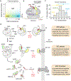
Fig. 2.. Membrane protein targeting to the endoplasmic reticulum.
| a | Targeting-sequence position and hydrophobicity influences the route taken to the ER membrane. b | Diagram showing the position of SRP on a translating ribosome. c | Histogram of all 235 predicted human TA proteins plotted by the hydrophobicity of their TMDs calculated using the Kyte-Doolittle (K-D) scale. The model EMC- and GET-dependent proteins SQS and VAMP2, respectively, are within the bins indicated. SEC61B can use either pathway, a property that seems to be shared by most TA proteins. d | Model illustrating the sequence of events that determine which targeting pathway is used by which type of substrate. Dedicated cytosolic targeting factors are yellow, general TMD-binding chaperones are green, and the initial interaction partner at the ER membrane in dark grey.
Fig. 3.
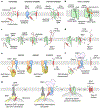
Fig. 3.. Membrane protein insertion at the endoplasmic reticulum.
| a | Comparison of unassisted, insertase-mediated, and channel-mediated TMD insertion. b | Schematic of the open Sec61 complex containing an aqueous channel for protein translocation and a lateral gate for membrane insertion of hydrophobic segments. c-g | Key steps during insertion of a Type I membrane protein by Sec61 (c), Type II membrane protein by Sec61 (d), TA protein by the GET complex (e), TA protein by EMC (f) and Type III membrane protein by EMC (g).
Fig. 4.
Fig. 4.. Structure and function of the Oxa1 superfamily insertases.
| a | Some examples of Oxa1 superfamily members (blue) found in the prokaryotic plasma membrane (YidC) and eukaryotic ER (GET1 and EMC3). Closeup views of the conserved 3-TMD core of each family member are shown below. The cartoon at right illustrates a cross-section at the plane of the vestibule where the membrane might be distorted or thinned. YidC is a single polypeptide, whereas GET1 forms an obligate complex with GET2 (orange), both of which participate in recruiting the targeting factor GET3 (gray). Only one of two GET1-GET2 complexes observed in the structure is shown; the other is behind the displayed complex facing the opposite direction. EMC3 is part of a 9-protein complex that includes EMC6 (orange), which is structurally and evolutionarily related to GET2. Other EMC subunits are shown in gray. Note that the membrane domains of EMC4, EMC7, and EMC10 probably reside in front of EMC3 in this view but were not resolved in this structure. PDB IDs: YidC, 3WO6; human GET complex, 6SO5; human EMC, 6WW7. b | Model for substrates favoured and dis-favoured for insertion by Oxa1 superfamily insertases such as YidC, the GET1-GET2 complex, and EMC. TA proteins with positive charges in the translocated tail are inserted into mitochondria. Signal anchors with a positively charged or lengthy N-terminal flanking domain are inserted by the Sec61 complex.
Fig. 5.
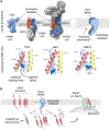
Fig. 5.. Biogenesis of multi-pass membrane proteins.
| a | The linear and folded states of the β1-adrenergic receptor (ADRB1, PDB ID 2VT4) illustrates how most of the hydrophilic side chains (blue, red and yellow) in the TMD become buried upon folding. b | Violin plots showing the hydrophobicity of TMDs in single-pass and multi-pass proteins. Hydrophobicity was calculated as the predicted energy of membrane insertion, where a negative value favours insertion and positive value disfavors insertion. c | The PAT complex engages and protects nascent TMDs with hydrophilic residues until they are buried in the protein interior upon folding. Substrates that are not shielded are potential targets for quality control (QC). Note that PAT complex shielding could occur regardless of the route of TMD insertion. d | Cryo-EM structure of the ribosome-bound multi-pass translocon (PDB ID 6W6L). The view from the ER lumen also indicates the potential positions of C20orf24 (adjacent to its likely interaction partner TMCO1), and Asterix (adjacent to its interaction partner CCDC47). e | Model for protein biogenesis by the multi-pass translocon. In the intermediate depicted, the first TMD is being held by the PAT complex until TMD2 and TMD3 are inserted as a unit using the hydrophilic groove of the TMCO1 complex. The lipid cavity may be the site where multiple substrate TMDs can fold protected from aggregation and inappropriate interactions. f | Hypothetical assembly factors temporarily shield hydrophilic regions of TMDs in the unassembled state. This may serve to stabilise substrates in the membrane until their assembly with interaction partners.

References

    1. von Heijne G The membrane protein universe: what’s out there and why bother? J Intern Med 261, 543–557, doi:10.1111/j.1365-2796.2007.01792.x (2007). - DOI - PubMed
    1. Xie K & Dalbey RE Inserting proteins into the bacterial cytoplasmic membrane using the Sec and YidC translocases. Nat Rev Microbiol 6, 234–244, doi:10.1038/nrmicro3595 (2008). - DOI - PubMed
    1. Shao S & Hegde RS Membrane protein insertion at the endoplasmic reticulum. Annu Rev Cell Dev Biol 27, 25–56, doi:10.1146/annurev-cellbio-092910-154125 (2011). - DOI - PMC - PubMed
    1. Baum DA & Baum B An inside-out origin for the eukaryotic cell. BMC Biol 12, 76, doi:10.1186/s12915-014-0076-2 (2014). - DOI - PMC - PubMed
    1. Wiedemann N & Pfanner N Mitochondrial Machineries for Protein Import and Assembly. Annu Rev Biochem 86, 685–714, doi:10.1146/annurev-biochem-060815-014352 (2017). - DOI - PubMed

Publication types

Substances

LinkOut - more resources