Review
. 2021 Apr;31(2):409-420.
doi: 10.1055/s-0041-1734225. Epub 2021 Aug 4.

Pulmonary Arterial Dilatation: Imaging Evaluation Using Multidetector Computed Tomography

Affiliations
Review

Pulmonary Arterial Dilatation: Imaging Evaluation Using Multidetector Computed Tomography

Sreenivasa Narayana Raju et al. Indian J Radiol Imaging. 2021 Apr.

Abstract

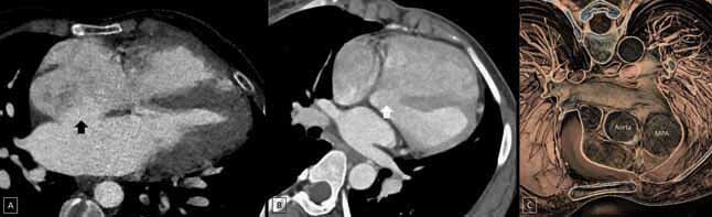
Pulmonary artery dilatation comprises a heterogeneous group of disorders. Early diagnosis is important as the presentation may be incidental, chronic, or acute and life threatening depending upon the etiology. Cross-sectional imaging plays an important role, with CT pulmonary angiography being regarded as the first line investigation in the evaluation of pulmonary artery pathologies. Moreover, effects of pulmonary artery lesions on proximal and distal circulation can also be ascertained with the detection of associated conditions. Special attention should also be given to the left main coronary artery and the trachea-bronchial tree as they may be extrinsically compressed by the dilated pulmonary artery. In context of an appropriate clinical background, CT pulmonary angiography also helps in treatment planning, prognostication, and follow-up of these patients. This review mainly deals with imaging evaluation of the pulmonary arterial dilatations on CT with emphasis on the gamut of etiologies in the adult as well as pediatric populations.

Keywords: cardiac shunt; pulmonary arterial hypertension; pulmonary artery dilatation; pulmonary thromboembolism; vasculitis.


Conflict of interest statement

Conflict of Interest None declared. Authors’ Contributions All authors contributed to the conception or design, literature search, drafting, critical revision, and final approval of the manuscript.

Figures

Fig. 1
Fig. 1
Flowchart showing the algorithmic approach to the etiological diagnosis of pulmonary artery dilatation on MDCT angiography in the pediatric population. ASD, atrial septal defect; MDCT, multidetector computed tomography; PDA, patent ductus arteriosus; VSD, ventricular septal defect.
Fig. 2
Fig. 2
Flowchart showing the algorithmic approach to the etiological diagnosis of pulmonary artery dilatation on MDCT angiography in the adult population. ASD, atrial septal defect; MDCT, multidetector computed tomography; PA, pulmonary artery; PDA, patent ductus arteriosus; PE, pulmonary embolism; VSD, ventricular septal defect.
Fig. 3
Fig. 3
Technique of measurement of main pulmonary artery (MPA). MPA measured 35.3 mm while the ascending aorta (AA) measured 20 mm.
Fig. 4
Fig. 4
( A ) Acute embolus ( white arrow ) in the left descending pulmonary artery forming acute angle with the wall of the artery. ( B ) Acute thrombus in the right pulmonary artery (RPA) and left pulmonary artery (LPA) placed eccentrically along the arterial wall. ( C ) Hypodense web ( black arrowhead ) in the right descending pulmonary artery. MPA, main pulmonary artery.
Fig. 5
Fig. 5
( A ) Axial section at the level of pulmonary valve and the aortic sinus showing thickening of the pulmonary valve leaflets ( black arrowhead ). ( B ) Axial section at the main pulmonary artery (MPA) bifurcation showing disproportionate dilatation of the MPA and left pulmonary artery (LPA). RPA, right pulmonary artery.
Fig. 6
Fig. 6
Multiplanar reconstruction (MPR) images ( A, C and D ) and volume-rendered image ( B ) show constriction ( black arrow ) at the level of the annulus of the main pulmonary artery (MPA) with dilatation of the MPA, left pulmonary artery (LPA), and right pulmonary artery (RPA) with hypoplastic pulmonary valve leaflets.
Fig. 7
Fig. 7
Four chamber view of the heart ( A ) showing atrial septal defect ( black arrow ). Oblique axial image ( B ) and virtual dissection image ( C ) of a different patient shows a ventricular septal defect (w hite arrow ) and dilated main pulmonary artery (MPA), respectively.
Fig. 8
Fig. 8
Multiplanar reconstruction image ( A ) and volume-rendered image ( B ) showing patent ductus arteriosus ( black arrow ) with dilated main pulmonary artery (MPA).
Fig. 9
Fig. 9
Multiplanar reconstruction image ( A ) and volume-rendered image ( B ) showing type I truncus arteriosus with common truncus channel ( black arrow ). Multiplanar reconstruction image ( C ) and volume rendered image ( D ) show Type B interrupted aortic arch with patent ductus arteriosus ( white arrow ) reforming the descending thoracic aorta (DTA). AA, ascending aorta; MPA, main pulmonary artery.
Fig. 10
Fig. 10
Volume-rendered images ( A and B ) showing pseudoaneurysms of the left descending pulmonary artery ( black arrow ) and the right descending pulmonary artery ( white arrow ), respectively.
Fig. 11
Fig. 11
( A ) Sagittal oblique images show dilated aortic sinus (*) and ascending aorta. ( B ) Oblique axial section showing dilated main pulmonary artery (MPA) and dilated ascending aorta.
Fig. 12
Fig. 12
Hypodense dissection flap ( black arrow ) in the right lower descending pulmonary artery with dilated right pulmonary artery (RPA).

References

    1. Escalon J G, Browne L P, Bang T J, Restrepo C S, Ocazionez D, Vargas D. Congenital anomalies of the pulmonary arteries: an imaging overview. Br J Radiol. 2019;92(10/93):2018–185. - PMC - PubMed
    1. Carter B W, Lichtenberger J P, III, Wu C C. Congenital abnormalities of the pulmonary arteries in adults. AJR Am J Roentgenol. 2014;202(04):W308–13. - PubMed
    1. McLoud T C, Boiselle P M. Thoracic radiology: imaging methods, radiographic signs, and diagnosis of chest disease. In: McLoud TC, Boiselle PM, eds. Thoracic Radiology: The Requisites. 2nd ed. Philadelphia, PA: Elsevier. 2010:8.
    1. Frazier A A, Galvin J R, Franks T J, Rosado-De-Christenson M L. From the archives of the AFIP: pulmonary vasculature: hypertension and infarction. Radiographics. 2000;20(02):491–524. - PubMed
    1. Wittram C. How I do it: CT pulmonary angiography. AJR Am J Roentgenol. 2007;188(05):1255–1261. - PubMed