. 2021 Sep 7:12:738172.
doi: 10.3389/fpls.2021.738172. eCollection 2021.

Dynamic Development of White Lupin Rootlets Along a Cluster Root

Affiliations

Dynamic Development of White Lupin Rootlets Along a Cluster Root

Tamara Le Thanh et al. Front Plant Sci. .

Abstract

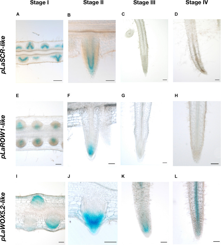
White lupin produces cluster roots in response to phosphorus deficiency. Along the cluster root, numerous short rootlets successively appear, creating a spatial and temporal gradient of developmental stages that constitutes a powerful biological model to study the dynamics of the structural and functional evolution of these organs. The present study proposes a fine histochemical, transcriptomic and functional analysis of the rootlet development from its emergence to its final length. Between these two stages, the tissue structures of the rootlets were observed, the course of transcript expressions for the genes differentially expressed was monitored and some physiological events linked to Pi nutrition were followed. A switch between (i) a growing phase, in which a normal apical meristem is present and (ii) a specialized phase for nutrition, in which the rootlet is completely differentiated, was highlighted. In the final stage of its determinate growth, the rootlet is an organ with a very active metabolism, especially for the solubilization and absorption of several nutrients. This work discusses how the transition between a growing to a determinate state in response to nutritional stresses is found in other species and underlines the fundamental dilemma of roots between soil exploration and soil exploitation.

Keywords: cluster root; determinate growth; mineral nutrition; rootlet; white lupin.


Conflict of interest statement

The authors declare that the research was conducted in the absence of any commercial or financial relationships that could be construed as a potential conflict of interest.

Figures

FIGURE 1
FIGURE 1
White lupin root system architecture. (A) Root system of a 16-days-old white lupin hydroponically grown in Pi deficiency conditions resulting in the development of several cluster roots each bearing a single cluster of rootlets (*). (B) A typical 7 cm-long cluster root displaying one cluster of rootlets. (C) Measurement of rootlet length: 5 rootlets were measured in 0.5 cm sections from the tip of the root to the end of the cluster (n = 21). Scale bars: (A–B) 1 cm.
FIGURE 2
FIGURE 2
Spatio-temporal evolution of rootlet development. (A) Division of a cluster into 5 stages of rootlet development. Calcofluor was used to stain 100-μm thin longitudinal sections of rootlets at (B) Stage I: pre-emergence. (C) Stage II: emergence. (D) Stage III: growing. (E) Stage IV: rootlets having reached their maximal length. (F) Stage V: fully differentiated rootlets. Red arrows indicate the closest root hair from the tip. Scale bars: (A) 1 cm, (B–F) 100 μm.
FIGURE 3
FIGURE 3
Anatomical study of the cluster root tip, and rootlet tips at different stages of development. DAPI was used to stain 8-μm thin longitudinal sections of (A) cluster root tip, and rootlet tips at (B) stage I, (C) stage II, (D) stage III, (E) stage IV, (F) stage V. Scale bars: (A–F) 50 μm.
FIGURE 4
FIGURE 4
Evolution of cortical cells in the rootlet tips from stage III to V. Calcofluor was used to stain 100-μm thin longitudinal sections of rootlets. Typical images of rootlet tips at (A) stage III, (B) stage IV, and (C) stage V, are shown. Cell lengths were measured with Cell-o-Tape along 300 μm following a cortical cell line starting from the organization center (D) Cell lengths of a cortical line from stage III to V (n = 11). (E) Schematic representation of the evolution of rootlet tip before and after the developmental shift. Dz, differentiation zone; Ez, elongation zone; Pm, proximal meristem; Scn, stem cell niche; Dm, distal meristem; Cl, Columella. Scale bars: (A–C) 50 μm.
FIGURE 5
FIGURE 5
Tissular expression patterns of three meristematic markers during rootlet development. The promoter activities of three genes typically expressed in the meristematic zone were studied with a GUS-reporter-based analysis on 80-μm longitudinal sections of transgenic hairy-root tips. Images from stages I to IV are shown for each promoter (A–D) pLaSCR-like:GUS, (E–H) pLaROW1-like:GUS, (I–L) pLaWOX5.2-like:GUS. Scale bars: (A–L) 100 μm.
FIGURE 6
FIGURE 6
Clustering analysis of the normalized expression profiles of the differentially expressed transcripts between S1 and S7 sections of the RNA-seq experiment. Normalized gene expression profiles come from our previous RNA-seq study (Hufnagel et al., 2020). The clustering was performed with the Coseq package in DIANE (Cassan et al., 2021). The numbers of genes clustered in each gene expression profile are the following: Profile 1: 1,792; Profile 2: 2,129; Profile 3: 411; Profile 4: 482; Profile 5: 584; Profile 6: 1,836. Below each profile, the cluster root sections from S1 to S7 of the RNA-seq analysis are presented.
FIGURE 7
FIGURE 7
Physiological activities along rootlet development. (A) 33P influx in 0.5 cm cluster root sections, root tip and link segment. Values are means ± SD of three biological replicates coming each from 6 cluster roots sampled across 6 white lupin plants (n = 3). (B) Citrate and (C) malate exudations quantified in 0.5 cm cluster root sections, root tip and link segment. Values are means ± SD of five biological replicates coming each from 6 cluster roots sampled across 6 white lupin plants (n = 5).

References

    1. Abdolzadeh A., Wang X., Veneklaas E. J., Lambers H. (2010). Effects of phosphorus supply on growth, phosphate concentration and cluster-root formation in three Lupinus species. Ann. Bot. 105 365–374. 10.1093/aob/mcp297 - DOI - PMC - PubMed
    1. Al-Ghazi Y., Muller B., Pinloche S., Tranbarger T. J., Nacry P., Rossignol M., et al. (2003). Temporal responses of Arabidopsis root architecture to phosphate starvation: evidence for the involvement of auxin signalling. Plant Cell. Environ. 26 1053–1066. 10.1046/j.1365-3040.2003.01030.x - DOI
    1. Balzergue C., Dartevelle T., Godon C., Laugier E., Meisrimler C., Teulon J.-M., et al. (2017). Low phosphate activates STOP1-ALMT1 to rapidly inhibit root cell elongation. Nat. Commun. 8:15300. - PMC - PubMed
    1. Bouain N., Doumas P., Rouached H. (2016). Recent advances in understanding the molecular mechanisms regulating the root system response to phosphate deficiency in Arabidopsis. Curr. Genom. 17:308. 10.2174/1389202917666160331201812 - DOI - PMC - PubMed
    1. Cassan O., Lèbre S., Martin A. (2021). Inferring and analyzing gene regulatory networks from multi-factorial expression data: a complete and interactive suite. BMC Genom. 22:387. 10.1186/s12864-021-07659-2 - DOI - PMC - PubMed