A Novel Method for Measuring the Pupil Diameter and Pupillary Light Reflex of Healthy Volunteers and Patients With Intracranial Lesions Using a Newly Developed Pupilometer
- PMID: 34557496
- PMCID: PMC8452878
- DOI: 10.3389/fmed.2021.598791
A Novel Method for Measuring the Pupil Diameter and Pupillary Light Reflex of Healthy Volunteers and Patients With Intracranial Lesions Using a Newly Developed Pupilometer
Abstract
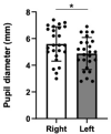
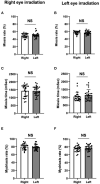
Background: Physicians currently measure the pupil diameter and the pupillary light reflex with visual observations using a ruler and a traditional penlight, leading to possibly inaccurate and subjective assessments. Although a mobile pupillometer has been developed and is available in clinical settings, this device can only assess one pupil at a time. Hence, an indirect pupillary light reflex, including those under irradiation to the opposite side of pupil, cannot be evaluated. Consequently, we have developed a new automatic mobile pupilometer, the Hitomiru®, with Hitomiru Co., Ltd. (Tokyo, Japan). This device is a two-glass type pupilometer with a video recording system. The pupil diameter and light reflex of both pupils can be measured simultaneously; therefore, both indirect and direct light reflexes can be assessed. Purpose: To evaluate the clinical ability of the Hitomiru® pupilometer to assess the pupil diameter and the pupillary light reflex of healthy volunteers and patients with intracranial lesions in an intensive care unit (ICU). Methods: Twenty-five healthy volunteers and five ICU patients with intracranial lesions on only the left side were assessed using the Hitomiru® pupilometer. The protocol was as follows: infrared light was applied to both pupils, followed by visible light to the right pupil, infrared light to both pupils, visible light to the left pupil, and then infrared light to both pupils. All the intervals were 2 s, and the dynamics of pupil diameters on both sides were continuously recorded. Results: The healthy adults had approximately 0.5 mm anisocoria, miosis was harder, and mydriasis was less with increased age. There were several differences in miosis rates, miosis times, and mydriasis rates between the healthy adults and the patients with intracranial lesions with both direct irradiation and indirect irradiation. Conclusions: The initial trial estimated and digitally recorded direct and indirect light reflexes, including rapidity of miosis after direct and indirect lights on, and mydriasis after direct and indirect lights off. The Hitomiru® pupilometer was a useful device to digitally record and investigate the relationship between pupil reflexes and intracranial diseases.
Keywords: digital record; direct and indirect irradiation; intracranial lesions; miosis; mobile type pupilometer; mydriasis; pupil diameter; pupil light reflexes.
Copyright © 2021 Kotani, Nakao, Yamada, Miyawaki, Mambo and Ono.
Conflict of interest statement
The authors declare that the research was conducted in the absence of any commercial or financial relationships that could be construed as a potential conflict of interest.
Figures


References
-
- Wilson S. Ectopia pupilliae in certain mesencephalic lesions. Brain. (1907) 29:524–36. 10.1093/brain/29.4.524 - DOI
LinkOut - more resources
Full Text Sources

