Review
. 2021 Sep 7:8:742596.
doi: 10.3389/fmolb.2021.742596. eCollection 2021.

Electromagnetic Field as a Treatment for Cerebral Ischemic Stroke

Affiliations
Review

Electromagnetic Field as a Treatment for Cerebral Ischemic Stroke

Amanda Moya Gómez et al. Front Mol Biosci. .

Abstract

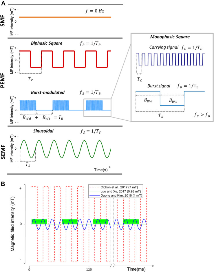
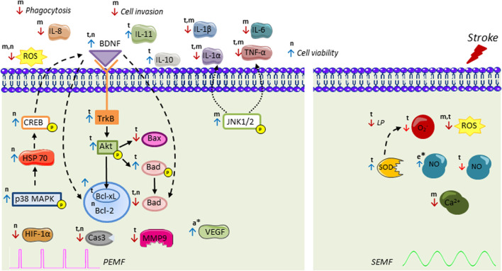
Cerebral stroke is a leading cause of death and adult-acquired disability worldwide. To this date, treatment options are limited; hence, the search for new therapeutic approaches continues. Electromagnetic fields (EMFs) affect a wide variety of biological processes and accumulating evidence shows their potential as a treatment for ischemic stroke. Based on their characteristics, they can be divided into stationary, pulsed, and sinusoidal EMF. The aim of this review is to provide an extensive literature overview ranging from in vitro to even clinical studies within the field of ischemic stroke of all EMF types. A thorough comparison between EMF types and their effects is provided, as well as an overview of the signal pathways activated in cell types relevant for ischemic stroke such as neurons, microglia, astrocytes, and endothelial cells. We also discuss which steps have to be taken to improve their therapeutic efficacy in the frame of the clinical translation of this promising therapy.

Keywords: cerebral ischemia; electromagnetic field; neuroprotection; neurorehabilitation; pulsed electromagnetic field; sinusoidal electromagnetic field; stroke.


Conflict of interest statement

The authors declare that the research was conducted in the absence of any commercial or financial relationships that could be construed as a potential conflict of interest.

Figures

FIGURE 1
FIGURE 1
Representation of different types of EMFs used for stroke treatment. (A) EMF classification used in this article based on waveform and frequency (inverse of the signal period). SMF does not change its magnitude and direction in time (f = 0 Hz). Time-varying EMFs are divided into PEMF and SEMF. PEMFs include non-sinusoidal EMFs. This category includes monophasic square (like the carrying and burst signals), biphasic square, and burst-modulated signals. Burst-modulated signals consist of a carrying signal with a higher frequency modulated by a burst signal with a burst width and burst wait interval at a lower frequency. In this representation, f S > f P > f B. (B) Three of the EMF waveforms used in the consulted literature. In red (Cichoń et al., 2017a), with a 7 mT/40 Hz biphasic square PEMF. In blue (Duong and Kim, 2016), with a 1 mT/50 Hz SEMF. In green (Luo and Xu, 2017), using a burst-modulated PEMF with a burst frequency of 15 Hz (burst width, 5 ms; burst wait, 60 ms) and 4.5 MHz carrying monophasic square signal. SMF: stationary magnetic field; PEMF: pulsed electromagnetic field; SEMF: sinusoidal electromagnetic field; MF: magnetic field; f: frequency; f P: pulse frequency; T P: pulse period; f C: carrying signal frequency; T C: carrying signal period; B Wd: burst width; B Wt: burst wait interval; T B: burst period; f B: burst frequency; f S: sinusoid frequency; T S: sinusoid period.
FIGURE 2
FIGURE 2
Intracellular signal transduction cascades activated by pulsed electromagnetic field (PEMF) and sinusoidal electromagnetic field (SEMF) in ischemic stroke. PEMF increases activation of p38 kinase cascade, followed by HSP70, CREB, BDNF recruitment leading to an increase of antiapoptotic Bcl-2 family and decrease in apoptotic Bad. PEMF also stimulates BDNF/TrkB/Akt pathway, thus increasing phospho-Bad that cannot bind to Bcl-xL and decreasing Bax, Bad, and Cas3. PEMF decreases MMP9, HIF-1α, ROS, and proinflammatory interleukins and upregulates anti-inflammatory IL-11 and IL-10. PEMF-induced downregulation of TNF-α and IL-1β is possibly mediated by JNK1/2. The molecular mechanisms of SEMF have been less studied. PEMF also exerts regulation on endocannabinoid system (AEA, 2-AG, MAGL, DAGL, NAPE-PLD, FAAH) with ERK signaling as an essential downstream mechanism. SEMF decreases superoxide (O2 ), intracellular Ca2+, ROS, NO, and LP level, while it increases SOD and NO levels. Results have been assessed in t, brain tissue; n, neuron-like; m, microglia; a, astrocytes; and e, endothelial cells. * indicates only EMF stimulation without stroke. BDNF: brain-derived neurotrophic factor; CREB: cAMP response element-binding protein; HSP70: heat-shock proteins of 70; Akt: protein kinase B; TrkB: tropomyosin-related kinase B; ROS: reactive oxygen species; HIF-1: hypoxia-induced factor 1; Cas3: Caspase-3; MMP9: matrix metalloproteinase 9; IL: interleukin; TNF-α: tumor necrosis factor α; JNK1/2: c-Jun-N terminal kinase; SOD: superoxide dismutase; NO: nitric oxide; LP: lipid peroxidation; VEGF: vascular endothelial grow factor; AEA: N-arachidonylethanolamide; 2-AG: 2-arachidonoylglycerol; DAGL: diacylglycerol lipase; MAGL: monoacylglycerol lipase; NAPE-PLD: N-acyl phosphatidylethanolamine phospholipase; FAAH: fatty acid amide hydrolase. Figure was created using Servier Medical Art (smart.servier.com).

References

    1. Akdag M. Z., Bilgin M. H., Dasdag S., Tumer C. (2007). Alteration of Nitric Oxide Production in Rats Exposed to a Prolonged, Extremely Low-Frequency Magnetic Field. Electromagn. Biol. Med. 26, 99–106. 10.1080/15368370701357866 - DOI - PubMed
    1. Albers G. W., Marks M. P., Kemp S., Christensen S., Tsai J. P., Ortega-Gutierrez S., et al. (2018). Thrombectomy for Stroke at 6 to 16 hours with Selection by Perfusion Imaging. N. Engl. J. Med. 378, 708–718. 10.1056/NEJMoa1713973 - DOI - PMC - PubMed
    1. Amki M. E., Wegener S. (2017). Improving Cerebral Blood Flow after Arterial Recanalization: a Novel Therapeutic Strategy in Stroke. Int. J. Mol. Sci. 18, 26–69. 10.3390/ijms18122669 - DOI - PMC - PubMed
    1. Balkaya M., Kröber J. M., Rex A., Endres M. (2013). Assessing post-stroke Behavior in Mouse Models of Focal Ischemia. J. Cereb. Blood Flow Metab. 33, 330–338. 10.1038/jcbfm.2012.185 - DOI - PMC - PubMed
    1. Bates K. A., Clark V. W., Meloni B. P., Dunlop S. A., Rodger J. (2012). Short-term Low Intensity PMF Does Not Improve Functional or Histological Outcomes in a Rat Model of Transient Focal Cerebral Ischemia. Brain Res. 1458, 76–85. 10.1016/j.brainres.2012.04.006 - DOI - PubMed

LinkOut - more resources