Case Reports
. 2022 Jan;52(1):144-147.
doi: 10.1007/s00247-021-05199-1. Epub 2021 Sep 24.

Electrical impedance tomography clues to detect pulmonary thrombosis in a teenager with COVID-19

Affiliations
Case Reports

Electrical impedance tomography clues to detect pulmonary thrombosis in a teenager with COVID-19

Flávia Andrea Krepel Foronda et al. Pediatr Radiol. 2022 Jan.

Abstract

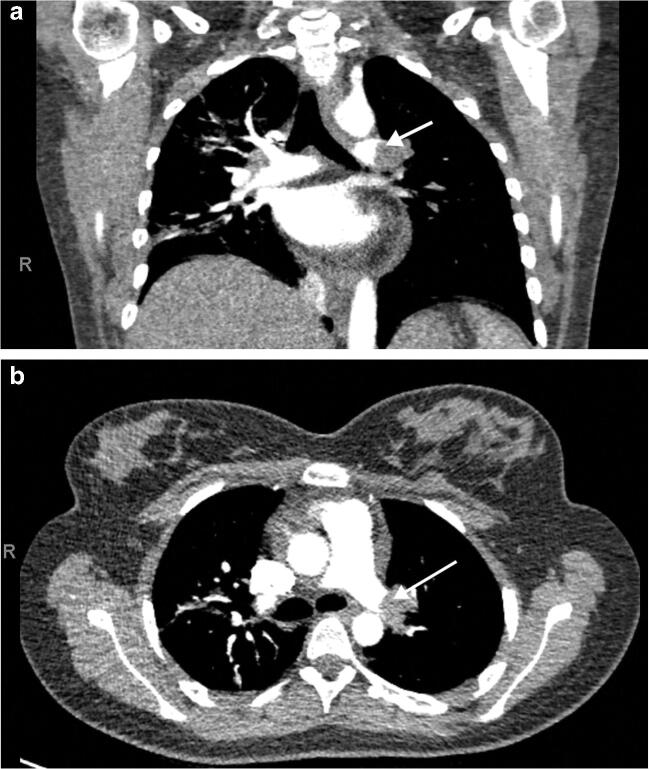
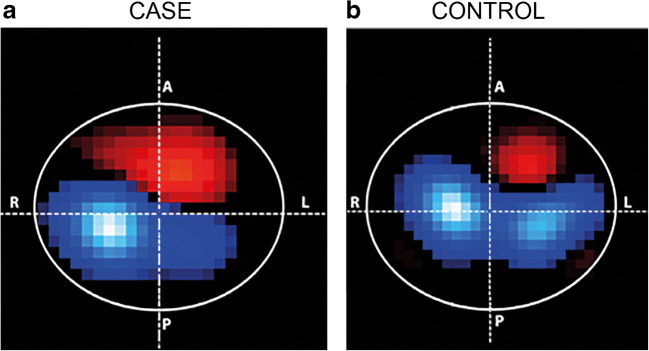
We report a case of pulmonary thrombosis in a teenager during a hypercoagulable state associated with COVID-19 (coronavirus disease 2019) caused by SARS-CoV-2 (severe acute respiratory syndrome coronavirus 2). A condition rare in children and adolescents, pulmonary thrombosis underdiagnosis likely increases morbidity and mortality. A pulmonary thrombosis diagnosis requires a high level of suspicion and relies on the combination of clinical presentation, D-dimer elevation, and computed tomography (CT) pulmonary angiography or ventilation/perfusion scans, imaging techniques that are difficult to perform. Electrical impedance tomography (EIT) has gained attention, as it provides real-time ventilation distribution analysis. In addition, lung pulsatility images can be obtained through this technique using electrocardiogram gating to filter out ventilation. In this case report, the reduced EIT pulsatility corresponded to the perfusion defect found on the CT scan, information that was obtained at the bedside without radiation or contrast exposure.

Keywords: Children; Coronavirus disease 2019; Electrical impedance tomography; Lung; Pulmonary thrombosis.


Conflict of interest statement

None

Figures

Fig. 1
Fig. 1
Computed tomography (CT) pulmonary angiography in a 15-year-old girl. a, b Coronal (a) and axial (b) images show a central filling defect in the left pulmonary artery, consistent with an occlusive thrombus (CT dose index: 7.46 mGy). The arrows indicate the thrombus
Fig. 2
Fig. 2
Axial electrical impedance tomography (EIT) pulsatility maps. The pulsatility images are electrocardiogram gated and filter out the ventilation component in the thoracic impedance variation, thus representing impedance variations related to the cardiac cycle. a, b The off-line image shows the heart in red and the pulsatility in the lungs in blue. Of note, the higher detected pulsatility in this EIT algorithm is displayed in white and light blue colors. An image from the 15-year-old girl (a) shows reduced pulsatility in the left lung, secondary to the pulmonary thrombosis. The dashed lines intersect at the center of the thorax. For comparison, we present an image obtained from another 15-year-old girl without ventilation or perfusion abnormalities (b). Note that the pulsatility in the right image is more evenly distributed between the right and left lungs. A anterior, L left, P posterior, R right
Fig. 3
Fig. 3
Axial electrical impedance tomography (EIT) imaging performed in four different positions: sitting, right lateral decubitus (RLD), supine and left lateral decubitus (LLD). The upper figures display EIT lung ventilation maps in scales of blue (the higher the tidal volume detected, the lighter the colors). Noticeably, ventilation distribution varies as the patient changes position. In the lower images, EIT electrocardiogram-gated pulsatility maps were not impacted by the four different studied positions. The pulsatility, also presented on a scale of blue to white, is always diminished in the left lung area. The heart position is in red

References

    1. Zaidi AU, Hutchins KK, Rajpurkar M. Pulmonary embolism in children. Front Pediatr. 2017;5:170. doi: 10.3389/fped.2017.00170. - DOI - PMC - PubMed
    1. Connors JM, Levy JH. Thromboinflammation and the hypercoagulability of COVID-19. J Thromb Haemost. 2020;18:1559–1561. doi: 10.1111/jth.14849. - DOI - PMC - PubMed
    1. Mitchell WB. Thromboinflammation in COVID-19 acute lung injury. Paediatr Respir Rev. 2020;35:20–24. - PMC - PubMed
    1. Kearon C, de Wit K, Parpia S, et al. Diagnosis of pulmonary embolism with D-dimer adjusted to clinical probability. N Engl J Med. 2019;381:2125–2134. doi: 10.1056/NEJMoa1909159. - DOI - PubMed
    1. Davies P, Yasin S, Gates S, et al. Clinical scenarios of the application of electrical impedance tomography in paediatric intensive care. Sci Rep. 2019;9:5362. doi: 10.1038/s41598-019-41774-1. - DOI - PMC - PubMed

Publication types