. 2021 Sep;129(9):95001.
doi: 10.1289/EHP9321. Epub 2021 Sep 24.

Key Characteristics of Cardiovascular Toxicants

Affiliations

Key Characteristics of Cardiovascular Toxicants

Lars Lind et al. Environ Health Perspect. 2021 Sep.

Abstract

Background: The concept of chemical agents having properties that confer potential hazard called key characteristics (KCs) was first developed to identify carcinogenic hazards. Identification of KCs of cardiovascular (CV) toxicants could facilitate the systematic assessment of CV hazards and understanding of assay and data gaps associated with current approaches.

Objectives: We sought to develop a consensus-based synthesis of scientific evidence on the KCs of chemical and nonchemical agents known to cause CV toxicity along with methods to measure them.

Methods: An expert working group was convened to discuss mechanisms associated with CV toxicity.

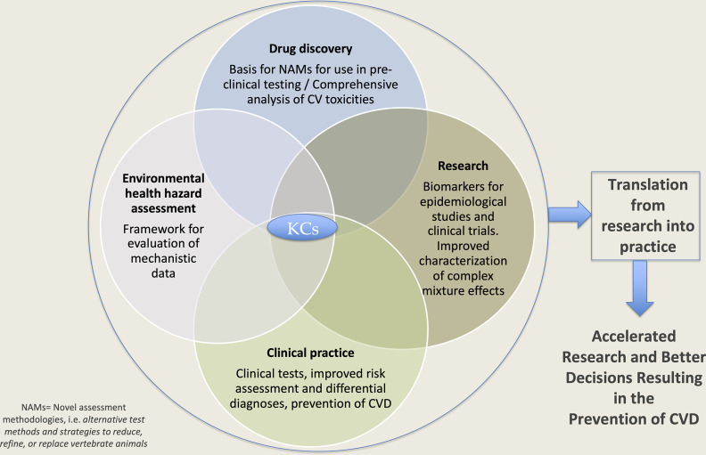
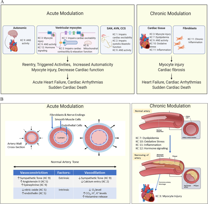
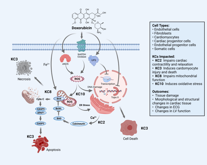
Results: The group identified 12 KCs of CV toxicants, defined as exogenous agents that adversely interfere with function of the CV system. The KCs were organized into those primarily affecting cardiac tissue (numbers 1-4 below), the vascular system (5-7), or both (8-12), as follows: 1) impairs regulation of cardiac excitability, 2) impairs cardiac contractility and relaxation, 3) induces cardiomyocyte injury and death, 4) induces proliferation of valve stroma, 5) impacts endothelial and vascular function, 6) alters hemostasis, 7) causes dyslipidemia, 8) impairs mitochondrial function, 9) modifies autonomic nervous system activity, 10) induces oxidative stress, 11) causes inflammation, and 12) alters hormone signaling.

Discussion: These 12 KCs can be used to help identify pharmaceuticals and environmental pollutants as CV toxicants, as well as to better understand the mechanistic underpinnings of their toxicity. For example, evidence exists that fine particulate matter [PM 2.5μm in aerodynamic diameter (PM2.5)] air pollution, arsenic, anthracycline drugs, and other exogenous chemicals possess one or more of the described KCs. In conclusion, the KCs could be used to identify potential CV toxicants and to define a set of test methods to evaluate CV toxicity in a more comprehensive and standardized manner than current approaches. https://doi.org/10.1289/EHP9321.


Figures

Figure 1 is a flow chart titled Key Characteristics comprising a Venn diagram. The Venn diagram has four circles, namely, Drug discovery; Basis for novel assessment methodologies for use in pre-clinical testing per comprehensive analysis of Cardiovascular toxicities; Research; Biomarkers for epidemiological studied and clinical trials. Improved characterization of complex mixture effects; Clinical practice; Clinical tests, improved risk assessment and differential diagnosis, prevention of cardiovascular disease; and Environmental health hazard assessment, Framework for evaluation of mechanistic data, interconnected to each other. The center of the Venn diagram is labeled key characteristics. The Venn diagram then leads to translation from research into practice. The translation from research into practice leads to Accelerated research and better decisions resulting in the prevention of cardiovascular disease.
Figure 1.
Utility of the key characteristics (KCs) of cardiovascular toxicants in research, drug discovery, hazard assessment, and clinical practice. An illustration of how the KCs could be used in different areas and how translation of the resulting information could lead to accelerated research, inform better regulatory decisions, improve clinical practice, and ultimately prevent CVD. Note: CV, cardiovascular; CVD, cardiovascular disease; NAM, novel assessment methodologies.
Figure 2A is a scientific illustration having two parts, namely, Acute modulation and Chronic modulation. In Acute modulation, there are two steps. Step 1: Autonomic, including key characteristic 9: Autonomic nervous system activity with an icon of a brain and heart; Ventricular myocytes, including key characteristic 3: Myocyte injury, key characteristic 9: Autonomic nervous system activity, key characteristic 12: Hormone signaling, key characteristic 1: impairs cardiac excitability, key characteristic 2: impairs cardiac contractility and relaxation, key characteristic 8: mitochondrial function; and sinoatrial node, avascular necrosis, and cardiac conduction system, including key characteristic 1: impairs cardiac excitability, key characteristic 2: impairs systolic and diastolic function, and key characteristic 9: Autonomic nervous system activity leads to Reentry, triggered activities, increased automaticity and myocyte injury and decreased cardiac function. Step 2: Reentry, triggered activities, increased automaticity and myocyte injury and decreased cardiac function leads to Acute heart failure, cardiac arrhythmias, and sudden cardiac death. In Chronic Modulation, there are two steps. Step 1: Cardiac tissue, including key characteristic 3: myocyte injury, key characteristic 7: dyslipidemia, key characteristic 9: Autonomic nervous system activity, key characteristic 10: Oxidative stress, key characteristic 11: inflammation with an icon of a heart and fibroblasts, including key characteristic 11: chronic inflammation with an image of fibroblasts leads to Myocyte injury and cardiac fibrosis. Step 2: Myocyte injury and cardiac fibrosis leads to Heart Failure, Cardiac Arrhythmias, and Sudden cardiac death. Figure 2B is a scientific illustration having two parts, namely, Acute modulation and Chronic modulation. In Acute Modulation, there are three scientific images of a cross-sectioned artery. The first image depicts the artery wall in cross section with arrows indicating constriction. The second image depicts the different layers of the artery. The outermost layer of the artery is known as fibroblasts and nerve endings, the center layer of the artery is known as smooth muscle cells, the innermost layer of the artery is known as endothelial cells, and the core of the artery is the lumen. The third image is an expanded image of the artery with arrows indicating dilation. Below, a tabular representation titled Normal Artery Tone has two rows and three columns, namely, Vasoconstriction, Factors, and Vasodilation. In Chronic Modulation, there are two steps. Step 1: Normal artery, including two scientific illustrations of an artery. The first image depicts the internal section of an artery labeled normal blood flow and the outer layer is labeled artery wall. The second image depicts the artery labeled artery cross-section and the outer layer of the artery is labeled artery wall. The normal artery includes key characteristic 7: Dyslipidemia, key characteristic 10: Oxidative Stress, key characteristic 11: Inflammation, and key characteristic 12: hormone signaling. Step 2: Narrowing of artery, including two scientific illustrations of an artery. The first image depicts the innermost layer and is labeled abnormal blood flow and the layer under the outer layer is labeled plaque. Due to Coronary artery blockage, key characteristic 3: Myocyte injury occurs with an icon of a heart. The second image of the artery depicts the inner part of the artery which is divided into two parts, namely, narrowed artery and plaque.
Figure 2.
Key characteristics (KCs) associated with cardiac and vascular dysfunction. A summary of how different KCs of cardiovascular toxicant could affect (A) the heart and (B) the vasculature in both the acute and chronic setting. Some of the detailed mechanisms are given, as well as some clinical end points. Note: ANS, autonomic nervous system; AVN, avascular necrosis; CCS, cardiac conduction system; CO2, carbon dioxide; H+, hydrogen ion; K+, potassium ion; O2, oxygen; SAN, sinoatrial node.
Figure 3 is a scientific illustration that has five steps. Step 1: Inhalation, fine particulate matter pointing toward the illustration of a human head and lungs. The lungs depict the main bronchi, bronchi, bronchioles, alveolar sacs, and alveolar capillaries. The response in the lungs depicts Hydrogen peroxide, Hydroxide, Peroxynitrite, reactive oxygen species, neutrophils, and macrophages, which induces oxidative stress and causes inflammation which leads to blood vessels. Step 2: Blood Vessels. The response in systemic circulation depicts Hydrogen peroxide, Hydroxide, Peroxynitrite, reactive oxygen species, interleukin-1 lowercase beta with an arrow pointing up, interleukin-6 with an arrow pointing up, tumor necrosis factor lowercase alpha with an arrow pointing up, and monocytes, neutrophils, C-reactive protein with an arrow pointing up, key characteristic 10 causes inflammation. Step 3: Inhalation leads to the Central nervous system with an icon of a brain with key characteristic 9 modifies autonomic nervous system activity to heart and vasculature, secondary to sensory nerve activation in the lung. Step 4: Blood Vessels and Central nervous system lead to key characteristic 12 alters hormone signaling, including hypothalamic pituitary adrenal axis, hypothalamic pituitary thyroid axis, renin-angiotensin system, and insulin and glucose homeostasis, vasodilation, where key characteristic 5 impacts endothelial and vascular function, impaired artery, normal artery, causes dyslipidemia, low-density lipoprotein with an arrow pointing up, high-density lipoprotein with an arrow pointing up, triglycerides with an arrow pointing up, and key characteristic 6 alters hemostasis, platelets with an arrow pointing up, von Willebrand factor with an arrow pointing up, and fibrinogen with an arrow pointing up. Step 5: Step 4 leads to modifies autonomic nervous system activity with an image of a heart, with modified autonomic nervous system activity to heart and vasculature. The following information is given: Clinical outcomes are ischemic heart disease, stroke, and heart failure.
Figure 3.
Key characteristics (KCs) associated with PM2.5 toxicity. A summary of how different KCs of fine particulate air pollution (PM2.5) could affect the heart and the vasculature. Some of the detailed mechanisms are given, as well as some clinical end points. Note: H2O2, hydrogen peroxide; OH, hydroxide; O2•−, reactive oxygen species; ONOO, peroxynitrite; PM2.5, particulate matter 2.5μm in aerodynamic diameter (fine particulate matter).
Figure 4 is a scientific illustration flowchart depicting key characteristics associated with doxorubicin cardiotoxicity having ten steps. Step 1: Doxorubicin with an icon of the chemical structure of doxorubicin leads to Nicotinamide adenine dinucleotide phosphate oxidase, Endothelial nitric oxide synthase 3, and Upright scutellars. Step 2: Nicotinamide adenine dinucleotide phosphate oxidase and Endothelial nitric oxide synthase 3 lead to Reactive oxygen species. Step 3: Upright scutellars leads to transcription, D N A damage, and Topoisomerase II. Step 4: Doxorubicin with Iron ion and impairs mitochondrial function leads to Reactive oxygen species, including Bcl-2-associated X (apoptosis regulator), Bcl2-like 1, and Bcl-2-associated agonist of cell death. Step 4: Reactive oxygen species with induces oxidative stress and Endoplasmic Reticulum Stress leads to transcription, D N A damage, and Topoisomerase 2. Step 5: Impairs mitochondrial function leads to Necrosis which induces cardiomyocyte injury and death. Step 6: Reactive oxygen species, including Bcl-2-associated X (apoptosis regulator), Bcl2-like 1, and Bcl-2-associated agonist of cell death leads to cytochrome complex, including Caspase 9, Apoptotic protease activating factor 1, Caspase 3. Step 7: Cytochrome complex, including Caspase 9, Apoptotic protease activating factor 1, Caspase 3 leads to Apoptosis which induces cardiomyocyte injury and death. Step 8: Transcription, D N A damage, and Topoisomerase 2 with calcium ions and impairs cardiac contractility and relaxation lead to calcineurin. Step 9: Calcineurin leads to Bcl-2-associated X (apoptosis regulator), Bcl2-like 1, and Bcl-2-associated agonist of cell death. Step 10: Transcription, D N A damage, and Topoisomerase 2 lead to induces cardiomyocyte injury and death which causes cell death with an icon of a cell. The following information is given: Cell types include endothelial cells, fibroblasts, cardiomyocytes, cardiac progenitor cells, endothelial progenitor cells, and somatic cells. The outcomes include tissue damage, morphological and structural changes in cardiac tissue, changes in electrocardiogram, and changes in left ventricular function.
Figure 4.
Key characteristics (KCs) associated with doxorubicin cardiotoxicity. A summary of how different KCs of doxorubicin could affect the heart and the vasculature. Some detailed mechanisms are given, as well as some clinical outcomes. Note: APAF1, apoptotic protease activating factor 1; Bad, Bcl-2-associated agonist of cell death; Bax, Bcl-associated X; BclXL, B-cell lymphoma-extra large; Ca2+ calcium ion; CASP3, caspase 3; CASP9, caspase 9; CytoC, cytochrome complex; ECG, electrocardiogram; eNOS, endothelial nitric oxide synthase; ER, estrogen receptor; Fe2+, iron ion; LV, left ventricular; NADPH, nicotinamide adenine dinucleotide phosphate; ROS, reactive oxygen species; Topo II, topoisomerase II; UPS, ubiquitin-proteasome system.

Comment in

References

    1. Abernethy DR, Schwartz JB. 1999. Calcium-antagonist drugs. N Engl J Med 341(19):1447–1457, PMID: 10547409, 10.1056/NEJM199911043411907. - DOI - PubMed
    1. Abrams CS.2006. Acquired qualitative platelet disorders. In: Williams Hematology. 7th ed. Lichtman MA, Beutler E, Kipps TJ, Seligsohn U, Kaushansky K, Prchal JT, eds. New York, NY: McGraw-Hill, 1833–1855.
    1. Abumrad NA, Davidson NO. 2012. Role of the gut in lipid homeostasis. Physiol Rev 92(3):1061–1085, PMID: 22811425, 10.1152/physrev.00019.2011. - DOI - PMC - PubMed
    1. Adelborg K, Larsen JB, Hvas A-M. 2021. Disseminated intravascular coagulation: epidemiology, biomarkers, and management. Br J Haematol 192(5):803–818, PMID: 33555051, 10.1111/bjh.17172. - DOI - PubMed
    1. Akahane M, Matsumoto S, Kanagawa Y, Mitoma C, Uchi H, Yoshimura T, et al. . 2018. Long-term health effects of PCBs and related compounds: a comparative analysis of patients suffering from Yusho and the general population. Arch Environ Contam Toxicol 74(2):203–217, PMID: 29256109, 10.1007/s00244-017-0486-6. - DOI - PubMed

Publication types

MeSH terms