Review
. 2022 Mar 2;61(3):913-925.
doi: 10.1093/rheumatology/keab721.

Synovial single-cell heterogeneity, zonation and interactions: a patchwork of effectors in arthritis

Affiliations
Review

Synovial single-cell heterogeneity, zonation and interactions: a patchwork of effectors in arthritis

Barbora Schonfeldova et al. Rheumatology (Oxford). .

Abstract

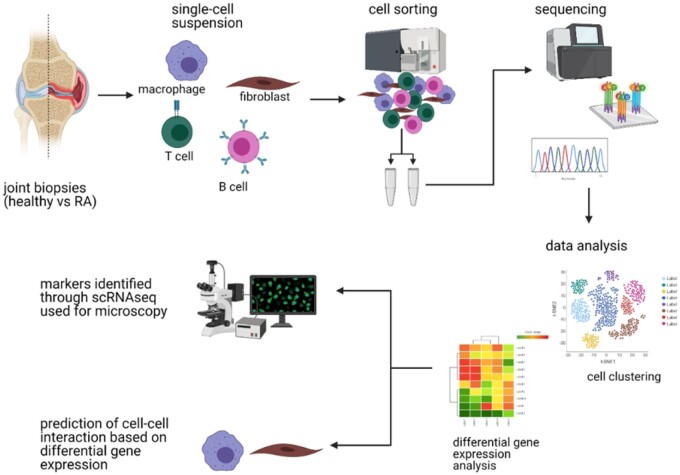
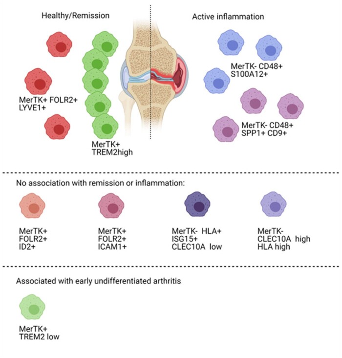
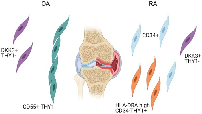
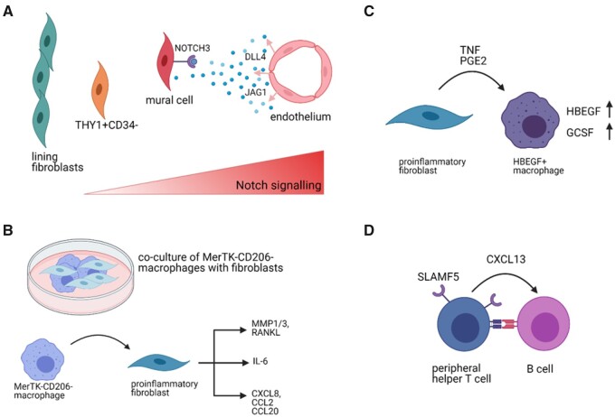
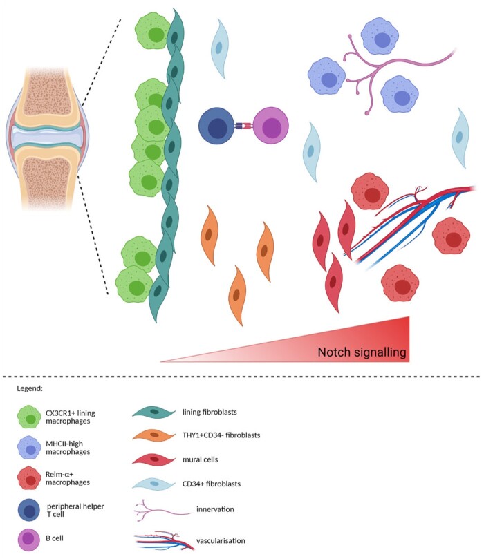
Despite extensive research, there is still no treatment that would lead to remission in all patients with rheumatoid arthritis as our understanding of the affected site, the synovium, is still incomplete. Recently, single-cell technologies helped to decipher the cellular heterogeneity of the synovium; however, certain synovial cell populations, such as endothelial cells or peripheral neurons, remain to be profiled on a single-cell level. Furthermore, associations between certain cellular states and inflammation were found; whether these cells cause the inflammation remains to be answered. Similarly, cellular zonation and interactions between individual effectors in the synovium are yet to be fully determined. A deeper understanding of cell signalling and interactions in the synovium is crucial for a better design of therapeutics with the goal of complete remission in all patients.

Keywords: RA; cellular localization; experimental arthritis; single-cell transcriptomics; synovium.


Figures

<sc>Fig</sc>. 1
Fig. 1
Simplified workflow of a sequencing experiment The sample acquisition (e.g. human or murine joint biopsies) is followed by the processing of the sample into a single-cell suspension. Population of interest is then sorted by FACS and prepared for sequencing (e.g. barcoding the mRNA to identify cell source, reverse transcription, preparation of cDNA library). Raw sequencing data need to be pre-processed and analysed. The findings from single-cell RNA sequencing experiments should be experimentally validated. Figure created with BioRender.com.
<sc>Fig</sc>. 2
Fig. 2
Macrophage populations identified in human synovium (based on [17]) Macrophage populations characterised as MerTKposFOLR2pos+LYVE1pos or MerTKposTREM2high were associated with remission, while MerTKnegCD48posS100A12pos or MerTKnegCD48posSPP1posCD9pos macrophages were preferentially found in actively inflamed synovium. Other populations had no clear association with either remission or inflammation. Interestingly, an increased number of MerTKposTREM2low macrophages was found in early undifferentiated arthritis. Figure created with BioRender.com.
<sc>Fig</sc>. 3
Fig. 3
Fibroblast populations identified in human synovium (based on [19]) CD55+THY1- lining fibroblast were found to be associated with OA while CD34+ and HLA-DRAhiCD34-THY1+ sublining fibroblasts were preferentially found in RA. Population of sublining fibroblasts defined as DKK3+THY1- was not clearly associated with either. Figure created with BioRender.com.
<sc>Fig</sc>. 4
Fig. 4
Examples of interactions identified in human or murine synovium (A) Arterial endothelial cells were found to release NOTCH3 ligands DLL4 and JAG1 that bind NOTCH3 receptor on mural cells and sublining fibroblasts. The Notch signalling gradient shapes the identity of fibroblasts where fibroblasts closer to the endothelium will develop into THY1+CD34- fibroblasts and cells further from it will become THY1- lining fibroblasts. (Based on [45]) (B) Co-culture of proinflammatory MerTKnegCD206neg macrophages with fibroblasts caused the fibroblasts to adopt a proinflammatory phenotype and secrete bone and cartilage destructing mediators (MMP1/3, RANKL) and inflammatory cytokines (IL6) or chemokines (CXCL8, CCL2, CCL20) [17]. (C) Proinflammatory fibroblasts were found to secrete TNF or PGE2, which induce expression of HBEGF and GCSF in macrophages, inducing pro-inflammatory phenotype [20]. (D) Peripheral helper T cells and B cells communicate in the synovium through multiple pathways, for example via secretion of CXCL13 by T cells [19, 37, 81] or via upregulation of SLAMF5 [37]. Figure created with BioRender.com.
<sc>Fig</sc>. 5
Fig. 5
Prediction of the zonation of immune and structural cells in the synovium CX3CR1+ lining macrophages and lining fibroblast create the ‘barrier’ layer of the synovium [18, 19]. Based on our analysis, MHCIIhigh macrophages could be positioned near the innervation, while RELM-α+ macrophages could be located near the vasculature, similarly to the lung interstitium [72]. Notch signalling axis is instructing the positioning of fibroblasts with mural cells closest to the vasculature, THY1+CD34- fibroblasts near the vasculature and mural cells with THY1-CD34- lining fibroblast at the opposing end [43, 45]. CD34+ fibroblasts are positioned both in the immediate sublining and deep interstitium [43]. Figure created with BioRender.com.

References

    1. Weyand CM, Goronzy JJ.. The immunology of rheumatoid arthritis. Nat Immunol 2021;22:10–8. - PMC - PubMed
    1. Smolen JS, Aletaha D, Barton A. et al. Rheumatoid arthritis. Nat Rev Dis Primers 2018;4:18001. - PubMed
    1. Schett G, Tanaka Y, Isaacs JD.. Why remission is not enough: underlying disease mechanisms in RA that prevent cure. Nat Rev Rheumatol 2021;17:135–44. - PubMed
    1. Orr C, Vieira-Sousa E, Boyle DL. et al. Synovial tissue research: a state-of-the-art review. Nat Rev Rheumatol 2017;13:463–75. - PubMed
    1. Tang X, Huang Y, Lei J, Luo H, Zhu X.. The single-cell sequencing: new developments and medical applications. Cell Biosci 2019;9:53. - PMC - PubMed