

Radiomics for detecting prostate cancer bone metastases invisible in CT: a proof-of-concept study
- PMID: 34559264
- PMCID: PMC8831270
- DOI: 10.1007/s00330-021-08245-6
Radiomics for detecting prostate cancer bone metastases invisible in CT: a proof-of-concept study
Abstract
Objectives: To investigate, in patients with metastatic prostate cancer, whether radiomics of computed tomography (CT) image data enables the differentiation of bone metastases not visible on CT from unaffected bone using 68 Ga-PSMA PET imaging as reference standard.
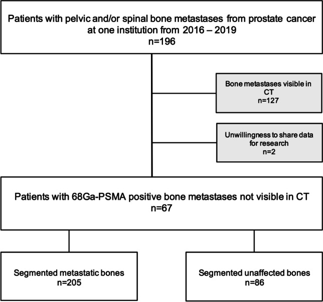
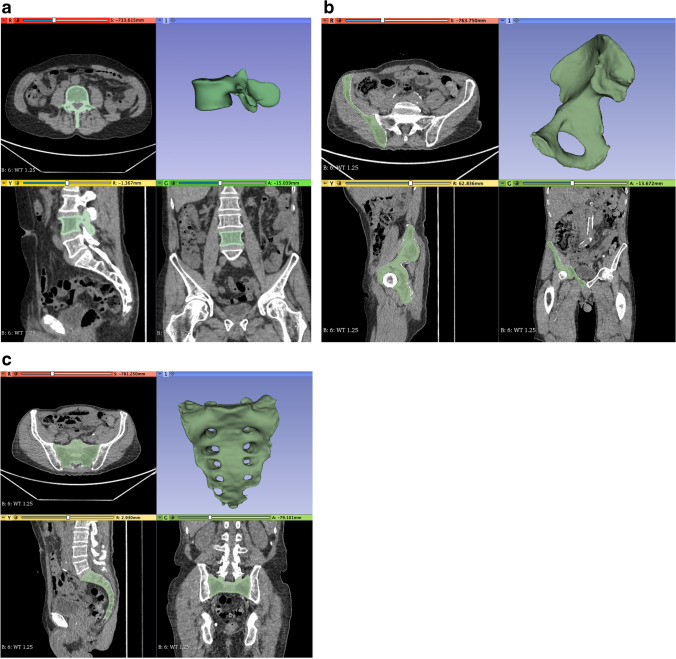
Methods: In this IRB-approved retrospective study, 67 patients (mean age 71 ± 7 years; range: 55-84 years) showing a total of 205 68 Ga-PSMA-positive prostate cancer bone metastases in the thoraco-lumbar spine and pelvic bone being invisible in CT were included. Metastases and 86 68 Ga-PSMA-negative bone volumes in the same body region were segmented and further post-processed. Intra- and inter-reader reproducibility was assessed, with ICCs < 0.90 being considered non-reproducible. To account for imbalances in the dataset, data augmentation was performed to achieve improved class balance and to avoid model overfitting. The dataset was split into training, test, and validation set. After a multi-step dimension reduction process and feature selection process, the 11 most important and independent features were selected for statistical analyses.
Results: A gradient-boosted tree was trained on the selected 11 radiomic features in order to classify patients' bones into bone metastasis and normal bone using the training dataset. This trained model achieved a classification accuracy of 0.85 (95% confidence interval [CI]: 0.76-0.92, p < .001) with 78% sensitivity and 93% specificity. The tuned model was applied on the original, non-augmented dataset resulting in a classification accuracy of 0.90 (95% CI: 0.82-0.98) with 91% sensitivity and 88% specificity.
Conclusion: Our proof-of-concept study indicates that radiomics may accurately differentiate unaffected bone from metastatic bone, being invisible by the human eye on CT.
Key points: • This proof-of-concept study showed that radiomics applied on CT images may accurately differentiate between bone metastases and metastatic-free bone in patients with prostate cancer. • Future promising applications include automatic bone segmentation, followed by a radiomics classifier, allowing for a screening-like approach in the detection of bone metastases.
Keywords: Bone metastases; Computed tomography; Prostate cancer; Radiomics; Texture analysis.
© 2021. Crown.
Conflict of interest statement
The authors of this manuscript declare no relationships with any companies whose products or services may be related to the subject matter of the article.
Figures


References
MeSH terms
Substances
LinkOut - more resources
Full Text Sources
Medical
Miscellaneous
