. 2022 Mar;49(4):1136-1147.
doi: 10.1007/s00259-021-05559-x. Epub 2021 Sep 24.

PET/CT imaging of head-and-neck and pancreatic cancer in humans by targeting the "Cancer Integrin" αvβ6 with Ga-68-Trivehexin

Affiliations

PET/CT imaging of head-and-neck and pancreatic cancer in humans by targeting the "Cancer Integrin" αvβ6 with Ga-68-Trivehexin

Neil Gerard Quigley et al. Eur J Nucl Med Mol Imaging. 2022 Mar.

Abstract

Purpose: To develop a new probe for the αvβ6-integrin and assess its potential for PET imaging of carcinomas.

Methods: Ga-68-Trivehexin was synthesized by trimerization of the optimized αvβ6-integrin selective cyclic nonapeptide Tyr2 (sequence: c[YRGDLAYp(NMe)K]) on the TRAP chelator core, followed by automated labeling with Ga-68. The tracer was characterized by ELISA for activities towards integrin subtypes αvβ6, αvβ8, αvβ3, and α5β1, as well as by cell binding assays on H2009 (αvβ6-positive) and MDA-MB-231 (αvβ6-negative) cells. SCID-mice bearing subcutaneous xenografts of the same cell lines were used for dynamic (90 min) and static (75 min p.i.) µPET imaging, as well as for biodistribution (90 min p.i.). Structure-activity-relationships were established by comparison with the predecessor compound Ga-68-TRAP(AvB6)3. Ga-68-Trivehexin was tested for in-human PET/CT imaging of HNSCC, parotideal adenocarcinoma, and metastatic PDAC.

Results: Ga-68-Trivehexin showed a high αvβ6-integrin affinity (IC50 = 0.047 nM), selectivity over other subtypes (IC50-based factors: αvβ8, 131; αvβ3, 57; α5β1, 468), blockable uptake in H2009 cells, and negligible uptake in MDA-MB-231 cells. Biodistribution and preclinical PET imaging confirmed a high target-specific uptake in tumor and a low non-specific uptake in other organs and tissues except the excretory organs (kidneys and urinary bladder). Preclinical PET corresponded well to in-human results, showing high and persistent uptake in metastatic PDAC and HNSCC (SUVmax = 10-13) as well as in kidneys/urine. Ga-68-Trivehexin enabled PET/CT imaging of small PDAC metastases and showed high uptake in HNSCC but not in tumor-associated inflammation.

Conclusions: Ga-68-Trivehexin is a valuable probe for imaging of αvβ6-integrin expression in human cancers.

Keywords: Carcinoma; Gallium-68; Integrins; Positron emission tomography.


Conflict of interest statement

N.G.Q., K.S., and J.N. are co-inventors of patents related to 68 Ga-Trivehexin. J.N. is shareholder of TRIMT GmbH (Radeberg, Germany), which is active in the field of radiopharmaceutical develeopment.

Figures
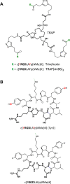
Fig. 1
Fig. 1
Chemical structures of peptides and conjugates. A Structure of the trimers TRAP(AvB6)3 and Trivehexin, which were synthesized by “click-chemistry” (i.e., copper(I) catalyzed azide-alkyne cycloaddition, CuAAC) conjugation of the TRAP chelator core (highlighted in boldface). B Structures of the peptides used, with their RGD sequences highlighted in boldface. On an atomic level, the structural differences merely consists of two additional oxygens of Tyr2 (sequence: c[YRGDLAYp(NMe)K]) (colored in red)
Fig. 2
Fig. 2
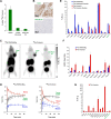
Preclinical development of 68 Ga-Trivehexin. Data were generated using H2009 (αvβ6+ human lung adenocarcinoma) or MDA-MB-231 (αvβ6 human triple-negative breast cancer) cell lines and murine xenografts thereof. A Cellular uptake of 68 Ga-Trivehexin with and without blocking with natGa-Trivehexin. Mean and SD of n = 4–6 biological repeats. B β6-integrin immunohistochemistry (IHC) confirms moderate αvβ6-integrin expression in H2009 and absence of αvβ6 in MDA-MB-231 tumor xenografts. C Static µPET images (maximum intensity projections), 75 min p.i., recording time 20 min. The same H2009-tumor bearing animal was used for both PET scans shown, with a 24-h recovery period. D Biokinetics of 68 Ga-labeled trimers, derived from dynamic PET scans (averages ± SD for 3 independent scans). E Biodistribution of trimers in H2009-bearing animals. Dark bars indicate controls (injected molar amounts of approx. 100 pmol, n = 5); light bars of the same color show blockade (50 nmol unlabeled, administered 10 min before the radioactive compound, n = 3). F Tumor-to-organ ratios derived from H2009 biodistribution data. G Biodistribution of 68 Ga-Trivehexin (105 ± 34 pmol) in MDA-MB-231-bearing animals (n = 5). – Numerical data for graphs shown in E, F, and G, including the exact values of injected amounts, are provided in the Supporting Information, Tables S1–3
Fig. 3:
Fig. 3:
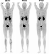
68 Ga-Trivehexin PET of patient #1, 172 MBq, total peptide amount: 5 nmol, start times p.i. denoted above anterior maximum intensity projections (MIPs) scaled to SUV 15. Apart from kidney and urinary bladder, a significant uptake is noted in the (empty) stomach, which however had disappeared 1.5 h after injection. PET at 1 h p.i. or later is thus preferred. For organ SUVs and dosimetry data, see Supporting Information, Tables S4 and S5
Fig. 4
Fig. 4
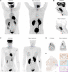
[18F]FDG and corresponding 68 Ga-Trivehexin PET of head-and-neck cancers (locations indicated by blue arrows, slice planes by blue dashed lines). All PET images are scaled to SUV 8, except D (to SUV 5). A PET (MIPs) of HNSCC patient #2 (m, 61 y, 70 kg; 331 MBq [18F]FDG 62 min p.i.; 142 MBq 68 Ga-Trivehexin, AM = 27 MBq/nmol, total peptide amount 5.3 nmol, 61 min p.i.) with locally advanced oropharyngeal carcinoma and several adjacent unilateral lymph node metastases. 68 Ga-Trivehexin showed uptake in the primary and metastases (SUVmax = 13.9 and 12.9, respectively) and in a lesser extent around a 7-week-old tracheostoma (red arrows, SUVmax = 5.8; cf. 10.2 for [18F]FDG). B Sagittal slices corresponding to dashed lines in A. C PET (MIPs) of patient #3 (f, 37 y, 61 kg; 286 MBq [18F]FDG 71 min p.i.; 135 MBq 68 Ga-Trivehexin, AM = 29 MBq/nmol, total peptide amount 4.7 nmol, 67 min p.i.) suffering from a solitary prelaryngeal cutaneous metastasis of a previously resected and irradiated left-sided parotideal adenocarcinoma. D Axial slices corresponding to dashed lines in C. SUVmax of the metastasis was 6.1 for [18F]FDG and 7.6 for 68 Ga-Trivehexin, metabolic tumor volume was 2.2 cm3. E β6-integrin IHC of excised tissue of the patient shown in C + D; magnifications are shown for the central tumor area (orange box), the infiltrative margin (cyan box), and non-tumor tissue (violet box). Bars represent 1 mm for overview and 100 µm for the magnifications, respectively
Fig. 5:
Fig. 5:
68 Ga-Trivehexin PET/CT of metastatic PDAC (confirmed by biopsy) in patient #4 (f, 80 y, 80 kg, 105 MBq, 120 min p.i.). PET is scaled to SUV 10 in all images. A Anterior and sagittal MIP, showing 68 Ga-Trivehexin uptake in the primary tumor (SUVmax = 9.8–10.9) and in 7 metastases. Selected metastases highlighted in panels BG are indicated (SUVmax of #1, #2, and #3 are 10.9, 10.0, and 9.9, respectively). B Coronal and sagittal PET/CT slices through metastasis #1. C Coronal and sagittal PET/CT slices through primaries and metastasis #2. D, E, F, G, axial PET/CT slices though primaries and metastases #1, #2, and #3, respectively

References

    1. Sontag S. Illness as metaphor. Farrar, Straus & Giroux, New York, 1978, ISBN: 978-0-374-17443-9.
    1. Mukherjee S. The emperor of all maladies: a biography of cancer. Scribner, New York, 2010, ISBN: 978-1-4391-0795-9.
    1. Virchow R. Die krankhaften Geschwülste. Berlin: August Hirschwald; 1863.
    1. Brooks PC, Clark RAF, Cheresh DA. Requirement Of Vascular Integrin αvβ3 For Angiogenesis. Science. 1994;264:569–571. - PubMed
    1. Avraamides CJ, Garmy-Susini B, Varner JA. Integrins in angiogenesis and lymphangiogenesis. Nat Rev Cancer. 2008;8:604–617. - PMC - PubMed