. 2021 Sep 24;7(39):eabc7371.
doi: 10.1126/sciadv.abc7371. Epub 2021 Sep 24.

UFMylation of MRE11 is essential for telomere length maintenance and hematopoietic stem cell survival

Affiliations

UFMylation of MRE11 is essential for telomere length maintenance and hematopoietic stem cell survival

Lara Lee et al. Sci Adv. .

Abstract

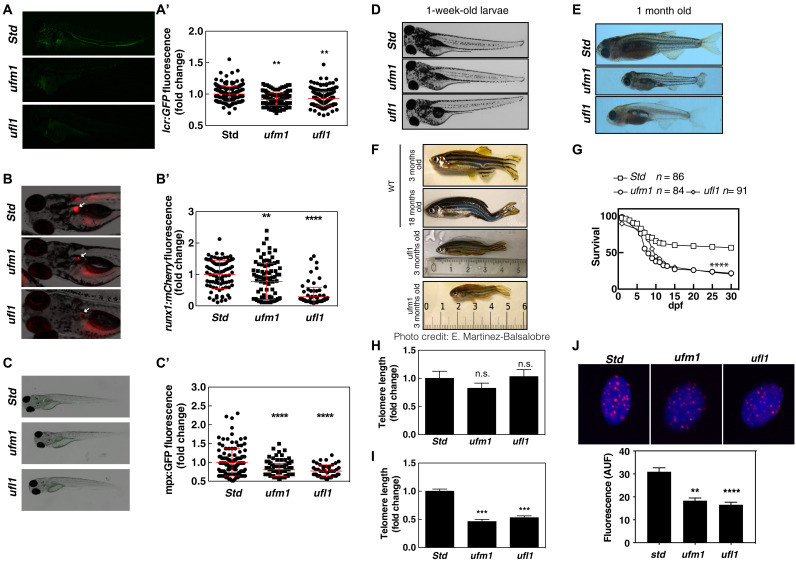
Ubiquitin-fold modifier 1 (UFM1) is involved in neural and erythroid development, yet its biological roles in these processes are unknown. Here, we generated zebrafish models deficient in Ufm1 and Ufl1 that exhibited telomere shortening associated with developmental delay, impaired hematopoiesis and premature aging. We further report that HeLa cells lacking UFL1 have instability of telomeres replicated by leading-strand synthesis. We uncover that MRE11 UFMylation is necessary for the recruitment of the phosphatase PP1-α leading to dephosphorylation of NBS1. In the absence of UFMylation, NBS1 remains phosphorylated, thereby reducing MRN recruitment to telomeres. The absence of MRN at telomeres favors the formation of the TRF2-Apollo/SNM1 complex consistent with the loss of leading telomeres. These results suggest that MRE11-UFMylation may serve as module to recruit PP1-α. Last, zebrafish expressing Mre11 that cannot be UFMylated phenocopy Ufm1-deficient zebrafish, demonstrating that UFMylation of MRE11 is a previously undescribed evolutionarily conserved mechanisms regulating telomere length.


Figures

Fig. 1.
Fig. 1.. UFM1 pathway prevents premature aging and telomere shortening.
Tg(lcr:eGFP) (A), Tg(runx1:GAL4; UAS:nfsb-mCherry) (B), and Tg(mpx:eGFP) (C) one-cell embryos were injected with standard control (Std), ufm1, or ufl1 sgRNA and recombinant Cas9. Representative images of green and red channel of whole larvae for the different treatments (C′) and quantitation of erythroid cells at 4 dpf (A′), HSPCs (B′) and neutrophils cells at 5 dpf are shown. White arrows indicated HSPCs in the kidney marrow of 5-dpf larvae (B). Each dot represents normalized fluorescence from a single larva, while the mean ± SEM for each group is also shown. A representative image of control and deficient ufm1 or ufl1 larval phenotype at 1-week- and 1-month-old fish is shown (D and E). Phenotypic characteristics of 3-month-old fish deficient in Ufm1 and Ufl1 are displayed with an old and young zebrafish. Larval survival curve (Kaplan-Meier representation) of genotype Ufm1- and Ufl1-deficient zebrafish compared with std. (F) Photo credit: Elena Martinez Balsalobre. Survival curve of fish deficient in Ufm1 and Ufl1. (G). Telomere length was determined by quantitative polymerase chain reaction (qPCR) in sorted erythroid cells (GFP) (H) or (GFP+) (I) of 6-dpf larvae. Telomere was determined by quantitative fluorescence in situ hybridization (qFISH) in GFP+ cells (J). The data are shown as the means ± SEM of two independent experiments. **P < 0.01; ***P < 0.001; ****P < 0.0001 according to Student’s t test and analysis of variance followed by Tukey multiple range test. n.s., not significant.
Fig. 2.
Fig. 2.. UFL1 KO cells have defect in telomere length.
(A) Representative image of chromosome metaphase spreads from WT and UFL1−/− cells. Black arrow: telomere fusion. (B) Quantification of the chromosomes breaks and fusions. (C) Quantification of the types of fusions. (D) Representative images of telomere FISH on metaphase spreads in HeLa and HeLa UFL1−/− cells. Blue, 4′,6-diamidino-2-phenylindole(DAPI)–stained chromosomes. Red dots, telomeres; white arrows, telomere loss. (E) Quantification of (D). (F) Representative image of CO-FISH labeling. (G) Quantification of the leading and lagging telomere losses. (H) Southern blot analysis of the terminal restriction fragment (TRF) lengths in indicated cells. Red lines indicate the peak signal intensity of the TRF smears. (I) Analysis of the individual telomere length distribution by telomere shortest length assay (TeSLA). Nine TeSLA PCRs were performed for each DNA sample. (J) Quantification of the TeSLA band distributions. *P < 0.1; **P < 0.01; ***P < 0.001; ****P < 0.0001 according to Student’s t test and analysis of variance followed by Tukey multiple range test. n.s., not significant.
Fig. 3.
Fig. 3.. UFL1 interacts with MRE11.
(A) Strategy for the identification of UFL1 binding proteins. (B) Biotinylated proteins are purified from cells expressing BirA-GFP or BirA-UFL1. The samples were then subjected to immunoblot with indicated antibodies. (C) List of the candidate target proteins identified by mass spectrometry. (D) Biotinylated proteins are purified from cells expressing BirA-GFP or BirA-UFL1. The samples were then subjected to immunoblot with indicated antibodies. (E) Biotinylated proteins are purified from cells expressing BirA-GFP or BirA-UFM1. The samples were then subjected to immunoblot with indicated antibodies.
Fig. 4.
Fig. 4.. MRE11 is UFMylated on K281-282.
(A) Recombinant His6-MRE11 were incubated with UFMylation enzymes and UFM1 in the presence of adenosine triphosphate and MgCl2 for 1 hour at 37°C. The reaction was stopped by the addition of 3× SDS loading buffer, and the reaction products were separated on a 4 to 12% NuPAGE SDS–polyacrylamide gel electrophoresis (SDS-PAGE) gel. Top: Western blot analysis of UFMylation of MRE11 WT and mutants using MRE11-specific antibody. Bottom: Ponceau-stained nitrocellulose blot as loading control. (B) Cells (106) are lysed in SDS, and after treatment or not with recombinant UFSP2, samples were subjected to immunoblot with indicated antibodies. (C) Protein alignment of ASC1 UFMylated site with MRE11 sequence. (D) Same as (A) with MRE11 fragment mutated in indicated residues. (E) GFP-tagged MRE11 WT and mutant were expressed in cells with 6HIS-UFM1 as indicated. Cell lysates were subjected to pull-down with NTA resins followed by immunoblot with the indicated antibodies.
Fig. 5.
Fig. 5.. UFL1 regulates the interaction of MRE11 and NBS1 with telomeres.
(A) Colocalization of MRE11 (MRE11 antibodies) and telomere (telomere probe) in the indicated cells. (B) Quantification of (A). (C) Protein lysates from the indicated cells were subjected to MRE11 immunoprecipitation. Immunoprecipitation with immunoglobulin G (IgG) was used as a negative control. After SDS-PAGE, samples were analyzed with the indicated antibodies. (D) GFP-tagged MRE11 WT and mutant were expressed in cells as indicated. Cell lysates were subjected to GFP TRAP followed by immunoblot with the indicated antibodies. (E) Protein lysates from the indicated cells were subjected to TRF2 or NBS1 immunoprecipitation. Immunoprecipitation with IgG was used as a negative control. After SDS-PAGE, samples were analyzed with the indicated antibodies. (F) NBS1 immunoprecipitation from WT or UFL1−/− cells were incubated with phosphatase or heat-inactivated phosphatase before Western blot analysis of NBS1. Intensities of the band along the indicated red line were quantified using Fiji and are represented on the graphs. (G) Same as (D). (H) Protein lysates from the indicated cells overexpressing Flag-Apollo were subjected to Flag or TRF2 immunoprecipitation. Immunoprecipitation with IgG was used as a negative control. After SDS-PAGE, samples were analyzed with the indicated antibodies.
Fig. 6.
Fig. 6.. UFMylation of MRE11 prevents premature aging and telomeres shortening in zebrafish.
(A and A′) Tg(lcr:eGFP) one-cell embryos were injected with standard control (Std) or mre11a sgRNA recombinant Cas9 and complemented with the indicated mRNA. Representative images of green channel of whole larvae for the different treatments and quantitation of erythroid cells at 4 dpf. Each dot represents normalized fluorescence from a single larva, while the mean ± SEM for each group is also shown. (B) Telomere length was determined in GFP+ cells by qFISH (B and B′) or qPCR (C). Larval survival curve (Kaplan-Meier representation) of genotype mre11a-deficient zebrafish compared with std (D). Representative images of control and deficient mre11a larval phenotype at 1-week- and 1-month-old fish are shown (E). Representative images of Tert-deficient larvae injected with mre11a phenotype at 8 dpf (F). Larval survival curve (Kaplan-Meier representation) of Tert-deficient one-cell embryos injected with mre11a gRNA (G).

References

    1. Hochstrasser M., Origin and function of ubiquitin-like proteins. Nature 458, 422–429 (2009). - PMC - PubMed
    1. Komatsu M., Chiba T., Tatsumi K., Iemura S. I., Tanida I., Okazaki N., Ueno T., Kominami E., Natsume T., Tanaka K., A novel protein-conjugating system for Ufm1, a ubiquitin-fold modifier. EMBO J. 23, 1977–1986 (2004). - PMC - PubMed
    1. Daniel J., Liebau E., The ufm1 cascade. Cell 3, 627–638 (2014). - PMC - PubMed
    1. Tatsumi K., Yamamoto-Mukai H., Shimizu R., Waguri S., Sou Y. S., Sakamoto A., Taya C., Shitara H., Hara T., Chung C. H., Tanaka K., Yamamoto M., Komatsu M., The Ufm1-activating enzyme Uba5 is indispensable for erythroid differentiation in mice. Nat. Commun. 2, 181 (2011). - PMC - PubMed
    1. Zhang M., Zhu X., Zhang Y., Cai Y., Chen J., Sivaprakasam S., Gurav A., Pi W., Makala L., Wu J., Pace B., Tuan-Lo D., Ganapathy V., Singh N., Li H., RCAD/Ufl1, a Ufm1 E3 ligase, is essential for hematopoietic stem cell function and murine hematopoiesis. Cell Death Differ. 22, 1922–1934 (2015). - PMC - PubMed

LinkOut - more resources