. 2021 Sep 30;184(20):5163-5178.e24.
doi: 10.1016/j.cell.2021.09.001. Epub 2021 Sep 23.

Lrp1 is a host entry factor for Rift Valley fever virus

Affiliations

Lrp1 is a host entry factor for Rift Valley fever virus

Safder S Ganaie et al. Cell. .

Abstract

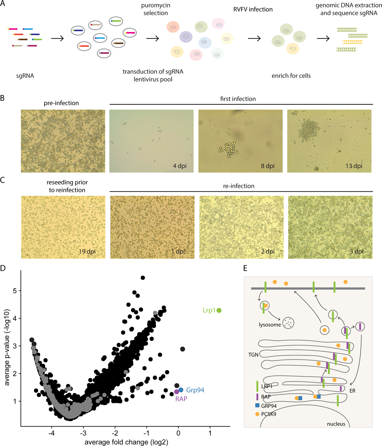
Rift Valley fever virus (RVFV) is a zoonotic pathogen with pandemic potential. RVFV entry is mediated by the viral glycoprotein (Gn), but host entry factors remain poorly defined. Our genome-wide CRISPR screen identified low-density lipoprotein receptor-related protein 1 (mouse Lrp1/human LRP1), heat shock protein (Grp94), and receptor-associated protein (RAP) as critical host factors for RVFV infection. RVFV Gn directly binds to specific Lrp1 clusters and is glycosylation independent. Exogenous addition of murine RAP domain 3 (mRAPD3) and anti-Lrp1 antibodies neutralizes RVFV infection in taxonomically diverse cell lines. Mice treated with mRAPD3 and infected with pathogenic RVFV are protected from disease and death. A mutant mRAPD3 that binds Lrp1 weakly failed to protect from RVFV infection. Together, these data support Lrp1 as a host entry factor for RVFV infection and define a new target to limit RVFV infections.

Keywords: CRISPR screen; Lrp1; Rift Valley fever virus; viral entry.


Conflict of interest statement

Declaration of interests H.W.V. is a founder of Casma Therapeutics and pierianDx and is employed by Vir Biotechnology. None of these companies funded the work reported here. Invention disclosures for method of use for Lrp1 interaction with RVFV Gn (G.K.A., A.L.H., D.W.L., H.W.V., and S.S.G.) and for the use of anti-Lrp1 antibodies by U Toronto (S.S.S., S.M., and G.K.A.) have been filed. Inclusion and diversity We worked to ensure sex balance in the selection of non-human subjects. We worked to ensure diversity in experimental samples through the selection of the cell lines. One or more of the authors of this paper self-identifies as an underrepresented ethnic minority in science. One or more of the authors of this paper self-identifies as a member of the LGBTQ+ community. While citing references scientifically relevant for this work, we also actively worked to promote gender balance in our reference list.

Figures

Figure 1.
Figure 1.. A pooled genome-scale CRISPR screen identifies Lrp1 and Lrp1-associated proteins RAP and Grp94 as critical proteins for RVFV infection.
A. Schematic of the CRISPR/Cas9 screen in BV2 cells. B. Light microscope images (4X) of BV2 cells before infection and at 4 different time points post-infection. C. At 18 dpi, surviving cells were reseeded into new flasks for reinfection on 19 dpi and imaged at 4X by light microscopy. D. Volcano plot analysis of the BV2 screen results of surviving cells from the initial infection at an MOI 0.1. E. Summary of key interactions that modulate Lrp1 surface presentation, including RAP and Grp94.
Figure 2.
Figure 2.. LRP1 is essential for RVFV infection of BV2 cells.
A. Western blot of BV2 Lrp1 knockout clones (Lrp1KO C3, Lrp1KO R1, Lrp1KO R2, Lrp1KO R4, Lrp1KO R5, and Lrp1KO R6), and partial knockout (Lrp1PKO R3) generated using either single gRNA or dual gRNA CRISPR/Cas9 approaches, as described in methods. B. BV2 wildtype (WT) and Lrp1KO clones were infected with RVFV ZH501 at a MOI of 0.1. After 18 hours, the cells were harvested for RNA extraction and subjected to q-RT-PCR analysis. Data shown are viral RNA (vRNA) titers normalized to wildtype BV2 cells. C. Microscopic images showing the WT and LRP1KO R4 cells infected with RVFV MP12GFP (MOI 5 for 6 hours) in fluorescence images (top panels) and DAPI-stained images (bottom panels). Images were taken at 20X magnification (For quantification, refer to Fig. S2H). D. Flow cytometry of WT, Lrp1PKO R3, and Lrp1KO R4 cells infected with RVFV MP12GFP. E. Corresponding analysis of flow cytometry histograms in D. F. Western blot of mouse embryonic fibroblasts (MEFs) from Lrp+/+ and Lrp1F/F mice infected with AdCre. G. Representative flow cytometry of MEFs Lrp1+/+ and LrpF/F cells infected for 5 days with AdCre and then infected with RVFV-MP12GFP at MOI of 1 for 15 hours. H. Corresponding analysis of flow cytometry histogram data in G.
Figure 3.
Figure 3.. RAP and GRP94 knockout reduce RVFV infection indirectly by modulating Lrp1 levels.
A. Western blot of BV2 knockout clones for RAP probed with an anti-Lrp1 antibody. B. BV2 knockout clones in A were infected with RVFV-MP12GFP at an MOI 1 for 15 hours. The cells were examined for virus infection (GFP) using flow cytometry. C. Quantitative analysis of flow data in B. Data are expressed as % infection relative to BV2 WT cells. D. Western blot of BV2 knockout clones for Grp94 probed with an anti-Lrp1 antibody. E. BV2 knockout clones in D were infected with RVFV-MP12GFP at an MOI 1 for 15 hours. The cells examined for virus infection (GFP) using flow cytometry. F. Quantification of the flow data in E. Data are expressed as % infection relative to BV2 WT cells. Experiments were done at least three times. ****, p<0.0001.
Figure 4.
Figure 4.. Lrp1 binds RVFV glycoprotein Gn.
A. LRP1 is comprised of four clusters, CLI, CLII, CLIII, and CLIV, and the cytoplasmic and transmembrane domains (left). Mini-domains of CLI, CLII, CLIII, and CLIV were generated as -Fc fusions (top right). Lentiviruses carrying either pLVX-empty vector or pLVX-expressing HA-tagged minidomains mini-LRP1 CLI, CLII, CLIII, and CLIV were also generated (bottom right). B. BV2 WT and Lrp1KO cells were transduced with lentiviruses carrying either pLVX-empty vector (EV) or pLVX-expressing mini-LRP1 CLI, CLII, CLIII, and CLIV prior to infection with RVFV MP12GFP. The bar graph shows the quantification of % infectivity. C. Cell surface expression of mini-LRP1 Clusters in BV2 Lrp1KO cells using flow cytometry for HA. Biolayer interferometry sensograms of RVFV Gn binding to immobilized: D. Fc control, E. Fc-hLrp1 CLII, and F. Fc-hLrp1 CLIV. Neutraliztion of RVFV MP12GFP infection in the presence of exogenous G. Fc control, H. hLrp1 CLII, and I. hLrp1 CLIV.
Figure 5.
Figure 5.. Lrp1 is critical for virus binding and internalization and anti-Lrp1 Abs inhibit RVFV infection.
To evaluate binding vs internalization, BV2 WT and BV2 Lrp1KO R4 cells were incubated with RVFV MP12GFP at 4°C for binding assay A, and 37°C for internalization assay B. After washings, bound viral particles were quantified by RT-qPCR analysis. Quantification of Alexa Fluor labeled viral particles binding with BV2 WT and BV2 Lrp1KO R4 cells C. at 4°C and D. at 37°C were evaluated and normalized to respective levels of BV2 WT cells. E. BV2 WT cells were pre-incubated for 1 hour with 2.5 µg/mL of hLrp1 CLII-specific (15409), CLIV-specific antibodies (15438) and bi-specific (15408 and 15430) and then infected with RVFV MP12GFP. Cells were analyzed for virus infection after 16 hours. Bar graph represents % cells infected after the antibody treatment, compared to the infection of untreated cells. F. Dose-response curve showing the inhibition of RVFV MP12GFP infection of BV2 cells (y-axis) with EC50 936 ± 78 ng/mL after treatment with serial dilutions of IgG 15408 (x-axis).
Figure 6.
Figure 6.. mRAPD3 competes with RVFV glycoprotein Gn for binding to Lrp1 and inhibits RVFV infection.
A. Domain organization of mouse RAP (mRAP) protein. BLI sensograms of mRAPD3 binding to immobilized B. LRP1 CLII immobilized C. LRP1 CLIV. D. mRAPD3 competition assay to assess relative binding of Gn to LRP1 CLIV in the presence of 1, 3, 6, or 10 μg/mL concentrations of mRAPD3. E. Flow cytometry data for BV2 cells infected with RVFV MP12GFP in the presence of increasing concentrations of mRAPD3. F. Analysis of relative infectivity as a function of mRAPD3 concentration. EC50 is 0.59 ± 0.2 μg/ml. BLI sensograms showing the binding of mRAPD3 and mutant mRAPD3 with G. LRP1 CLII and H. LRP1 CLIV. I. RVFV MP12GFP infection of BV2 cells in presence of mRAPD3 and mutant mRAPD3. J. Cell lines from different species were infected with RVFV-MP12GFP at an MOI 1 in the absence (−) or presence (+) of 5 μg/mL of mRAPD3 (10x EC50). Infection was assessed 15 hpi by flow cytometry. K. Mouse (BV2) and human (HepG2 and SH-SY5Y) cell lines were infected with RVFV ZH501 at an MOI 1 in the absence (−) or presence (+) of mRAPD3. Infection was assessed at 18 hpi by RT-qPCR on cell supernatants and intracellular flow cytometry for viral Gn protein.
Figure 7.
Figure 7.. mRAP binding to Lrp1 protects mice from intracranial infection of RVFV ZH501.
A. Survival of mice infected intracranially with 10 pfu of RVFV ZH501 in absence or presence of 215 μg of recombinant mRAPD3 protein, 210 μg of mutant mRAPD3, 250 μg of control protein (Ebola VP30). In a second experiment, 3 mice/group were euthanized at 3 dpi, and liver, spleen, brain and serum were harvested at necropsy and assessed for B. viral RNA levels by qRT-PCR or C. infectious virus by plaque assay. Heatmaps show average log-transformed titer for each tissue (indicated by the number in each cell of the heatmap) and are also represented visually by the color shading in the legend. X through the cell indicates samples that were not available for analysis. Pathology in D. liver and E. brain tissue was assessed by immunofluorescence for viral antigen using an anti-NP antibody (top panels) or H&E staining (lower panels) in presence or absence of the indicated proteins. Images were taken at 20X magnification. The liver and brain tissues shown in D and E are from respective animals; IF and H&E are from the same tissues. Additional representative animals and controls are shown in Figure S7.

Comment in

References

    1. ALBE JR, BOYLES DA, WALTERS AW, KUJAWA MR, MCMILLEN CM, REED DS & HARTMAN AL 2019. Neutrophil and macrophage influx into the central nervous system are inflammatory components of lethal Rift Valley Fever encephalitis in rats. PLoS Pathog, In press. - PMC - PubMed
    1. BLASI E, RADZIOCH D, DURUM SK & VARESIO L 1987. A murine macrophage cell line, immortalized by v-raf and v-myc oncogenes, exhibits normal macrophage functions. Eur J Immunol, 17, 1491–8. - PubMed
    1. BRUSTOLIN M, TALAVERA S, NUNEZ A, SANTAMARIA C, RIVAS R, PUJOL N, VALLE M, VERDUN M, BRUN A, PAGES N & BUSQUETS N 2017. Rift Valley fever virus and European mosquitoes: vector competence of Culex pipiens and Stegomyia albopicta (= Aedes albopictus). Med Vet Entomol, 31, 365–372. - PubMed
    1. BU G 2001. The roles of receptor-associated protein (RAP) as a molecular chaperone for members of the LDL receptor family. Int Rev Cytol, 209, 79–116. - PubMed
    1. BU G & SCHWARTZ AL 1998. RAP, a novel type of ER chaperone. Trends Cell Biol, 8, 272–6. - PubMed

Publication types

MeSH terms