. 2021 Nov:193:114781.
doi: 10.1016/j.bcp.2021.114781. Epub 2021 Sep 21.

Probing the molecular basis for signal transduction through the Zinc-Activated Channel (ZAC)

Affiliations

Probing the molecular basis for signal transduction through the Zinc-Activated Channel (ZAC)

Nawid Madjroh et al. Biochem Pharmacol. 2021 Nov.

Abstract

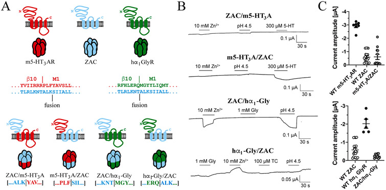
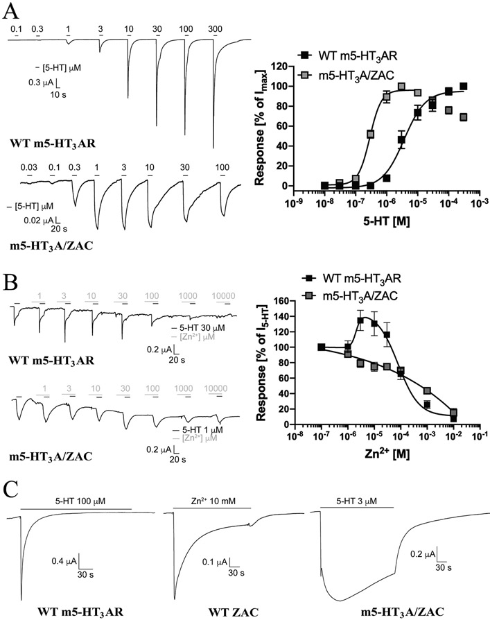
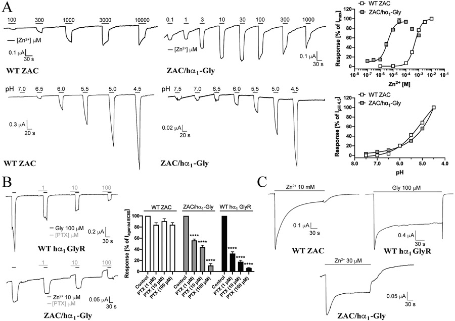
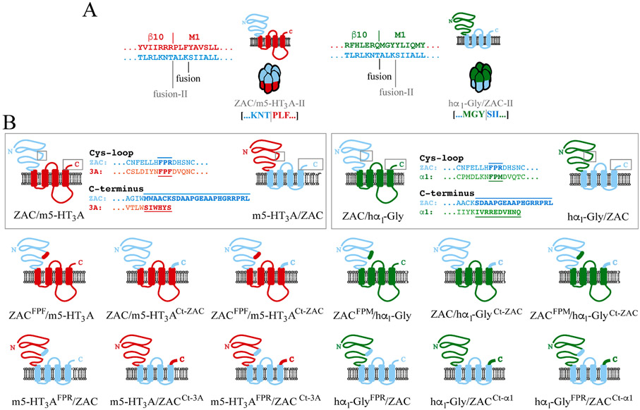
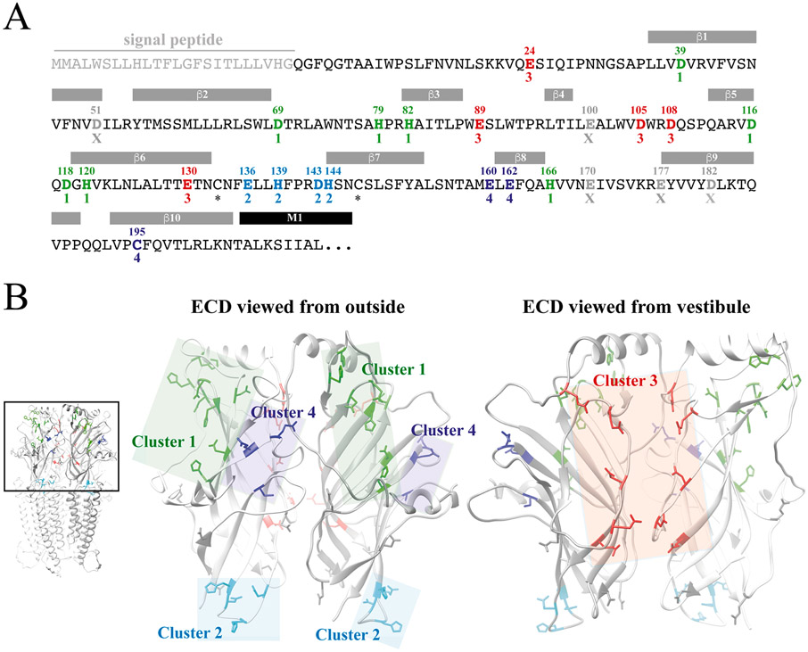
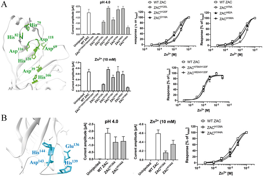
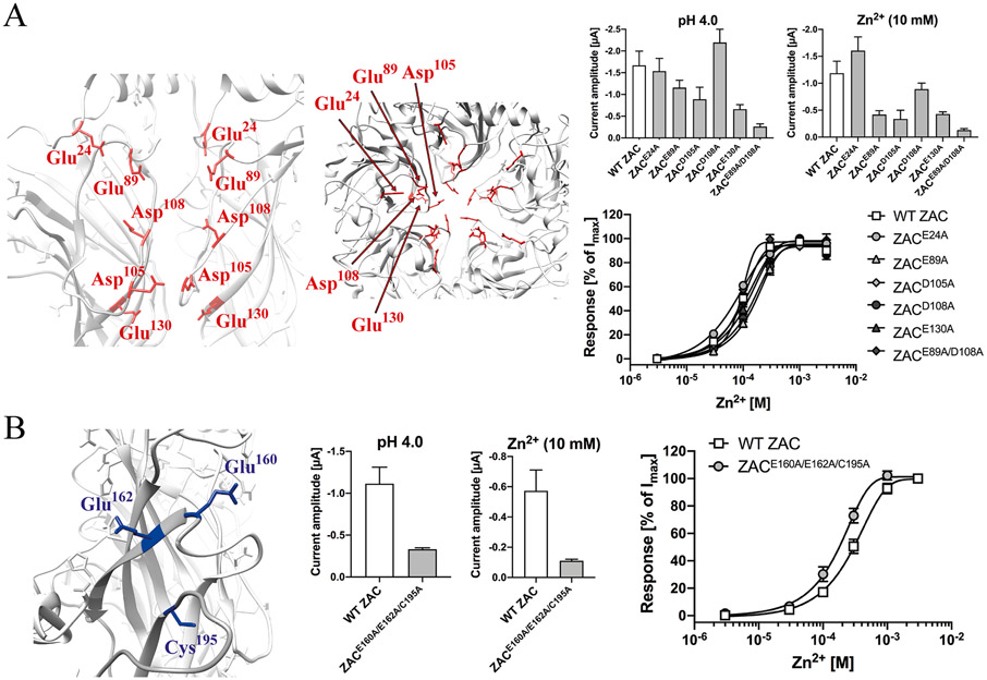
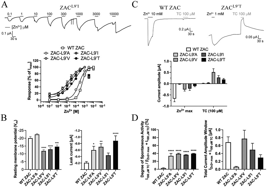
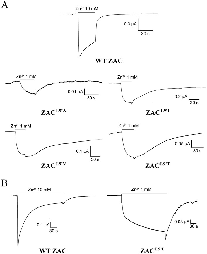
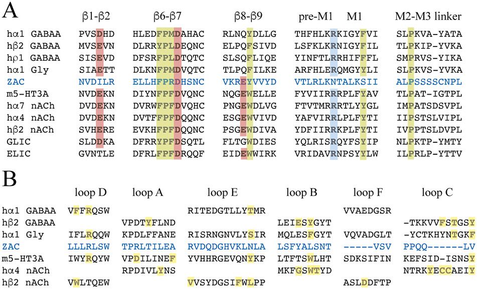
The molecular basis for the signal transduction through the classical Cys-loop receptors (CLRs) has been delineated in great detail. The Zinc-Activated Channel (ZAC) constitutes a so far poorly elucidated fifth branch of the CLR superfamily, and in this study we explore the molecular mechanisms underlying ZAC signaling in Xenopus oocytes by two-electrode voltage clamp electrophysiology. In studies of chimeric receptors fusing either the extracellular domain (ECD) or the transmembrane/intracellular domain (TMD-ICD) of ZAC with the complementary domains of 5-HT3A serotonin or α1 glycine receptors, serotonin and Zn2+/H+ evoked robust concentration-dependent currents in 5-HT3A/ZAC- and ZAC/α1-Gly-expressing oocytes, respectively, suggesting that Zn2+ and protons activate ZAC predominantly through its ECD. The molecular basis for Zn2+-mediated ZAC signaling was probed further by introduction of mutations of His, Cys, Glu and Asp residues in this domain, but as none of the mutants tested displayed substantially impaired Zn2+ functionality compared to wild-type ZAC, the location of the putative Zn2+ binding site(s) in the ECD was not identified. Finally, the functional importance of Leu246 (Leu9') in the transmembrane M2 α-helix of ZAC was investigated by Ala, Val, Ile and Thr substitutions. In concordance with findings for this highly conserved residue in classical CLRs, the ZACL9'X mutants exhibited left-shifted agonist concentration-response relationships, markedly higher degrees of spontaneous activity and slower desensitization kinetics compared to wild-type ZAC. In conclusion, while ZAC is an atypical CLR in terms of its (identified) agonists and channel characteristics, its signal transduction seems to undergo similar conformational transitions as those in the classical CLR.

Keywords: Agonist binding; Chimeric subunits; Cys-loop receptor (CLR); Leu9′ residue; Pentameric ligand-gated ion channel (pLGIC); Zinc-Activated Channel (ZAC).


Figures

Fig. 1.
Fig. 1.. Chimeric receptors fusing the ECDs and TMD-ICDs of ZAC, m5-HT3AR and hα1 GlyR.
A. Topologies of WT m5-HT3A, WT ZAC and WT hα1 GlyR subunits and chimeric ZAC/m5-HT3A, m5-HT3A/ZAC, ZAC/hα1-Gly and hα1-Gly/ZAC subunits and illustration of the pentameric complexes assembled from them. The amino acid sequences of the ECD-into-TMD sequences in the three WT subunit proteins are given. The borders between β10 (ECD) and M1 (TMD) reported for m5-HT3AR [11] and hα1 GlyR [15] and the fusion points in the four chimeras (“fusion”) are indicated. B. Functionalities of the constructed chimeric receptors. Representative traces from the testing of the putative agonists at ZAC/m5-HT3A-, m5-HT3A/ZAC-, ZAC/hα1-Gly- and hα1-Gly/ZAC-expressing oocytes by TEVC electrophysiology. C. Averaged agonist-evoked current amplitudes in oocytes expressing the functional m5-HT3A/ZAC (top) and ZAC/hα1-Gly (bottom) chimeras and their respective parent receptors. Saturating agonist concentrations for the different receptors were used: 300 μM 5-HT (WT m5-HT3AR), 3 μM 5-HT (m5-HT3A/ZAC), 10 mM Zn2+ (WT ZAC), 30 μM Zn2+ (ZAC/hα1-Gly), and 1 mM Gly (WT hα1 GlyR). The averaged data are given as means ± S.E.M. and are based on data from recordings performed on the receptors expressed in at least two diffent oocyte batches (n = 5–19).
Fig. 2.
Fig. 2.. Functional properties exhibited by the chimeric m5-HT3A/ZAC receptor.
A. Agonist properties displayed by 5-HT at m5-HT3A/ZAC. Representative traces for 5-HT-evoked currents in WT m5-HT3AR- and m5-HT3A/ZAC-expressing oocytes (left) and averaged concentration-response relationships displayed by 5-HT at WT m5-HT3AR and m5-HT3A/ZAC (means ± S.E.M., n = 7–9) (right). B. Modulatory properties displayed by Zn2+ at m5-HT3A/ZAC. Representative traces for the Zn2+-mediated modulation of 5-HT (EC80)-evoked currents in WT m5-HT3AR- and m5-HT3A/ZAC-expressing oocytes (left) and averaged concentration-effect relationships displayed by Zn2+ at the 5-HT (EC80)-mediated currents through WT m5-HT3AR and m5-HT3A/ZAC (means ± S.E.M., n = 7–8) (right). 5-HT (30 μM) and 5-HT (1 μM) were used for WT m5-HT3AR and m5-HT3A/ZAC, respectively. C. Representative traces of the currents evoked by 4 min-applications of saturating agonist concentrations at WT m5-HT3AR-, WT ZAC- and m5-HT3A/ZAC-expressing oocytes. 5-HT (100 μM), Zn2+ (10 mM) and 5-HT (3 μM) were used at WT m5-HT3AR, WT ZAC and m5-HT3A/ZAC, respectively.
Fig. 3.
Fig. 3.. Functional properties exhibited by the chimeric ZAC/hα1-Gly receptor.
A. Agonist properties displayed by Zn+ and H+ at ZAC/α1-Gly. Representative traces for Zn2+- and H+-evoked currents in WT ZAC- and ZAC/α1-Gly-expressing oocytes (left), and averaged concentration-response relationships displayed by Zn2+ and H+ at WT ZAC and ZAC/α1-Gly [means ± S.E.M., n = 8–10 (Zn2+) and n = 8–11 (H+)] (right). B. Antagonist properties displayed by picrotoxin (PTX) at ZAC/hα1-Gly. Representative traces for picrotoxin-mediated inhibition of Gly (EC90)-evoked currents in WT hα1 GlyR and Zn2+ (EC90)-evoked currents in ZAC/α1-Gly-expressing oocytes (left), and averaged concentration-inhibition relationships displayed by picrotoxin at WT hα1 GlyR, WT ZAC and ZAC/hα1-Gly (means ± S.E.M., n = 9–12) (right). Zn2+ (1 mM), Zn2+ (10 μM) and Gly (100 μM) were used for WT ZAC, ZAC/α1-Gly and WT hα1 GlyR, respectively. C. Representative traces of the currents evoked by 4 min-application of saturating agonist concentrations at WT hα1 GlyR, WT ZAC- and ZAC/α1-Gly-expressing oocytes. Zn2+ (10 mM), Zn2+ (30 μM) and Gly (100 μM) were used as agonist contrations for WT ZAC, ZAC/α1-Gly and WT hα1 GlyR, respectively. The trace for WT ZAC is the same as that shown in Fig. 2C.
Fig. 4.
Fig. 4.. The alternative ZAC/m5-HT3A-II and hα1-Gly/ZAC-II chimeras and the ECD/TMD-ICD chimeras with Cys-loop and/or C-terminus modifications.
A. Topologies of the chimeric ZAC/m5-HT3A-II and hα1-Gly/ZAC-II subunits and illustration of the pentameric complexes assembled from them. The amino acid sequences of the ECD-into-TMD sequences in the three WT subunit proteins are given, and the alternative fusion points in ZAC/m5-HT3A-II and hα1-Gly/ZAC-II (“fusion-II”) compared to those in the original ZAC/m5-HT3A and hα1-Gly/ZAC chimeras (“fusion”) are indicated. B. Schematic outline of the modifications made to the PFX-motif in the Cys-loop and in the C-terminal in the ECD-parts and the TMD/ICD-parts of the ZAC/m5-HT3A, m5-HT3A/ZAC, ZAC/hα1-Gly and hα1-Gly/ZAC chimeras.
Fig. 5.
Fig. 5.. Candidate Zn2+-binding residues in the extracellular domain of ZAC.
A. Amino acid sequence of the ZAC ECD. The indicated signal peptide, the β-sheet β1-β10 and the M1 α-helix segments in ZAC are predicted based on amino acid sequence aligment of the ZAC and m5-HT3A subunits and these segments in the m5-HT3AR cryo-EM structure (PDB ID: 6HIN) [11]. The 25 candidate Zn2+-binding residues in the ZAC ECD are given in bold with their residue numbers above. The candidate Zn2+-binding residues in the four defined clusters are given (Cluster 1: green; Cluster 2: cyan; Cluster 3: red; Cluster 4: dark-blue), with the five candidate residues not included in a cluster given in grey (“X”), and the two cysteines forming the Cys-loop are indicated with asterisks. B. Homology model of ZAC based on the cryo-EM structure of m5-HT3AR (PDB ID: 6HIN). The pentameric ZAC complex (left) and the ECD for two neighbouring subunits in the ZAC complex viewed from the outside (middle) and from the vestibule (right). The candidate Zn2+-binding residues in this domain defined as Cluster 1 (green), Cluster 2 (cyan), Cluster 3 (red) and Cluster 4 (dark-blue) are indicated in the ECD dimer, with the five candidate residues not included in a cluster shown in grey.
Fig. 6.
Fig. 6.. Probing the importance of candidate Zn2+-binding residues in Clusters 1 and 2 of the ZAC ECD for Zn2+-mediated ZAC activation.
A. Cluster 1. Left: Cluster 1 residues (in green, detail of ZAC homology model). Middle: Averaged IpH 4.0 and I10 mM Zn2+ values recorded from oocytes expressing WT ZAC and various ZAC mutants [means ± S.E.M., H+: n = 5–8 (mutants), n = 14 (WT); Zn2+: n = 6–8 (mutants), n = 16 (WT)]. Right: Averaged concentration-response relationships displayed by Zn2+ at oocytes expressing WT ZAC and various ZAC mutants [Top graphs: means ± S.E.M., n = 6–8 (mutants), n = 14 (WT). Bottom graph: means ± S.E.M., n = 6–8]. B. Cluster 2. Left: Cluster 2 residues (in cyan, detail of ZAC homology model). Middle: Averaged IpH 4.0 and I10 mM Zn2+ values recorded from oocytes expressing WT ZAC and various ZAC mutants [means ± S.E.M., H+: n = 6–8; Zn2+: n = 6–7]. Right: Averaged concentration-response relationships displayed by Zn2+ at oocytes expressing WT ZAC, ZACH139A and ZACH144A [means ± S.E.M., n = 7–8 (mutants), n = 14 (WT)].
Fig. 7.
Fig. 7.. Probing the importance of candidate Zn2+-binding residues in Clusters 3 and 4 of the ZAC ECD for Zn2+-mediated ZAC activation.
A. Cluster 3. Left: Cluster 3 residues (in red, detail of ZAC homology model). Right, top: Averaged IpH 4.0 and I10 mM Zn2+ values recorded from oocytes expressing WT ZAC and various ZAC mutants [means ± S.E.M., n = 5–8]. Right, bottom: Averaged concentration-response relationships displayed by Zn2+ at oocytes expressing WT ZAC and various ZAC mutants [means ± S.E.M., n = 5–8 (mutants), n = 12 (WT)]. B. Cluster 4. Left: Cluster 4 residues (in dark-blue, detail of ZAC homology model). Middle: Averaged IpH 4.0 and I10 mM Zn2+ values recorded from oocytes expressing WT ZAC and various ZAC mutants [means ± S.E.M., n = 5–6]. Right: Averaged concentration-response relationships displayed by Zn2+ at oocytes expressing WT ZAC and ZACE160A/E162A/C195 [means ± S.E.M., n = 5–6]. WT ZAC- and ZACE160A/E162A/C195-oocytes were injected with 1.84 ng and 3.68 ng cRNA, respectively.
Fig. 8.
Fig. 8.. Functional importance of the Leu9′ residue in ZAC.
A. Representative traces for Zn2+-evoked currents in ZACL9′I-expressing oocytes (top), and averaged concentration-response relationships exhibited by Zn2+ at WT ZAC, ZACL9′A, ZACL9′V, ZACL9′I and ZACL9′T (means ± S.E.M., n = 6–8) (bottom). B. Resting membrane potentials (left) and leak currents (right) recorded from oocytes expressing WT ZAC, ZACL9′A, ZACL9′T, ZACL9′V and ZACL9′I. Data are given as mean ± S.E.M. values (n = 40–60). C. Representative traces for Zn2+- and TC (100 μM)-evoked currents in WT ZAC- and ZACL9′I-expressing oocytes (top), and averaged current amplitudes evoked by a saturating concentration of Zn2+ and by TC (100 μM) in WT ZAC-, ZACL9′A-, ZACL9′V-, ZACL9′I-, and ZACL9′T-oocytes (means ± S.E.M., n = 6–8) (bottom). 10 mM Zn2+ were used for WT ZAC and 1 mM Zn2+ were used for ZACL9′A, ZACL9′V, ZACL9′I and ZACL9′T. D. Degrees of spontaneous activity [defined as: I100 uM TC/ (IZn2+ max + I100 uM TC)] (left) and total current amplitude windows (defined as: IZn2+ max + I100 uM TC) (right) exhibited by WT ZAC, ZACL9′A, ZACL9′V, ZACL9′I and ZACL9′T expressed in oocytes.
Fig. 9.
Fig. 9.. Signalling characteristics exhibited by the ZACL9′X mutants.
A. Representative traces of currents evoked by saturating Zn2+ concentrations in WT ZAC-, ZACL9′A-, ZACL9′V-, ZACL9′I- and ZACL9′T-expressing oocytes. 10 mM Zn2+ was used for WT ZAC and 1 mM Zn2+ was used for the ZACL9′X mutants, respectively. B. Representative traces of currents evoked by sustained application of saturating Zn2+ concentrations in WT ZAC- and ZACL9′I-expressing oocytes. 10 mM Zn2+ and 1 mM Zn2+ were used for WT ZAC and for ZACL9′I, respectively.
Fig. 10.
Fig. 10.. Key residues involved in signal transduction through the CLR.
A. Residues involved in ECD/TMD cross-talk in the classical CLR. Alignment of the amino acid sequences of the β1-β2, β6-β7 (Cys-loop) and β8-β9 loops, the pre-M1/M1 segments and the M2-M3 linkers in ZAC, selected classical CLRs and the prokaryotic CLRs GLIC and ELIC. The conservation of key residues for the ECD/TMD cross-talk are indicated (negatively charged or charge-neutral, polar residues in red and positively charged residues blue, structural residues in green). B. Residues involved in orthosteric agonist binding to the classical CLR. Alignment of the amino acid sequences of loops A-F in ZAC and selected classical CLRs. The residues in the loops directly involved in orthosteric agonist binding to m5-HT3AR [12], hα4β2 nAChR [14], hα1β2γ2 GABAAR [13] and hα1 GlyR [10] are indicated in bold and highlighted in yellow.

Similar articles

Cited by

  • Cryo-EM structure of the zinc-activated channel (ZAC) in the Cys-loop receptor superfamily.
    Jin F, Lin YY, Wang RC, Xie TX, Zhao Y, Shen C, Sheng D, Ichikawa M, Yu Y, Wang J, Hattori M. Jin F, et al. Proc Natl Acad Sci U S A. 2024 Oct 29;121(44):e2405659121. doi: 10.1073/pnas.2405659121. Epub 2024 Oct 23. Proc Natl Acad Sci U S A. 2024. PMID: 39441630 Free PMC article.
  • Zinc, Copper, and Calcium: A Triangle in the Synapse for the Pathogenesis of Vascular-Type Senile Dementia.
    Kawahara M, Tanaka KI, Kato-Negishi M. Kawahara M, et al. Biomolecules. 2024 Jun 28;14(7):773. doi: 10.3390/biom14070773. Biomolecules. 2024. PMID: 39062487 Free PMC article. Review.
  • Recent Insight into Lipid Binding and Lipid Modulation of Pentameric Ligand-Gated Ion Channels.
    Ananchenko A, Hussein TOK, Mody D, Thompson MJ, Baenziger JE. Ananchenko A, et al. Biomolecules. 2022 Jun 10;12(6):814. doi: 10.3390/biom12060814. Biomolecules. 2022. PMID: 35740939 Free PMC article. Review.
  • VHH Nanobody Versatility against Pentameric Ligand-Gated Ion Channels.
    Nemecz D, Nowak WA, Nemecz Á. Nemecz D, et al. J Med Chem. 2024 Jun 13;67(11):8502-8518. doi: 10.1021/acs.jmedchem.4c00231. Epub 2024 Jun 3. J Med Chem. 2024. PMID: 38829690 Free PMC article. Review.
  • The Concise Guide to PHARMACOLOGY 2023/24: Ion channels.
    Alexander SPH, Mathie AA, Peters JA, Veale EL, Striessnig J, Kelly E, Armstrong JF, Faccenda E, Harding SD, Davies JA, Aldrich RW, Attali B, Baggetta AM, Becirovic E, Biel M, Bill RM, Caceres AI, Catterall WA, Conner AC, Davies P, De Clerq K, Delling M, Di Virgilio F, Falzoni S, Fenske S, Fortuny-Gomez A, Fountain S, George C, Goldstein SAN, Grimm C, Grissmer S, Ha K, Hammelmann V, Hanukoglu I, Hu M, Ijzerman AP, Jabba SV, Jarvis M, Jensen AA, Jordt SE, Kaczmarek LK, Kellenberger S, Kennedy C, King B, Kitchen P, Liu Q, Lynch JW, Meades J, Mehlfeld V, Nicke A, Offermanns S, Perez-Reyes E, Plant LD, Rash L, Ren D, Salman MM, Sieghart W, Sivilotti LG, Smart TG, Snutch TP, Tian J, Trimmer JS, Van den Eynde C, Vriens J, Wei AD, Winn BT, Wulff H, Xu H, Yang F, Fang W, Yue L, Zhang X, Zhu M. Alexander SPH, et al. Br J Pharmacol. 2023 Oct;180 Suppl 2(Suppl 2):S145-S222. doi: 10.1111/bph.16178. Br J Pharmacol. 2023. PMID: 38123150 Free PMC article.

References

    1. Taly A, Corringer P-J, Guedin D, Lestage P, Changeux J-P, Nicotinic receptors: allosteric transitions and therapeutic targets in the nervous system, Nat Rev Drug Discov 8 (9) (2009) 733–750. - PubMed
    1. Bertrand D, Lee C-H, Flood D, Marger F, Donnelly-Roberts D, Esbenshade TA, Therapeutic Potential of α7 Nicotinic Acetylcholine Receptors, Pharmacol Rev 67 (4) (2015) 1025–1073. - PubMed
    1. Grupe M, Grunnet M, Bastlund JF, Jensen AA, Targeting α4β2 nicotinic acetylcholine receptors in central nervous system disorders: perspectives on positive allosteric modulation as a therapeutic approach, Basic Clin Pharmacol Toxicol 116 (3) (2015) 187–200. - PubMed
    1. Walstab J, Rappold G, Niesler B, 5-HT3 receptors: role in disease and target of drugs, Pharmacol Ther 128 (1) (2010) 146–169. - PubMed
    1. Fakhfouri G, Rahimian R, Dyhrfjeld-Johnsen J, Zirak MR, Beaulieu J-M, Witkin JM, 5-HT3 receptor antagonists in neurologic and neuropsychiatric disorders: The iceberg still lies beneath the surface, Pharmacol Rev 71 (3) (2019) 383–412. - PubMed

Publication types