. 2021 Sep 24;40(1):301.
doi: 10.1186/s13046-021-02107-1.

MEST promotes lung cancer invasion and metastasis by interacting with VCP to activate NF-κB signaling

Affiliations

MEST promotes lung cancer invasion and metastasis by interacting with VCP to activate NF-κB signaling

Yang Wang et al. J Exp Clin Cancer Res. .

Abstract

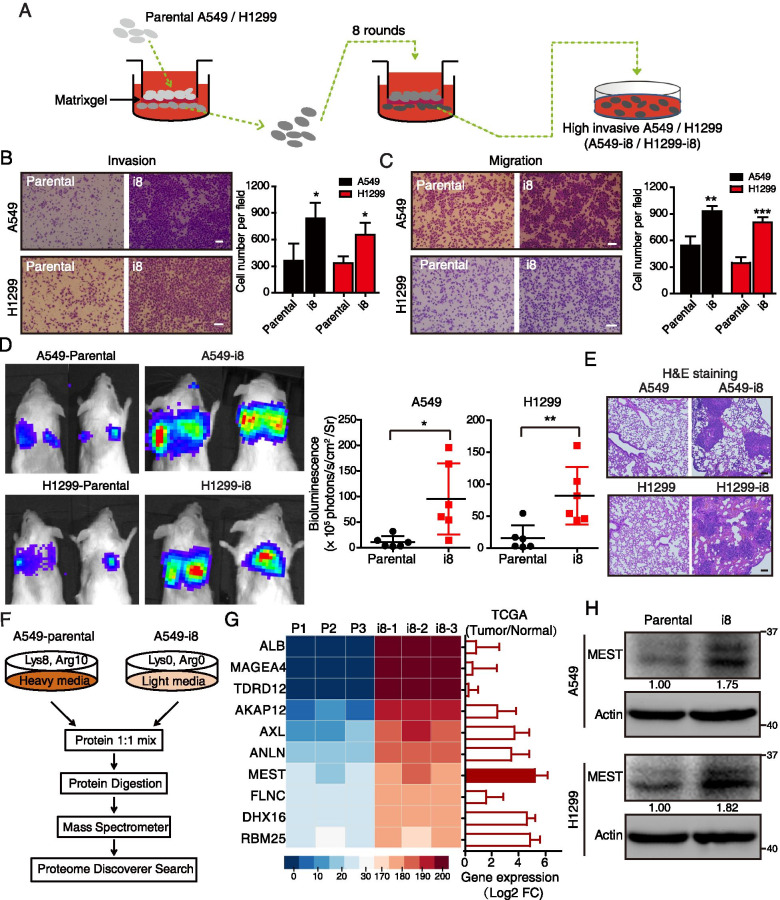
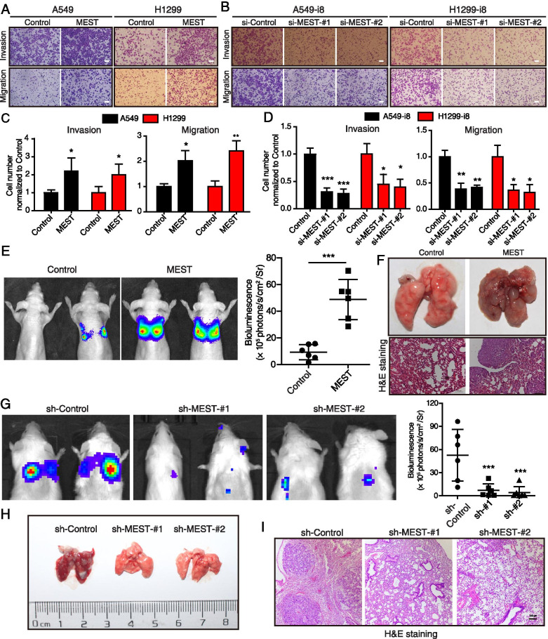
Background: Cell invasion is a hallmark of metastatic cancer, leading to unfavorable clinical outcomes. In this study, we established two highly invasive lung cancer cell models (A549-i8 and H1299-i8) and identified mesoderm-specific transcript (MEST) as a novel invasive regulator of lung cancer. We aim to characterize its biological function and clinical significance in lung cancer metastasis.

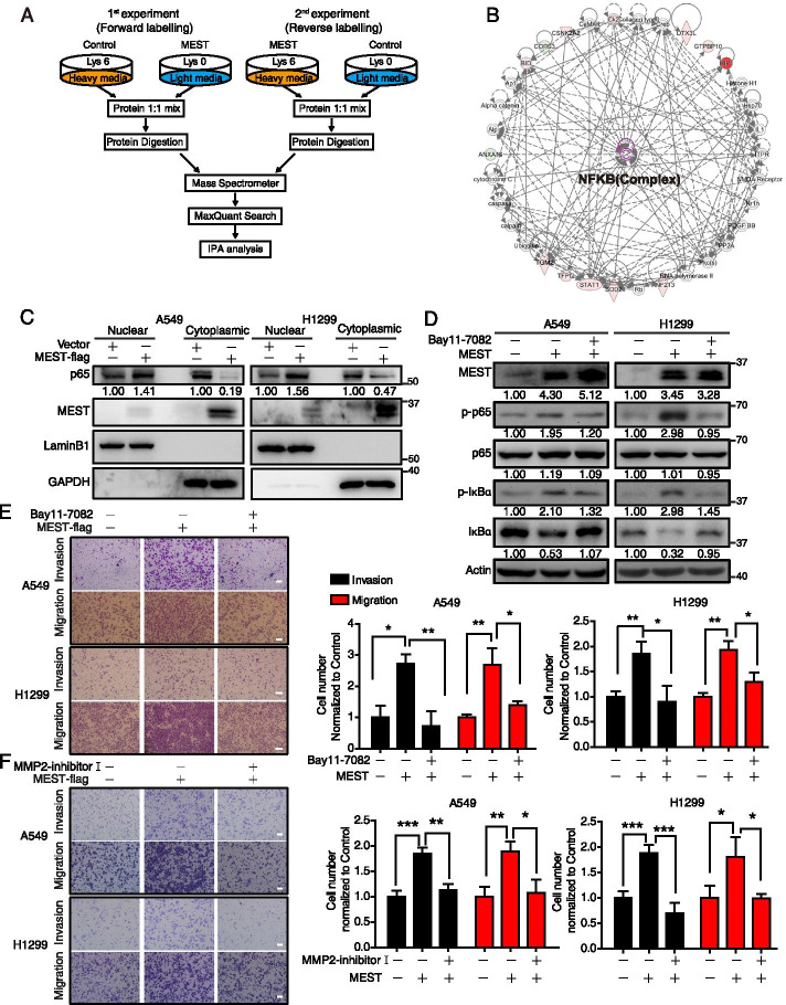
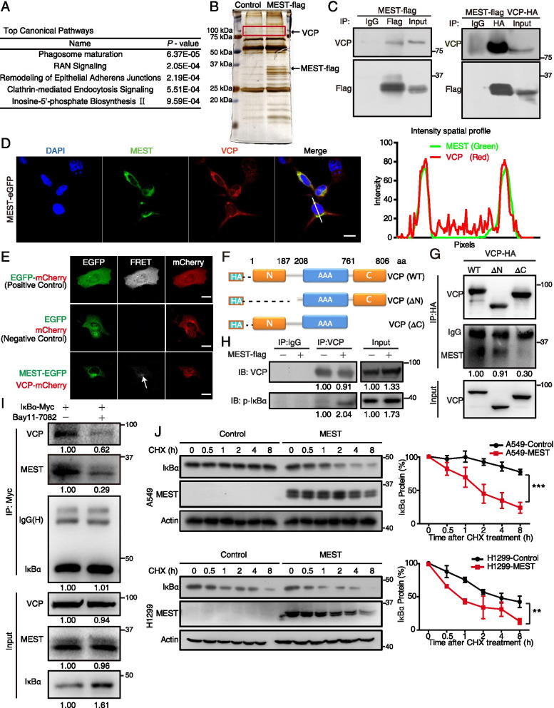
Methods: Transwell invasion assay was performed to establish high-invasive lung cancer cell model. Immunohistochemistry (IHC) was used to detect MEST expression in tumor tissues. Mass spectrometry and bioinformatic analyses were used to identify MEST-regulated proteins and binding partners. Co-immunoprecipitation assay was performed to detect the interaction of MEST and VCP. The biological functions of MEST were investigated in vitro and in vivo. Immunofluorescence staining was conducted to explore the colocalization of MEST and VCP.

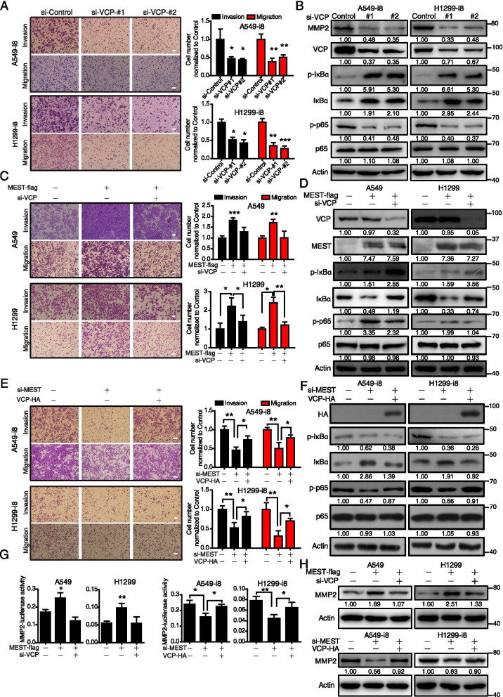
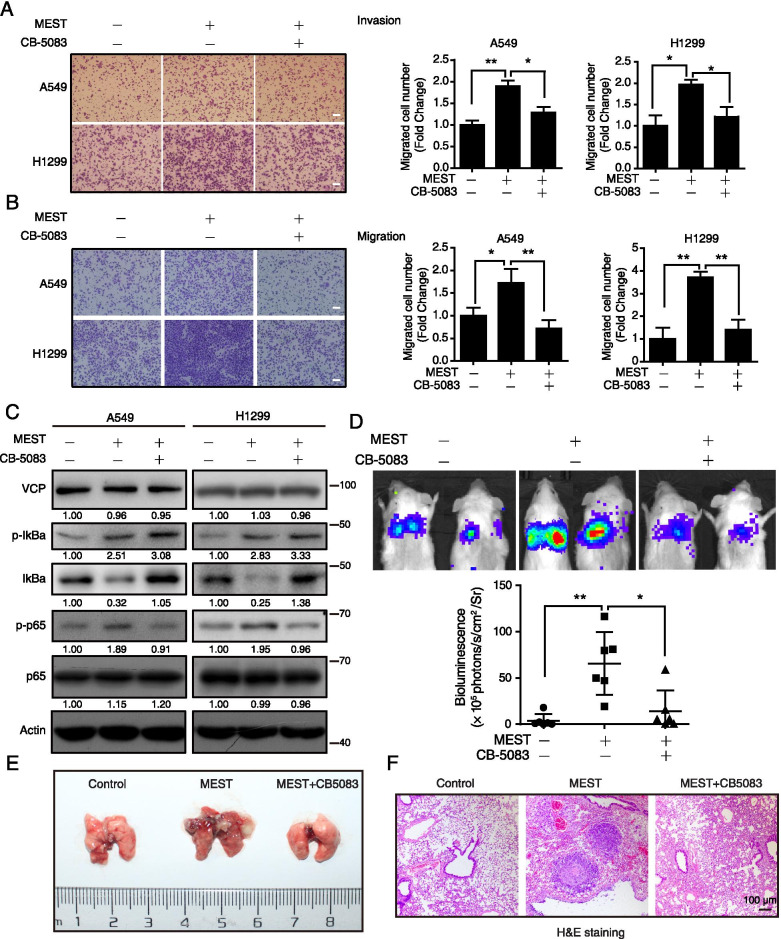
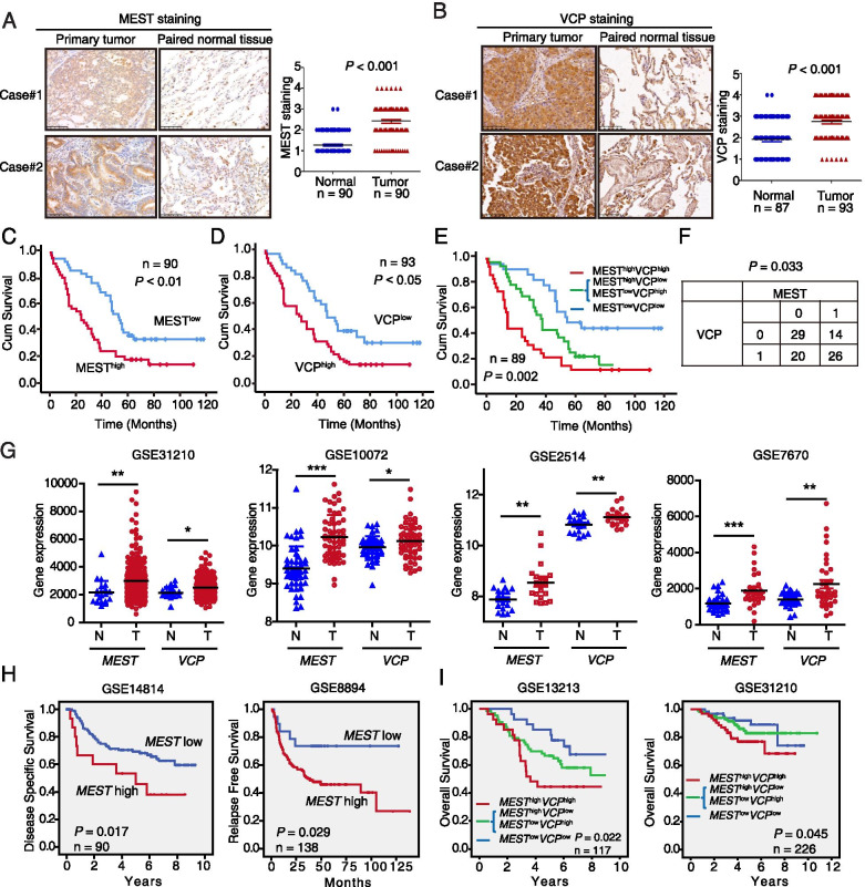
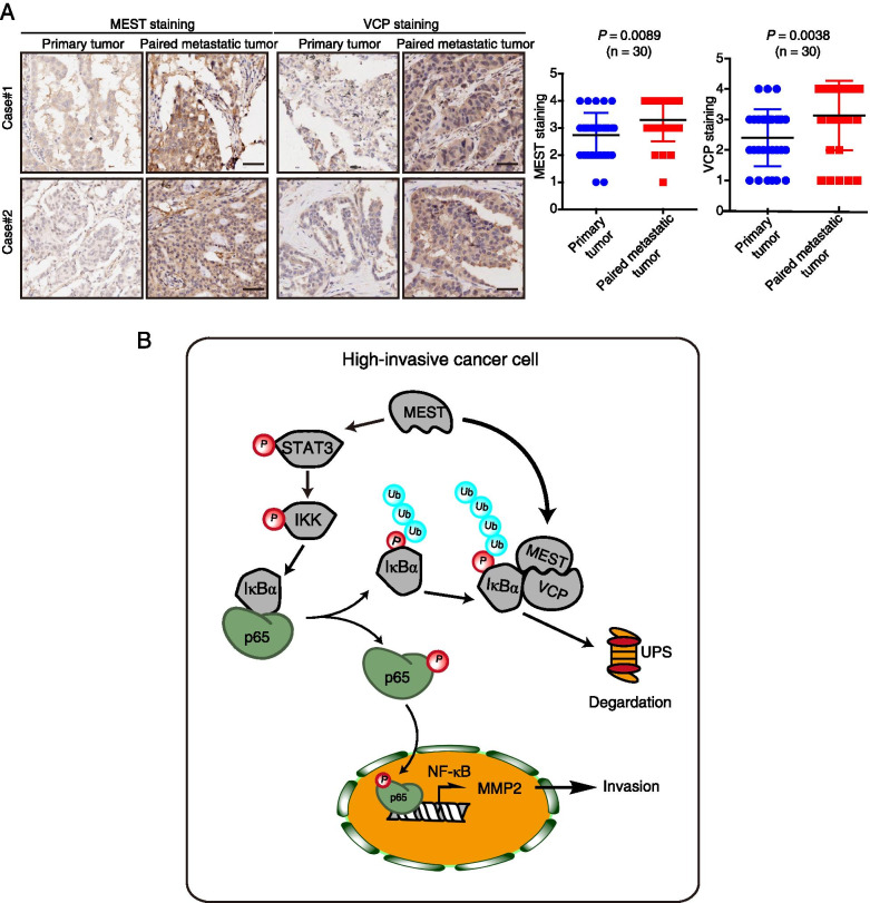
Results: MEST overexpression promoted metastasis of lung cancer cells in vivo and in vitro by activating NF-κB signaling. MEST increased the interaction between VCP and IκBα, which accelerated IκBα degradation and NF-κB activation. Such acceleration was abrogated by VCP silencing, indicating that MEST is an upstream activator of the VCP/IκBα/NF-κB signaling pathway. Furthermore, high expressions of MEST and VCP were associated with poor survival of lung cancer patients.

Conclusion: Collectively, these results demonstrate that MEST plays an important role in driving invasion and metastasis of lung cancer by interacting with VCP to coordinate the IκBα/NF-κB pathway. Targeting the MEST/VCP/IκBα/NF-κB signaling pathway may be a promising strategy to treat lung cancer.

Keywords: Lung cancer; MEST; Metastasis; NF-κB; VCP.


Conflict of interest statement

The corresponding author and all the co-authors have agreed to the publication of the manuscript to Journal of Experimental and Clinical Cancer Research as a research article and declare that they have no conflict of interest as to the results presented.

The authors declare that they have no competing interests.

Figures

Fig. 1
Fig. 1
SILAC-based proteomics identifies MEST as an invasion regulator in lung cancer cells. A Diagram depicting the establishment of highly invasive cell lines (A549-i8 and H1299-i8) from lung cancer cells via eight rounds of invasion selection. B, C The abilities of invasion (B) and migration (C) of A549-i8 and H1299-i8 cell lines were determined by transwell assays, as compared to their parental cell lines, respectively. Scale bar, 100 μm. D NCG mice were transplanted with luciferase-labeled A549-i8 cells or H1299-i8 cells, or their corresponding luciferase-labeled parental cells (2 × 106 cells per mouse) via tail vein injection (n = 6); mice were then visualized 1 month after transplantation by using an IVIS 200 Imaging System. Lungs harvested after imaging were histologically analyzed by H&E staining (E). Scale bar, 100 μm. F Scheme for the identification of differentially expressed proteins by SILAC-based quantitative proteomics. G The expression levels of the top 10 differentially expressed proteins in A549-i8 cells are represented by heatmap. Their expressions in lung adenocarcinoma and normal tissue were compared by using TCGA dataset (n = 503), showing that MEST is highly expressed in clinical lung cancer tissues, as indicated by red bars. H The expression of MEST in A549-i8 and H1299-i8 cells and in their parental cells was assessed by western blot analysis
Fig. 2
Fig. 2
MEST promotes invasion and migration of lung cancer cells. A, B A549 and H1299 cells were overexpressed with MEST, while A549-i8 and H1299-i8 cells were transfected with two siRNAs against MEST (si-MEST#1 and si-MEST#2, 100 nM), their invasion and migration abilities were measured by transwell assay. Scale bar, 100 μm. All data are representative of three independent experiments (C, D). Bars, S.D. *P < 0.05, **P < 0.01, ***P < 0.001 (Student’s t-test). E–F Nude mice were transplanted with luciferase-labeled cells with or without MEST overexpression (2 × 106 cells per mouse) via tail vein injection (n = 6); mice were then visualized 1 month after transplantation by using an IVIS 200 Imaging System. Lungs harvested after imaging are shown. Note that only MEST-overexpressing A549 cells formed large and more metastatic nodules in the lungs. The pulmonary metastases in the mouse model were histologically analyzed by H&E staining (F). Scale bar, 100 μm. G-I NCG mice were transplanted with the luciferase-labeled A549-i8 cells expressing two shRNA against MEST (1 × 106 cells per mouse) via tail vein injection (n = 6); the mice were visualized 1.5 months post-transplantation by using an IVIS 200 Imaging System (G). Lungs harvested after imaging are shown (H). Note that only the control group cells formed large and more metastatic nodules in the lungs. The pulmonary metastases in the mouse model were histologically analyzed by H&E staining (I). Scale bar, 100 μm
Fig. 3
Fig. 3
SILAC quantitative proteomics characterizes MEST-regulated NF-κB pathway in lung cancer. A The scheme of two independent SILAC experiments, including forward labeling and reverse labeling. MEST-regulated proteins were identified by two independent SILAC experiments, and the differentially expressed proteins were analyzed by Ingenuity Pathway Analysis (B). MEST-driven functional signaling networks indicate involvement in the NF-κB signaling pathway. Red, upregulated proteins; green, downregulated proteins; white, speculative proteins. C MEST overexpression promotes nuclear translocation of NF-κB subunits (p65) in both A549 and H1299 cells. LaminB1 and GAPDH were used as markers for nucleus and cytoplasma, respectively. D A549 and H1299 overexpressing MEST were treated with either 2.5 μM of Bay11-7082 or DMSO, as indicated. Expression of NF-κB markers (including p-p65, p65, p-IκBα, and IκBα) was assessed by western blot analysis. Invasion and migration of treated cells were measured by transwell assay (E). F A549 and H1299 cells overexpressing MEST were treated with either 25 μM of MMP2 inhibitor I or DMSO; their invasion and migration abilities were determined by transwell assay. Scale bar, 100 μm. All data are representative of three independent experiments. Bars, S.D. *P < 0.05, **P < 0.01, ***P < 0.001 (Student’s t-test)
Fig. 4
Fig. 4
MEST interacts with VCP and accelerates IκBα degradation. A H1299 cells were transfected with MEST-FLAG expression plasmids, and MEST-binding proteins were identified by co-immunoprecipitation coupled with LC/MS analyses. A list of candidate proteins was uploaded for Ingenuity Pathway Analysis, which revealed that the top canonical pathways involved membrane trafficking. B Silver staining proteins coimmunoprecipitated with MEST show a specific 97 kDa band corresponding to VCP. C H1299 cells were transfected with MEST-FLAG and VCP-HA expression plasmids. The interaction between MEST and VCP was demonstrated by co-immunoprecipitation assays. D A549 cells transfected with MEST-eGFP (green) were stained with VCP antibody (red) for 48 h, and co-localization of MEST and VCP in the perinuclear area was imaged by using a confocal microscope; intensity spatial profiles are plotted. Scale bar, 20 μm. E Fluorescence resonance energy transfer experiment showed the energy transfers from MEST-eGFP (donor) to VCP-mCherry (acceptor). Expression of an eGFP-mCherry fusion protein is a positive control, and co-expression of eGFP-N1 and mCherry-N1 is a negative control. Excitation wavelength is 488 nm, and collection wavelength is 600 nm. Scale bar, 20 μm. F Schematics for the structural domains of VCP and for the N-terminal deletion (ΔN) and C-terminal deletion (ΔC) mutants. The blue line represents the AAA domain of VCP. G The interactions of VCP-truncation mutants (WT, ΔN, and ΔC) with MEST were detected by co-immunoprecipitation assay. H MEST increases the binding of VCP to p-IκBα. H1299 cells were transfected either with or without MEST-FLAG expression plasmids for 48 h; cell lysates were incubated with anti-VCP antibody, and expression of p-IκBα was assessed by western blot analysis. I Bay11-7082 abolishes interactions between MEST, VCP, and IκBα. A549 cells expressing Myc-tagged IκBα were treated with Bay11-7082 (2.5 μM) for 12 h. Cell lysates were incubated with anti-Myc antibody for co-immunoprecipitation assay, and VCP and MEST were detected by western blot analysis. J MEST-overexpressing H1299 cells, MEST-overexpressing A549 cells, and their respective vector control cells were treated with cycloheximide (CHX; 50 μg/mL). The cell lysates were collected at the indicated time points and compared for IκBα expression by western blot analysis. IκBα signals are quantified by densitometry, and the degradation rate is shown as the ratio of IκBα expression level at each time point to the original expression level (0 h). The half-life (t1/2) of IκBα was 0.9 and 3.6 h in MEST-overexpressing H1299 cells and corresponding vector control cells, respectively; t1/2 values were 1.8 and 28.6 h in MEST-overexpressing A549 cells and vector control cells, respectively
Fig. 5
Fig. 5
MEST and VCP promote lung cancer migration and invasion via activating the NF-κB pathway. A A549-i8 and H1299-i8 cells were transfected with two anti-VCP siRNAs, and the migration and invasion abilities were determined by using a transwell assay; MMP2 and NF-κB markers (p-IκBα, IκBα, p-p65, and p65) were analyzed by western blot analysis (B). C MEST-overexpressing A549 and H1299 cells were transfected with si-VCP and scramble si-RNA for 24 h. Transwell assay indicates that knockdown of VCP inhibits cell migration and invasion enhanced by MEST in A549 and H1299 cells. Expression of NF-κB markers (p-IκBα, IκBα, p-p65, and p65) were analyzed by western blot analysis (D). E A549-i8 and H1299-i8 were transfected with VCP-HA plasmid or si-MEST for 24 h, as indicated. Transwell assay shows that knockdown of MEST in A549-i8 and H1299-i8 cells decreases cell migration and invasion, and that transfection with VCP-expression plasmid partially restores this effect. The corresponding NF-κB markers (p-IκBα, IκBα, p-p65, and p65) were analyzed by western blot analysis (F). G The indicated cell lines were co-transfected with MMP2 promoter-driven luciferase reporter, with pRL-TK (loading control), as well as with either the indicated plasmids or siRNA. Luciferase activity was measured (n = 3). H Western blot analysis indicates that MMP2 expression is regulated by MEST and VCP. Bars, SD. *, P < 0.05; **, P < 0.01; ***, P < 0.001 compared with control cells unless otherwise indicated. Scale bar, 100 μm
Fig. 6
Fig. 6
Inhibition of VCP suppresses the pro-metastatic effect of MEST. A, B MEST-overexpressing A549 and H1299 cells were treated with either 0.5 μM of CB-5083 or DMSO for 48 h and subjected to transwell assays to detect their invasion (A) and migration (B) abilities. Scale bar, 100 μm. C The NF-κB markers including p-IκBα, IκBα, p-p65, and p65 were determined by western blot analysis. D-F NCG mice were transplanted with luciferase-labeled cells that either with or without MEST overexpression (1 × 106 cells per mouse) via tail vein injection (n = 6). The indicated treatment group or control group was orally administered CB-5083 (30 mg kg−1) or vehicle, respectively, every 2 days. Mice were visualized 1.5 months after transplantation by using an IVIS 200 Imaging System (D). Lungs harvested after imaging are shown (E). Note that CB-5083 suppresses the metastatic nodules formed by MEST-overexpressing A549 cells in the lungs. Pulmonary metastases in the mouse model were histologically analyzed by H&E staining (F); scale bar, 100 μm
Fig. 7
Fig. 7
Overexpression of MEST and VCP are associated with poor prognosis in lung cancer patients. A Two representative immunohistochemistry (IHC) images of MEST expression in lung cancer tissues and in corresponding adjacent non-tumor lung tissues. Differences in MEST expression scores between lung cancer and corresponding adjacent non-tumor tissues are shown (n = 90). B Two representative IHC images of VCP expression in lung cancer tissues and in corresponding adjacent non-tumor lung tissues. Differences in VCP expression scores between lung cancer (n = 93) and non-cancerous tissues (n = 87) are shown. Kaplan–Meier survival analysis of lung cancer patients according to the expression of MEST (C) and VCP (D) by using median expression level as the cut-off point for survival analyses. High MEST and VCP expressions are significantly associated with shorter survival (E), and statistical significance was calculated by log-rank test (P = 0.002). F The correlation between the immunostaining intensity of the proteins MEST and VCP was determined by Fisher exact test. G MEST and VCP are highly expressed in lung cancer tissues as compared with non-tumor tissues. Analysis of MEST and VCP expression by using the GEO database. Plots derived from gene expression data in GEO (GSE31210, GSE10072, GSE2514, and GSE7670) comparing the expression of MEST and VCP gene in normal lung and lung cancer tissues. H Kaplan–Meier survival curves based on GEO datasets (GSE14814 and GSE8894) show the overall survival of patients with either high or low MEST expression levels. I Kaplan–Meier plots based on GEO datasets indicated that MESThigh/VCPhigh (red) is associated with worse overall survival in lung cancer patients compared with other groups. Survival curves of patients with tumor MEST and VCP expression above the median (green) and patients with tumor MEST and VCP expression below the median (blue) are indicated. Higher MEST expression predicts shorter overall survival in two lung cancer-associated GEO databases (GSE31210, GSE13213)
Fig. 8
Fig. 8
The expression of MEST and VCP in metastatic cancer and a schematic diagram of the action mechanism of MEST. A Two representative IHC images of MEST and VCP expression in primary lung cancer tissues and in corresponding metastatic tumor tissues. Differences in MEST and VCP expression scores between lung cancer and metastatic tumor tissues are shown (n = 30). Scale bar, 50 μm. B Working model of MEST on promotion of cancer metastasis. MEST is upregulated in highly invasive lung cancer cells, it interacts with VCP and increases VCP to capture IκBα for degradation, leading to the nuclear translocation of p65 to activate the NF-κB pathway for cancer metastasis. In addition, MEST can induce IκBα phosphorylation via the STAT3/IKK pathway, mediating NF-κB signaling activation at multiple levels

Similar articles

Cited by

References

    1. Miller KD, Siegel RL, Lin CC, Mariotto AB, Kramer JL, Rowland JH, Stein KD, Alteri R, Jemal A. Cancer treatment and survivorship statistics, 2016. CA Cancer J Clin. 2016;66:271–289. doi: 10.3322/caac.21349. - DOI - PubMed
    1. Siegel RL, Miller KD, Jemal A. Cancer statistics, 2016. CA Cancer J Clin. 2016;66:7–30. doi: 10.3322/caac.21332. - DOI - PubMed
    1. Hu Y, Yu X, Xu G, Liu S. Metastasis: an early event in cancer progression. J Cancer Res Clin Oncol. 2017;143:745–757. doi: 10.1007/s00432-016-2279-0. - DOI - PMC - PubMed
    1. Popper HH. Progression and metastasis of lung cancer. Cancer Metastasis Rev. 2016;35:75–91. doi: 10.1007/s10555-016-9618-0. - DOI - PMC - PubMed
    1. Schafer M, Werner S. Cancer as an overhealing wound: an old hypothesis revisited. Nat Rev Mol Cell Biol. 2008;9:628–638. doi: 10.1038/nrm2455. - DOI - PubMed

MeSH terms