. 2022 Apr 14;59(4):2100664.
doi: 10.1183/13993003.00664-2021. Print 2022 Apr.

Protease activity sensors enable real-time treatment response monitoring in lymphangioleiomyomatosis

Affiliations

Protease activity sensors enable real-time treatment response monitoring in lymphangioleiomyomatosis

Jesse D Kirkpatrick et al. Eur Respir J. .

Abstract

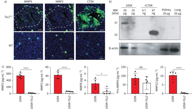
Background: Biomarkers of disease progression and treatment response are urgently needed for patients with lymphangioleiomyomatosis (LAM). Activity-based nanosensors, an emerging biosensor class, detect dysregulated proteases in vivo and release a reporter to provide a urinary readout of disease. Because proteases are dysregulated in LAM and may directly contribute to lung function decline, activity-based nanosensors may enable quantitative, real-time monitoring of LAM progression and treatment response. We aimed to assess the diagnostic utility of activity-based nanosensors in a pre-clinical model of pulmonary LAM.

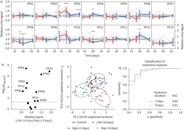
Methods: Tsc2-null cells were injected intravenously into female nude mice to establish a mouse model of pulmonary LAM. A library of 14 activity-based nanosensors, designed to detect proteases across multiple catalytic classes, was administered into the lungs of LAM mice and healthy controls, urine was collected, and mass spectrometry was performed to measure nanosensor cleavage products. Mice were then treated with rapamycin and monitored with activity-based nanosensors. Machine learning was performed to distinguish diseased from healthy and treated from untreated mice.

Results: Multiple activity-based nanosensors (PP03 (cleaved by metallo, aspartic and cysteine proteases), padjusted<0.0001; PP10 (cleaved by serine, aspartic and cysteine proteases), padjusted=0.017)) were differentially cleaved in diseased and healthy lungs, enabling strong classification with a machine learning model (area under the curve (AUC) 0.95 from healthy). Within 2 days after rapamycin initiation, we observed normalisation of PP03 and PP10 cleavage, and machine learning enabled accurate classification of treatment response (AUC 0.94 from untreated).

Conclusions: Activity-based nanosensors enable noninvasive, real-time monitoring of disease burden and treatment response in a pre-clinical model of LAM.


Conflict of interest statement

Conflict of interest: J.D. Kirkpatrick reports grants from the Ludwig Fund for Cancer Research, during the conduct of the study. In addition, J.D. Kirkpatrick has a patent pending (Lung protease nanosensors and uses thereof; PCT/US2019/052868, filed 25 September 2019). Conflict of interest: A.P. Soleimany has nothing to disclose. Conflict of interest: J.S. Dudani has nothing to disclose. Conflict of interest: H-J. Liu has nothing to disclose. Conflict of interest: H.C. Lam has nothing to disclose. Conflict of interest: C. Priolo has nothing to disclose. Conflict of interest: E.P. Henske has nothing to disclose. Conflict of interest: S.N. Bhatia reports other support from the Howard Hughes Medical Institute and National Institute of Environmental Health Sciences, grants from the Ludwig Fund for Cancer Research and grants from the Koch Institute Marble Center for Cancer Nanomedicine, during the conduct of the study; other support from Vertex Pharmaceuticals, Glympse Bio, Maverick Therapeutics, Satellite Bio, CEND Rx and Moderna Therapeutics, outside the submitted work. In addition, S.N. Bhatia has a patent pending (Lung protease nanosensors and uses thereof; PCT/US2019/052868, filed 25 September 2019).

Figures

FIGURE 1
FIGURE 1
Tsc2 deficiency results in aberrant protease expression. a) Immunofluorescence staining (green) of matrix metalloproteinases (MMP) 9 and 2 and cathepsin K (CTSK) in representative primary lesions (outlined in white) that formed spontaneously in the kidneys of Tsc2+/− mice (top) compared with kidneys from healthy control mice (bottom). Blue: 4′,6-diamidino-2-phenylindole (nuclei). Scale bars: 200 μm. b) Western blot against mouse CTSK in 105K cell lysates, recombinant mouse CTSK (rCTSK), and healthy mouse kidney and lung. β-actin immunostaining is shown for each sample. Quantity of protein loaded into each lane is noted. c) Expression (mean±sd), by multiplexed protein assay, of MMPs in conditioned media from 105K cells and 105K cells with retroviral re-introduction of Tsc2 (n=5). ns: nonsignificant; *: p<0.05; ****: p<0.0001 by the two-tailed t-test. WT: wild-type; MW: molecular weight.
FIGURE 2
FIGURE 2
PP03 is cleaved by aspartic proteases in Tsc2-deficient lesions at acidic pH. a) Fluorescence fold change of PP01–PP14 after 30 min of incubation with 105K tumour homogenates diluted in pH 5.25 or pH 7.5 buffer. *: padjusted<0.05; **: padjusted<0.01; ***: padjusted<0.001; ****: padjusted<0.0001 by two-tailed t-test followed by adjustment for multiple hypotheses with Holm–Šídák correction. b) Fluorescence fold change of PP03 after 30 min of incubation with homogenates diluted in pH 5.25 or pH 7.5 buffer with or without pepstatin (“Pep”). ns: nonsignificant; **: p<0.01. c) Substrate cleavage after 30 min in homogenates diluted in pH 5.25 buffer incubated with or without inhibitors against metalloproteases (marimastat), serine proteases (4-(2-aminoethyl)benzenesulfonyl fluoride hydrochloride (AEBSF)), cysteine proteases (E64) or aspartic proteases (pepstatin), relative to uninhibited homogenates. d) Fluorescence increase over time of PP03 incubated with homogenates of 105K cell tumours at pH 5.25 with or without protease inhibitors. e) Fluorescence increase over time of PP01–PP14 incubated with napsin A aspartic peptidase (NAPSA). PP03 is shown in red.
FIGURE 3
FIGURE 3
Activity-based nanosensors discriminate lymphangioleiomyomatosis (LAM) mice from healthy controls. a) Schematic of approach. b) Mean-scaled urinary reporter concentrations in LAM mice and healthy controls were compared at 14 days (LAM: n=19; control: n=9) and 18 days (LAM: n=19; control: n=10) after disease induction and −log10(padjusted) was plotted against fold change between LAM and control. Significance was calculated by the two-tailed t-test followed by adjustment for multiple hypotheses with Holm–Šídák correction. Dotted line is at padjusted=0.05. c) Principal component analysis (PCA) of urinary reporter output of LAM mice and healthy controls at 14 and 18 days after disease induction. d) A random forest classifier was trained on urinary reporters from a subset of LAM mice and healthy controls at both 14 days (LAM: n=5; control: n=5) and 18 days (LAM: n=5; control: n=5). Receiver operating characteristic curves show performance of this classifier in discriminating LAM mice from healthy controls in an independent test cohort at both 14 days (LAM: n=14; control: n=4) and 18 days (LAM: n=14; control: n=5) days. LC: liquid chromatography; MS: mass spectrometry; AUC: area under the curve.
FIGURE 4
FIGURE 4
Activity-based nanosensors enable rapid assessment of drug response in lymphangioleiomyomatosis (LAM). a) Control-normalised urinary reporter signal for each of the 14 activity-based nanosensors PP01–PP14. Thin lines show activity-based nanosensor trajectories of each mouse over time, while thick lines are averages over all mice. Red lines represent LAM mice (n=19) prior to rapamycin treatment and blue lines represent LAM mice treated with 3 mg·kg−1 rapamycin (3–4 times per week). Grey lines represent healthy control mice (n=10). *: p<0.05; **: p<0.01; ***: p<0.001; ****: p<0.0001 from control. Error bars are sd. For clarity, PP14 is presented on a larger scale y-axis. b) Volcano plot showing the significance (−log10(padjusted)) and fold change of each urinary reporter in LAM mice 18 days after 105K-Luc cell injection (“LAM (18 days)”) relative to LAM mice after 2 days of rapamycin treatment (“Rap (+2 days)”). Dotted line is at padjusted=0.05. c) Mean-scaled urinary reporter concentrations were normalised to matched controls at each time-point and subjected to principal component analysis. d) Two random forest classifiers were trained on urinary reporters from a subset of LAM (18 days) mice (n=10) and either Rap (+2 days) (n=10) or Rap (+8 days) (n=10) mice. Receiver operating characteristic curves show performance of these classifiers in discriminating Rap (+2 days) (n=9) and Rap (+8 days) (n=9) mice from untreated LAM (18 days) (n=9) mice in independent, held-out test cohorts from the same experiment. AUC: area under the curve.

Comment in

References

    1. Henske EP, McCormack FX. Lymphangioleiomyomatosis – a wolf in sheep's clothing. J Clin Invest 2012; 122: 3807–3816. doi:10.1172/JCI58709 - DOI - PMC - PubMed
    1. Guo M, Yu JJ, Perl AK, et al. . Single cell transcriptomic analysis identifies a unique pulmonary lymphangioleiomyomatosis cell. Am J Respir Crit Care Med 2020; 202: 1373–1387. doi:10.1164/rccm.201912-2445OC - DOI - PMC - PubMed
    1. Hayashi T, Fleming MV, Stetler-Stevenson WG, et al. . Immunohistochemical study of matrix metalloproteinases (MMPs) and their tissue inhibitors (TIMPs) in pulmonary lymphangioleiomyomatosis (LAM). Hum Pathol 1997; 28: 1071–1078. doi:10.1016/S0046-8177(97)90061-7 - DOI - PubMed
    1. Matsui K, Takeda K, Yu ZX, et al. . Role for activation of matrix metalloproteinases in the pathogenesis of pulmonary lymphangioleiomyomatosis. Arch Pathol Lab Med 2000; 124: 267–275. doi:10.5858/2000-124-0267-RFAOMM - DOI - PubMed
    1. Lee PS, Tsang SW, Moses MA, et al. . Rapamycin-insensitive up-regulation of MMP2 and other genes in tuberous sclerosis complex 2-deficient lymphangioleiomyomatosis-like cells. Am J Respir Cell Mol Biol 2010; 42: 227–234. doi:10.1165/rcmb.2009-0050OC - DOI - PMC - PubMed

Publication types

LinkOut - more resources