. 2021 Oct;297(4):101218.
doi: 10.1016/j.jbc.2021.101218. Epub 2021 Sep 23.

An atomistic model of the coronavirus replication-transcription complex as a hexamer assembled around nsp15

Affiliations

An atomistic model of the coronavirus replication-transcription complex as a hexamer assembled around nsp15

Jason K Perry et al. J Biol Chem. 2021 Oct.

Abstract

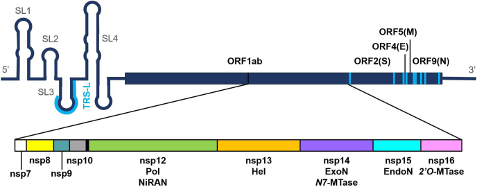
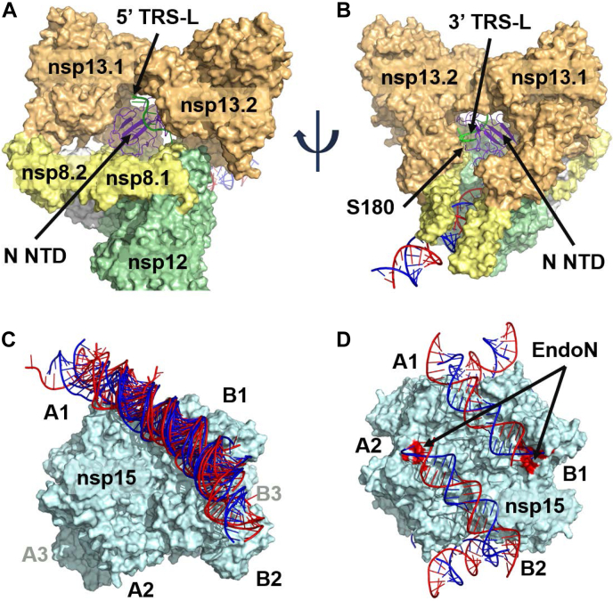
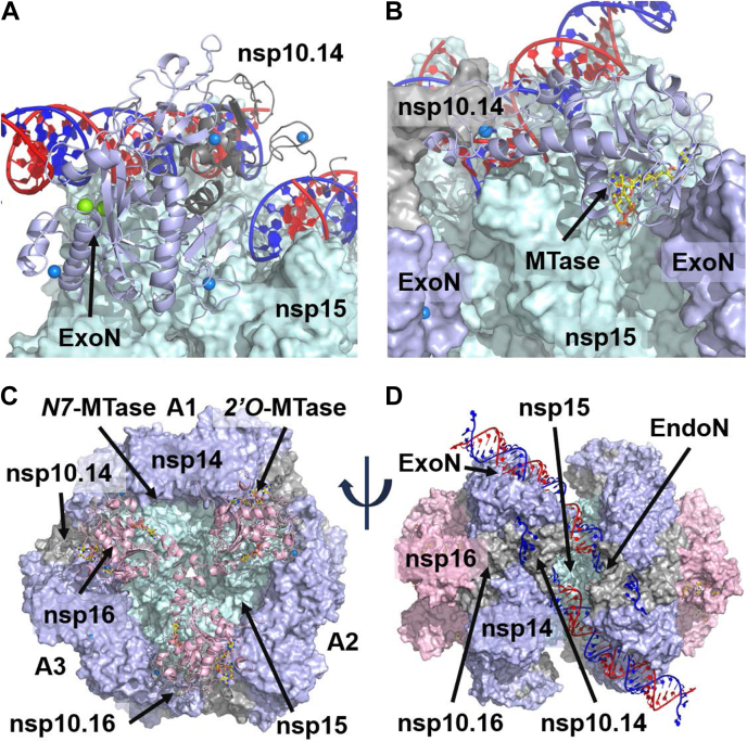
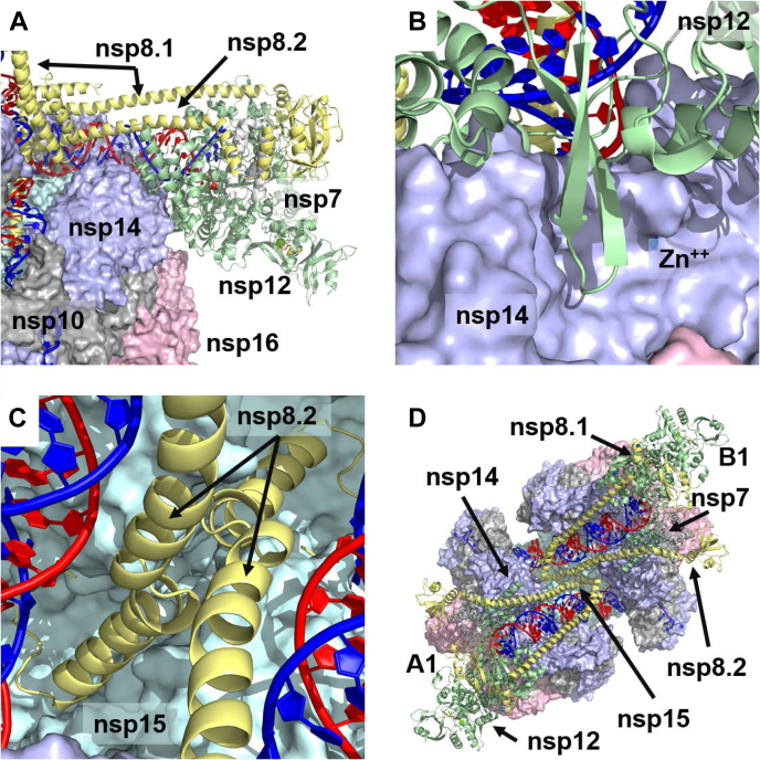
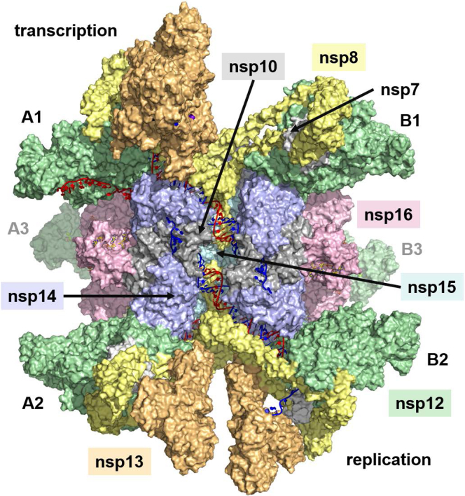
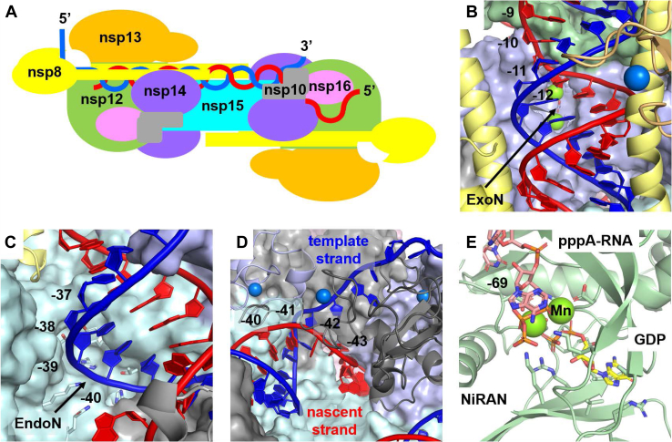
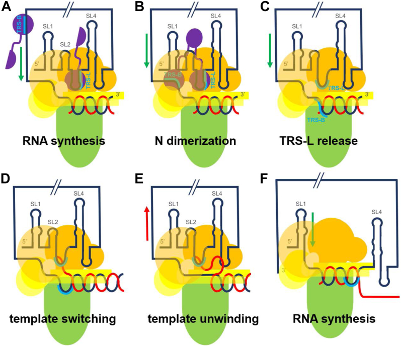
The SARS-CoV-2 replication-transcription complex is an assembly of nonstructural viral proteins that collectively act to reproduce the viral genome and generate mRNA transcripts. While the structures of the individual proteins involved are known, how they assemble into a functioning superstructure is not. Applying molecular modeling tools, including protein-protein docking, to the available structures of nsp7-nsp16 and the nucleocapsid, we have constructed an atomistic model of how these proteins associate. Our principal finding is that the complex is hexameric, centered on nsp15. The nsp15 hexamer is capped on two faces by trimers of nsp14/nsp16/(nsp10)2, which then recruit six nsp12/nsp7/(nsp8)2 polymerase subunits to the complex. To this, six subunits of nsp13 are arranged around the superstructure, but not evenly distributed. Polymerase subunits that coordinate dimers of nsp13 are capable of binding the nucleocapsid, which positions the 5'-UTR TRS-L RNA over the polymerase active site, a state distinguishing transcription from replication. Analysis of the viral RNA path through the complex indicates the dsRNA that exits the polymerase passes over the nsp14 exonuclease and nsp15 endonuclease sites before being unwound by a convergence of zinc fingers from nsp10 and nsp14. The template strand is then directed away from the complex, while the nascent strand is directed to the sites responsible for mRNA capping. The model presents a cohesive picture of the multiple functions of the coronavirus replication-transcription complex and addresses fundamental questions related to proofreading, template switching, mRNA capping, and the role of the endonuclease.

Keywords: SARS-CoV-2; coronavirus; molecular modeling; nsp15; proofreading; protein structure; structure model; viral replication; viral transcription.


Conflict of interest statement

Conflict of interest The authors declare that they have no conflicts of interest with the contents of this article.

Figures

Figure 1
Figure 1
Organization of the SARS-CoV-2 genome. The genome is divided into multiple open reading frames (ORFs), with ORF1ab containing the nonstructural proteins (nsps) required for RNA replication (nsp7–nsp16). The structured 5′-UTR leader contains a transcription regulatory sequence (TRS-L), which is repeated throughout the genome, each instance preceding an ORF (indicated in blue). During negative-strand synthesis, shorter transcripts may be generated when the template switches from one of the TRS locations in the body of the genome to the TRS-L location in the 5′-UTR, effectively skipping over the regions in between.
Figure 2
Figure 2
Significant findings from protein–protein docking.A and B, the N NTD bound to a TRS 10 nt oligo (PDB: 7ACT) docks into the void between the two nsp13 subunits of the polymerase complex. The TRS oligo is positioned over the polymerase active site, parallel to the template, with its 5′ end exposed on the entrance side of the polymerase and its 3′ end exposed on the exit side. The C-term S180 residue of the N NTD is also exposed on the exit side of the polymerase, indicating full-length N could bind to the complex unobstructed. C, multiple examples of dsRNA docked across the nsp15 hexamer, spanning subunit A1 to B1/B2. D, six dsRNA double helices can be symmetrically arranged around the hexamer: three directed from A → B, and three directed in an antiparallel fashion from B → A. Each dsRNA passes over an EndoN active site, colored in red.
Figure 3
Figure 3
Formation of the nsp15/nsp14/nsp16/nsp10 complex.A, the nsp14 ExoN NTD and nsp10 were manually positioned to interact with the dsRNA on the nsp15 hexamer, following the observed Lassa ExoN interaction with dsRNA. Nsp10 is positioned such that its two zinc fingers are over an antiparallel dsRNA, just past the nsp15 EndoN site. Six ExoN/nsp10 subunits can be arranged around the nsp15 hexamer. B, the nsp14 MTase CTD was docked to the nsp15/ExoN/nsp10 hexameric complex. The CTD zinc finger is positioned over an antiparallel dsRNA, opposite the nsp10 associated with another nsp14 subunit. The binding mode reflects a significant conformational change between the two domains of nsp14. C, Nsp16/nsp10 is docked to the nsp15/nsp14/nsp10 hexameric complex. Nsp10 is positioned between two nsp14 subunits, while three nsp16 subunits meet in the middle of the nsp15 trigonal face. D, the full nsp15/nsp14/nsp16/(nsp10)2 hexamer.
Figure 4
Figure 4
Binding of the polymerase to the nsp15/nsp14/nsp16 complex.A, binding of the polymerase is largely through nsp12 interactions with the conformationally altered face of nsp14. Much of this binding comes from the C-term helices of nsp12 (residues 855–923) interacting with the MTase domain of nsp14. B, the nsp12 beta hairpin (residues 815–831) sits in the cleft between the two domains of nsp14, in close proximity to a zinc finger. C, the short N-term helix of nsp8.2 (residues 12–28) extends to interact with nsp15, with the N-term residues (1–11) sitting under the dsRNA just ahead of the EndoN site. D, view of a pair of polymerases bound to the nsp15/nsp14/nsp16 complex. One polymerase complex is associated with the A1 nsp14 subunit, while the other is associated with the B1 subunit. Their nsp8.2 subunits meet in the middle where they interact with nsp15.
Figure 5
Figure 5
The complete replication/transcription complex, with a stoichiometry of six nsp15, six nsp14, six nsp16, six nsp12, six nsp13, six nsp7, 12 nsp8, 12 nsp10, and 2 N proteins. The six nsp13 subunits are arranged across nsp12 pairs in 2/0 (A1/B1), 1/1 (A2/B2), and 0/2 (A3/B3) stoichiometries. The polymerase complexes with two associated nsp13 subunits (A1 and B3) bind the N protein with the TRS-L oligo and are responsible for template switching during negative-strand synthesis (transcription). The two polymerase complexes with a single nsp13 subunit (A2 and B2) are responsible for replication.
Figure 6
Figure 6
Details of some key functions.A, schematic representation of the RNA path. dsRNA makes its way from the nsp12 polymerase, across the nsp14 ExoN and nsp15 EndoN. It is separated into template (blue) and nascent (red) strands at nsp10, and the nascent strand is directed to the NiRAN and two MTase sites. B, detail of dsRNA exiting the polymerase and passing over the ExoN. Nucleotides of the nascent strand are numbered starting from the 3′ primer position (−1), where nucleotide −12 is seen to pass over the ExoN site. The dsRNA is expected to shift into the ExoN active site when encountering a prematurely terminated nascent strand. C, detail of the dsRNA passing over the EndoN site, where nucleotides −39 and −40 of the template strand are best situated for potential cleavage. D, detail of strand separation occurring at the convergence of two zinc fingers from nsp10.14 and one from nsp14 CTD. Strand separation occurs across the base pairs −41 to −43. The template strand is directed away from the complex, while the nascent strand is funneled down to the capping sites. E, detail of the NiRAN site. The first capping step occurs when the NiRAN site transfers GDP to the 5′ pppA-RNA, releasing a pyrophosphate. This occurs at the terminal nucleotide −69 in our model.
Figure 7
Figure 7
Model of template switching [nsp12 (green), nsp13 (orange), nsp8 (yellow), N protein (purple), template RNA (black), nascent RNA (red), TRS (blue)].A, the 5′-UTR coordinates to the nsp13 dimer, with TRS-L bound N protein positioned above the polymerase active site. RNA synthesis begins on the 3′ end of the template. B, synthesis continues until the N protein dimerizes with another N protein bound to TRS-B on the template. C, the N proteins release the RNA. D, the complementary TRS-B of the nascent strand recouples with TRS-L. E, the shift in RNA position triggers nsp13 template backtracking, unwinding the dsRNA. F, once fully unwound, synthesis continues on the 5′ leader, starting from the TRS-L.

References

    1. V'Kovski P., Kratzel A., Steiner S., Stalder H., Thiel V. Coronavirus biology and replication: Implications for SARS-CoV-2. Nat. Rev. Microbiol. 2021;19:155–170. - PMC - PubMed
    1. Hillen H.S., Kokic G., Farnung L., Dienemann C., Tegunov D., Cramer P. Structure of replicating SARS-CoV-2 polymerase. Nature. 2020;584:154–156. - PubMed
    1. Lehmann K.C., Gulyaeva A., Zevenhoven-Dobbe J.C., Janssen G.M., Ruben M., Overkleeft H.S., van Veelen P.A., Samborskiy D.V., Kravchenko A.A., Leontovich A.M., Sidorov I.A., Snijder E.J., Posthuma C.C., Gorbalenya A.E. Discovery of an essential nucleotidylating activity associated with a newly delineated conserved domain in the RNA polymerase-containing protein of all nidoviruses. Nucleic Acids Res. 2015;43:8416–8434. - PMC - PubMed
    1. Slanina H., Madhugiri R., Bylapudi G., Schultheiss K., Karl N., Gulyaeva A., Gorbalenya A.E., Linne U., Ziebuhr J. Coronavirus replication-transcription complex: Vital and selective NMPylation of a conserved site in nsp9 by the NiRAN-RdRp subunit. Proc. Natl. Acad. Sci. U. S. A. 2021;118 - PMC - PubMed
    1. Yan L., Ge J., Zheng L., Zhang Y., Gao Y., Wang T., Huang Y., Yang Y., Gao S., Li M., Liu Z., Wang H., Li Y., Chen Y., Guddat L.W. Cryo-EM structure of an extended SARS-CoV-2 replication and transcription complex reveals an intermediate state in cap synthesis. Cell. 2021;184:184–193.e110. - PMC - PubMed

Publication types

MeSH terms

Substances

LinkOut - more resources