Review
. 2021 Aug 27;11(9):299.
doi: 10.3390/bios11090299.

Biodegradable Metal Organic Frameworks for Multimodal Imaging and Targeting Theranostics

Affiliations
Review

Biodegradable Metal Organic Frameworks for Multimodal Imaging and Targeting Theranostics

Xiangdong Lai et al. Biosensors (Basel). .

Abstract

Though there already had been notable progress in developing efficient therapeutic strategies for cancers, there still exist many requirements for significant improvement of the safety and efficiency of targeting cancer treatment. Thus, the rational design of a fully biodegradable and synergistic bioimaging and therapy system is of great significance. Metal organic framework (MOF) is an emerging class of coordination materials formed from metal ion/ion clusters nodes and organic ligand linkers. It arouses increasing interest in various areas in recent years. The unique features of adjustable composition, porous and directional structure, high specific surface areas, biocompatibility, and biodegradability make it possible for MOFs to be utilized as nano-drugs or/and nanocarriers for multimodal imaging and therapy. This review outlines recent advances in developing MOFs for multimodal treatment of cancer and discusses the prospects and challenges ahead.

Keywords: biodegradable materials; metal ion nodes; metal-organic framework; multimode imaging; theranostic nano-platforms.


Conflict of interest statement

The authors declare no conflict of interest.

Figures

Figure 1
Figure 1
Schematic diagram of MOFs as nano-drugs and nanocarriers for multimodal theranostic, typically comprising a suitable and effective combination of CT, CDT, RT, RDT, PTT, PDT, MDT, MTT, gas therapy and gene therapy, and imaging of FI, MRI, PAI, CTI and PTI.
Figure 2
Figure 2
(A) B16F10 cellular uptake of Cu-TBP or H4TBP at different time-points after incubation with equivalent TBP concentrations of 20 mM observed by confocal imaging. Free H4TBP emits red fluorescence. Scale bar, 50 μm. (B) Synergy of Cu-TBP mediated radical therapy stimulated by hormone, light and checkpoint blockade immunotherapy. Reprinted with permission from Ref. [54]. Copyright 2019, Elsevier.
Figure 3
Figure 3
Mn-Zr MOF generates abundant ROS of ·OH and a high microwave thermal conversion efficiency after exposure to MW irradiation, resulting in efficiently inhibiting the cancerous cell growth through the synergic effect of MDT and MTT. Reprinted with permission from Ref. [55]. Copyright 2018, American Chemical Society.
Figure 4
Figure 4
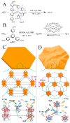
The synthesis methods, morphologies and structures of Hf12-Ir MOF nanolayer (A,C) and Hf6-Ir MOF nanolayer (B,D) and X-ray induced ROS generation. Reprinted with permission from Ref. [56]. Copyright 2018, American Chemical Society.
Figure 5
Figure 5
(A) MRI of the Cu-TCPP aqueous solution with different concentrations. (B) Plots of the 1/T1 value of the Cu-TCPP under concentration dependence. (C) mouse MRI before and after intratumoral injection of the Cu-TCPP. Red circles indicate the position of the tumor. Reprinted with permission from Ref. [41]. Copyright 2018, Ivyspring International Publisher.
Figure 6
Figure 6
(A) Fluorescence imaging of SGC-7901 tumor-bearing model mice after intravenous injection of different materials at 7 time points. Unit of scale bar: (p/s/cm2/sr)/(mW/cm2). (B) Ex vivo Fluorescence imaging of tumor, heart, lung, liver, spleen, and kidney in sequence in SGC-7901 tumor-bearing model mice after intravenous injection of G-BHM at 5 different time points. (C) Time-dependent in vivo integrated FL intensity for different materials (top) and in different organs (bottom). (D) PA imaging of SGC-7901 tumor after injection of G-BHM at different time points. (E) Stereoscopic PA images, and white arrow represents tumor zone. (F) PA signal intensity variation corresponding to part (D). Reprinted with permission from Ref. [66]. Copyright 2019, Elsevier Inc.
Figure 7
Figure 7
The structure of Dox@MOF-Au-PEG and the underlying of O2-generating synergistic chemoradiotherapy Reprinted with permission from Ref. [67]. Copyright 2019, Wiley-VCH Verlag GmbH & Co. KGaA, Weinheim.
Figure 8
Figure 8
siRNA/Zr-FeP MOF mediates PTT at a lower temperature and PDT for cancer. Reprinted with permission from Ref. [46]. Copyright 2018, Elsevier B.V.
Figure 9
Figure 9
(A) the preparation of Cypate@MIL-53/PEG-Transferrin MOF composite and (B) its bioapplication for PDT and PTT. Reprinted with permission from Ref. [70]. Copyright 2019, American Chemical Society.
Figure 10
Figure 10
(A) Infrared thermal imaging and (B) tumor temperature of 4T1-tumor model mice after intravenous injection with PBAM under 808 nm laser (1 W cm−2). White circle: tumor tissue. (C) Tumor PAI and (D) Corresponding PAI signal intensity of 4T1-tumor model mice after intravenous injection with PBAM. (E) Tumor PA signal intensity and corresponding PAI of 4T1-tumor model mice after subcutaneous injection with PBAM. (F) MRI of 4T1-tumor model mice after subcutaneous injection with PBAM. White circle: tumor tissue. (G) Schematic of lymphatic metastasis tumor model. (H) PAI of lymph nodes with or without metastasis at different time points after injection with PBAM. White circle: the lymph nodes in left leg. Red circle: the lymph nodes in right leg. (I) Corresponding PAI signal intensity of lymph nodes with or without metastasis. (J) MRI of lymph nodes with or without metastasis at different time points after injection with PBAM. Reprinted with permission from Ref. [20]. Copyright 2020, WILEY-VCH Verlag GmbH & Co. KGaA, Weinheim.

Similar articles

Cited by

References

    1. Allmani C., Matsuda T., Di Carlo V., Harewood R., Matz M., Niksic M., Bonaventure A., Valkov M., Johnson C.J., Esteve J., et al. Global surveillance of trends in cancer survival 2000–14 (CONCORD-3): Analysis of individual records for 37 513 025 patients diagnosed with one of 18 cancers from 322 population-based registries in 71 countries. Lancet. 2018;391:1023–1075. doi: 10.1016/S0140-6736(17)33326-3. - DOI - PMC - PubMed
    1. Li S., Jiang Q., Liu S., Zhang Y., Tian Y., Song C., Wang J., Zou Y., Anderson G.J., Han J.Y., et al. A DNA nanorobot functions as a cancer therapeutic in response to a molecular trigger in vivo. Nat. Biotechnol. 2018;36:258–264. doi: 10.1038/nbt.4071. - DOI - PubMed
    1. Cho K., Wang X., Nie S., Chen Z., Shin D.M. Therapeutic Nanoparticles for Drug Delivery in Cancer. Clin. Cancer Res. 2008;14:1310. doi: 10.1158/1078-0432.CCR-07-1441. - DOI - PubMed
    1. Goel S., Ferreira C.A., Chen F., Ellison P.A., Siamof C.M., Barnhart T.E., Cai W. Activatable Hybrid Nanotheranostics for Tetramodal Imaging and Synergistic Photothermal/Photodynamic Therapy. Adv. Mater. 2018;30:1704367. doi: 10.1002/adma.201704367. - DOI - PMC - PubMed
    1. Wang H., Agarwal P., Zhao G., Ji G., Jewell C.M., Fisher J.P., Lu X., He X. Overcoming Ovarian Cancer Drug Resistance with a Cold Responsive Nanomaterial. ACS Cent. Sci. 2018;4:567–581. doi: 10.1021/acscentsci.8b00050. - DOI - PMC - PubMed

Substances