

Periostin expression and its supposed roles in benign and malignant thyroid nodules: an immunohistochemical study of 105 cases
- PMID: 34563225
- PMCID: PMC8465710
- DOI: 10.1186/s13000-021-01146-8
Periostin expression and its supposed roles in benign and malignant thyroid nodules: an immunohistochemical study of 105 cases
Abstract
Background: Thyroid tumors are often difficult to histopathologically diagnose, particularly follicular adenoma (FA) and follicular carcinoma (FC). Papillary carcinoma (PAC) has several histological subtypes. Periostin (PON), which is a non-collagenous extracellular matrix molecule, has been implicated in tumor invasiveness. We herein aimed to elucidate the expression status and localization of PON in thyroid tumors.
Method: We collected 105 cases of thyroid nodules, which included cases of adenomatous goiter, FA, microcarcinoma (MIC), PAC, FC, poorly differentiated carcinoma (PDCa), and undifferentiated carcinoma (UCa), and immunohistochemically examined the PON expression patterns of these lesions.
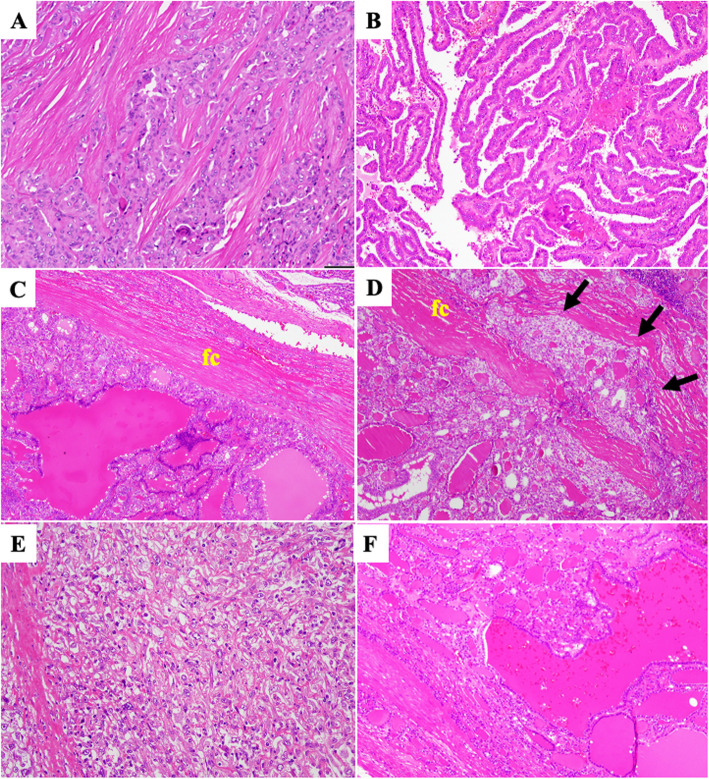
Results: Stromal PON deposition was detected in PAC and MIC, particularly in the solid/sclerosing subtype, whereas FA and FC showed weak deposition on the fibrous capsule. However, the invasive and/or extracapsular regions of microinvasive FC showed quite strong PON expression. Except for it, we could not find any significant histopathological differences between FA and FC. There were no other significant histopathological differences between FA and FC. Although PDCa showed a similar PON expression pattern to PAC, UCa exhibited stromal PON deposition in its invasive portions and cytoplasmic expression in its carcinoma cells. Although there was only one case of UCa, it showed strong PON immunopositivity. PAC and MIC showed similar patterns of stromal PON deposition, particularly at the invasive front.
Conclusions: PON may play a role in the invasion of thyroid carcinomas, particularly PAC and UCa, whereas it may act as a barrier to the growth of tumor cells in FA and minimally invasive FC.
© 2021. The Author(s).
Conflict of interest statement
The authors declare that they have no competing interests related to the present study.
Figures


Similar articles
-
Challenge in the Pathological Diagnosis of the Follicular- Patterned Thyroid Lesions.Asian Pac J Cancer Prev. 2021 Oct 1;22(10):3365-3376. doi: 10.31557/APJCP.2021.22.10.3365. Asian Pac J Cancer Prev. 2021. PMID: 34711014 Free PMC article.
-
[Expression of cytokeratin19, galectin-3 and HBME-1 in thyroid lesions and their differential diagnoses].Zhonghua Bing Li Xue Za Zhi. 2004 Jun;33(3):212-6. Zhonghua Bing Li Xue Za Zhi. 2004. PMID: 15256110 Chinese.
-
Cyclin D1 in well differentiated thyroid tumour of uncertain malignant potential.Diagn Pathol. 2015 Apr 18;10:32. doi: 10.1186/s13000-015-0262-8. Diagn Pathol. 2015. PMID: 25907675 Free PMC article.
-
[Update on thyroid immunocytochemistry and its value for managing patients with thyroid nodules].Ann Pathol. 2006 Oct;26(5):340-5. doi: 10.1016/s0242-6498(06)70738-6. Ann Pathol. 2006. PMID: 17255921 Review. French.
-
Primary thyroid biphasic synovial sarcoma and synchronous papillary carcinoma: report of a remarkable case.Pathologica. 2018 Sep;110(2):106-110. Pathologica. 2018. PMID: 30546147 Review.
Cited by
-
Identifying key genes of classic papillary thyroid cancer in women aged more than 55 years old using bioinformatics analysis.Front Endocrinol (Lausanne). 2022 Sep 2;13:948285. doi: 10.3389/fendo.2022.948285. eCollection 2022. Front Endocrinol (Lausanne). 2022. PMID: 36120433 Free PMC article.
-
Spatial Transcriptomics in a Case of Follicular Thyroid Carcinoma Reveals Clone-Specific Dysregulation of Genes Regulating Extracellular Matrix in the Invading Front.Endocr Pathol. 2024 Jun;35(2):122-133. doi: 10.1007/s12022-024-09798-0. Epub 2024 Jan 27. Endocr Pathol. 2024. PMID: 38280140 Free PMC article.
-
Effect of hypoxia‑HIF‑1α‑periostin axis in thyroid cancer.Oncol Rep. 2024 Apr;51(4):57. doi: 10.3892/or.2024.8716. Epub 2024 Feb 23. Oncol Rep. 2024. PMID: 38391012 Free PMC article.
References
-
- Rosai J, Albores Saavedra J, Asioki S, Bolach ZW, Bogdova T, Chen H. Papillary carcinoma. In: Lloyd RV, Osamura RY, Kloppel G, Rosai J, editors. WHO classification of Tumours of endocrine organs. 4th ed. Lyon: IARC Press. 2017:81–91.
-
- LiVolsi V, Abdulkader Nalib I, Baloch ZW, Bartolazzi A, Chan JKC, DeLellis RA. Follicular thyroidal carcinoma. In: Lloyd RV, Osamura RY, Kloppel G, Rosai J, editors. WHO classification of Tumours of endocrine organs. 4th ed. Lyon: IARC Press. 2017:92–5.
-
- Horiuchi K, Amizuka N, Takeshita S, Takamatsu H, Katsuura M, Ozawa H, et al. Identification and characterization of a novel protein, periostin, with restricted expression to periosteum and periodontal ligament and increased expression by transforming growth factor beta. J Bone Miner Res. 1999;14(7):1239–1249. doi: 10.1359/jbmr.1999.14.7.1239. - DOI - PubMed
MeSH terms
Substances
Supplementary concepts
Grants and funding
LinkOut - more resources
Full Text Sources
Medical
Miscellaneous
