Review
. 2021 Sep 25;25(1):29.
doi: 10.1186/s40824-021-00229-3.

Polyphenols-loaded electrospun nanofibers in bone tissue engineering and regeneration

Affiliations
Review

Polyphenols-loaded electrospun nanofibers in bone tissue engineering and regeneration

Iruthayapandi Selestin Raja et al. Biomater Res. .

Abstract

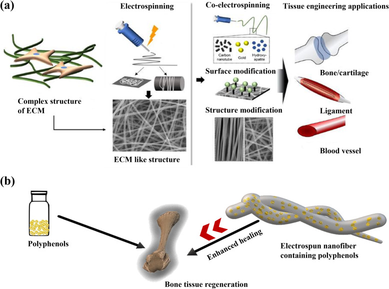
Bone is a complex structure with unique cellular and molecular process in its formation. Bone tissue regeneration is a well-organized and routine process at the cellular and molecular level in humans through the activation of biochemical pathways and protein expression. Though many forms of biomaterials have been applied for bone tissue regeneration, electrospun nanofibrous scaffolds have attracted more attention among researchers with their physicochemical properties such as tensile strength, porosity, and biocompatibility. When drugs, antibiotics, or functional nanoparticles are taken as additives to the nanofiber, its efficacy towards the application gets increased. Polyphenol is a versatile green/phytochemical small molecule playing a vital role in several biomedical applications, including bone tissue regeneration. When polyphenols are incorporated as additives to the nanofibrous scaffold, their combined properties enhance cell attachment, proliferation, and differentiation in bone tissue defect. The present review describes bone biology encompassing the composition and function of bone tissue cells and exemplifies the series of biological processes associated with bone tissue regeneration. We have highlighted the molecular mechanism of bioactive polyphenols involved in bone tissue regeneration and specified the advantage of electrospun nanofiber as a wound healing scaffold. As the polyphenols contribute to wound healing with their antioxidant and antimicrobial properties, we have compiled a list of polyphenols studied, thus far, for bone tissue regeneration along with their in vitro and in vivo experimental biological results and salient observations. Finally, we have elaborated on the importance of polyphenol-loaded electrospun nanofiber in bone tissue regeneration and discussed the possible challenges and future directions in this field.

Keywords: Bone tissue regeneration; Drug loading; Electrospun nanofiber; Polyphenols.


Conflict of interest statement

The authors declare that they have no competing interests.

Figures

Fig. 1
Fig. 1
Demonstration of molecular signaling pathways of polyphenols involved in bone tissue regeneration. ROS- reactive oxygen species; p53- tumor suppressor; Gpx-1- glutathione peroxidase 1; SOD- superoxide dismutase; RANKL- receptor activator of nuclear factor kappa-Β ligand; NF-κB- nuclear factor kappa-light-chain-enhancer of activated B cells; NFATc1- nuclear factor of activated T cells 1; c-Fos- proto-oncogene; MAPKs- mitogen-activated protein kinases; MMPs- matrix metalloproteinases; ECM- extracellular matrix [13]
Fig. 2
Fig. 2
Electrospun nanofibers imitating extracellular matrix (ECM). a Major types of electrospinning and post-modification of electrospun nanofibers for the application of bone tissue engineering are demonstrated [31]. b Though polyphenols alone can help bone tissue regeneration, electrospun nanofiber containing polyphenols shows enhanced wound healing due to the sustained release of bioactive molecules from the scaffold
Fig. 3
Fig. 3
Classification of bone tissue cells. Osteogenic cells, osteocytes, osteoclasts, and osteoblast are the primary bone cells involved in bone remodeling and formation [48]
Fig. 4
Fig. 4
In vitro mineralization assay of bone marrow mesenchymal stem cells (BMSCs) during osteogenic differentiation following treatment with curcumin (CR) and all-trans-retinoic acid (ATRA) groups. BMSCs in the osteogenic medium were indicated by the OM group. Alkaline phosphatase (ALP), Alizarin red (ALZ), and von Kossa (VK) staining results were obtained after 1, 3, and 4 weeks of post-induction, respectively. Quantification of staining intensity was performed with ImageJ software. The level of calcium deposition reflects the extent of mineralization, which was higher in the CR group than in the OM and ATRA groups. ATRA group did not show any symptoms of mineralization. The data were represented as the mean ± standard deviation (n = 3). The statistical significance was defined using a one-way analysis of variance, followed by Tukey’s post hoc test. *p < 0.05, **p < 0.01 and, ***p < 0.001 [59]
Fig. 5
Fig. 5
Analyses of in vitro cell viability and calcium deposition of catecholamine containing collagen nanofiber. A Electrospun nanofibrous mat was prepared from the composite of collagen (8% w/v), dopamine (10% w/w of collagen), and 20 mM CaCl2 in 90% HFIP. The brown coloration in the mats is due to the formation of polydopamine by electrochemical oxidation. Intensified brown color in mat and precipitation of CaCO3 occurred by the subsequent exposure of the mat to (NH4)2CO3 vapors. The nanofibrous mat exhibited excellent mechanical properties, surface wettability, fluorescence, and osteoblast cell proliferation and differentiation. B Human fetal osteoblastic cell line (hFob) viability was quantified from live/dead cell ratio cultured on various collagen scaffolds and tissue culture plate (TCP) (Mean ± SD, n = 3). C Calcium deposition on various collagen mats by ARS (Alizarin Red S) staining with scale bar = 50 μm. (a) TCP, (b) Pristine collagen mats (ES-Coll), (c) As-spun collagen mats with DA and 20 mM Ca2+ (Coll-DA-Ca), (d) As-spun collagen mats with NE and 20 mM Ca2+ (Coll-NE-Ca), (e) Collagen mats after (NH4)2CO3 exposure (Coll-pDA-Ca), and (f) Coll-pNE-Ca [102]
Fig. 6
Fig. 6
In vivo bone regeneration of polycaprolactone (PCL), catechin-loaded PCL (PCL-Cat), hADSC transplanted PCL (PCL-hADSC), and Cat-hADSC transplanted PCL (PCL-Cat-hADSC) scaffolds in critical-sized calvarial bone defect mouse model. A Micro-CT images of the defect site have been seen with a scale bar of 1 mm on 8 weeks of post-transplantation. Quantification of bone coverage area (%) and bone volume (%) has been shown in (B) and (C), respectively (n = 10, **p < 0.01 vs. no treatment group; #p < 0.05 and ##p < 0.01 vs. PCL group; +p < 0.05 vs. PCL-Cat group). D Colorized mineral map of the cross-sectioned micro-CT images with scale bar 1 μm. The defect region is indicated with white arrowheads. (E) Goldner’s Trichrome staining of each group (left, 1 mm) with their expanded images (right, 100 μm). Black arrowheads indicate the defect region [96]

References

    1. Scalbert A, Manach C, Morand C, Rémésy C, Jiménez L. Dietary polyphenols and the prevention of diseases. Crit Rev Food Sci Nutr. 2005;45:287–306. doi: 10.1080/1040869059096. - DOI - PubMed
    1. Rah DK, Han D-W, Baek HS, Hyon S-H, Park BY, Park J-C. Protection of rabbit kidney from ischemia/reperfusion injury by green tea polyphenol pretreatment. Arch Pharm Res. 2007;30:1447–1454. doi: 10.1007/BF02977370. - DOI - PubMed
    1. Nicolin V, De Tommasi N, Nori SL, Costantinides F, Berton F, Di Lenarda R. Modulatory effects of plant polyphenols on bone remodeling: a prospective view from the bench to bedside. Front Endocrinol. 2019;10:494. doi: 10.3389/fendo.2019.00494. - DOI - PMC - PubMed
    1. Park YH, Han D-W, Suh H, Ryu GH, Hyon SH, Cho BK, Park JC. Protective effects of green tea polyphenol against reactive oxygen species-induced oxidative stress in cultured rat calvarial osteoblast. Cell Biol Toxicol. 2003;19:325–337. doi: 10.1023/B:CBTO.0000004986.51081.c5. - DOI - PubMed
    1. Rah DK, Han DW, Baek HS, Hyon SH, Park JC. Prevention of reactive oxygen species-induced oxidative stress in human microvascular endothelial cells by green tea polyphenol. Toxicol Lett. 2005;155:269–275. doi: 10.1016/j.toxlet.2004.10.002. - DOI - PubMed