Review
. 2021 Aug 30;9(3):36.
doi: 10.3390/jdb9030036.

Regeneration in Reptiles Generally and the New Zealand Tuatara in Particular as a Model to Analyse Organ Regrowth in Amniotes: A Review

Affiliations
Review

Regeneration in Reptiles Generally and the New Zealand Tuatara in Particular as a Model to Analyse Organ Regrowth in Amniotes: A Review

Lorenzo Alibardi et al. J Dev Biol. .

Abstract

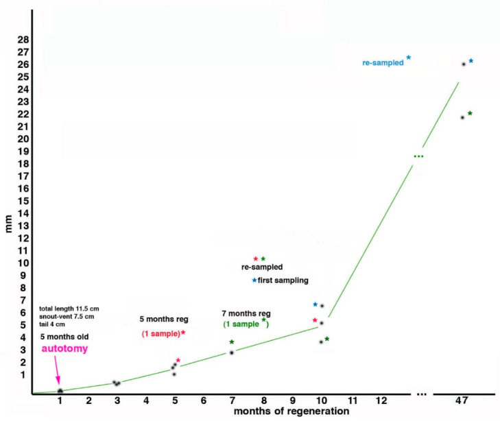
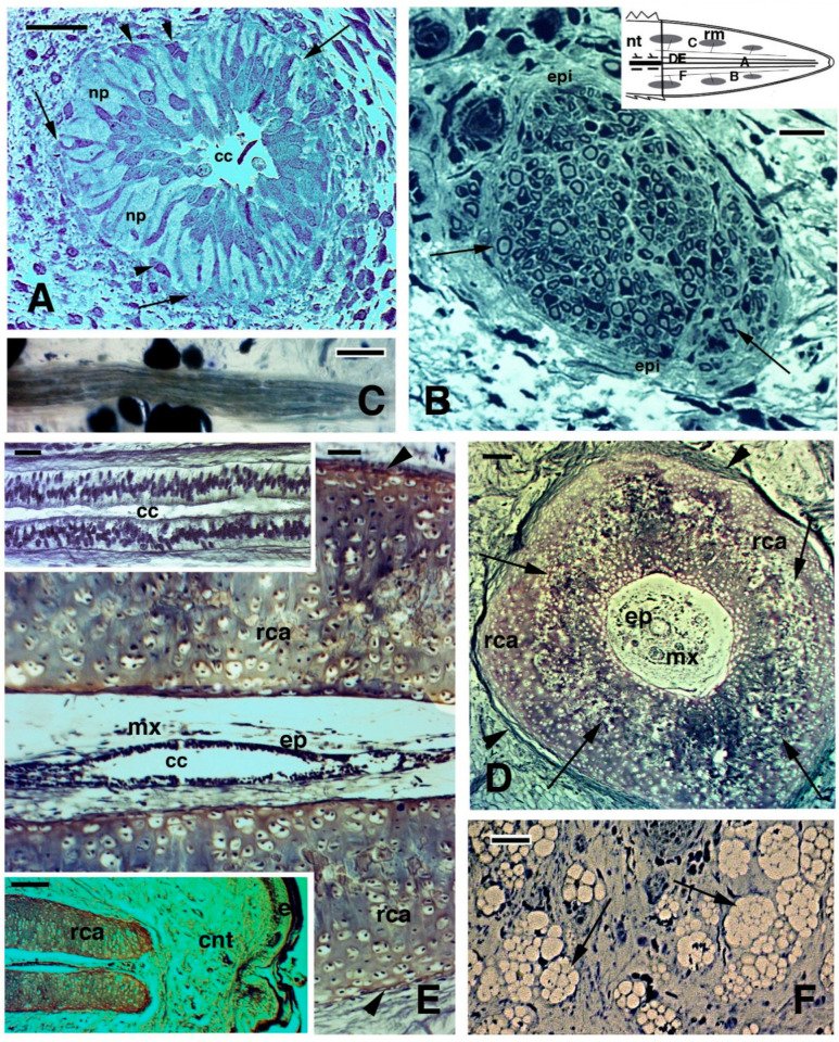
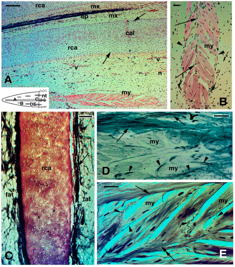
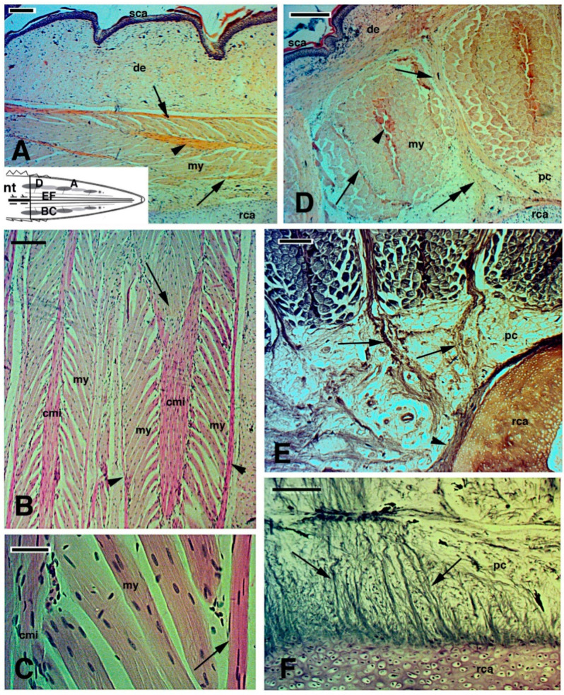
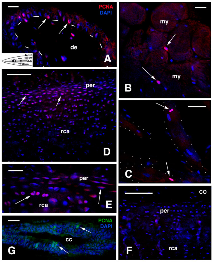
The ability to repair injuries among reptiles, i.e., ectothermic amniotes, is similar to that of mammals with some noteworthy exceptions. While large wounds in turtles and crocodilians are repaired through scarring, the reparative capacity involving the tail derives from a combined process of wound healing and somatic growth, the latter being continuous in reptiles. When the tail is injured in juvenile crocodilians, turtles and tortoises as well as the tuatara (Rhynchocephalia: Sphenodon punctatus, Gray 1842), the wound is repaired in these reptiles and some muscle and connective tissue and large amounts of cartilage are regenerated during normal growth. This process, here indicated as "regengrow", can take years to produce tails with similar lengths of the originals and results in only apparently regenerated replacements. These new tails contain a cartilaginous axis and very small (turtle and crocodilians) to substantial (e.g., in tuatara) muscle mass, while most of the tail is formed by an irregular dense connective tissue containing numerous fat cells and sparse nerves. Tail regengrow in the tuatara is a long process that initially resembles that of lizards (the latter being part of the sister group Squamata within the Lepidosauria) with the formation of an axial ependymal tube isolated within a cartilaginous cylinder and surrounded by an irregular fat-rich connective tissue, some muscle bundles, and neogenic scales. Cell proliferation is active in the apical regenerative blastema, but much reduced cell proliferation continues in older regenerated tails, where it occurs mostly in the axial cartilage and scale epidermis of the new tail, but less commonly in the regenerated spinal cord, muscles, and connective tissues. The higher tissue regeneration of Sphenodon and other lepidosaurians provides useful information for attempts to improve organ regeneration in endothermic amniotes.

Keywords: Sphenodon; Squamata; autotomy; lepidosauria; microscopy; morphogenesis; reptilia; rhynchocephalia; tail.


Conflict of interest statement

The authors declare that there is no conflict of interest.

Figures

Figure 1
Figure 1
(A), Schematic cladogramme indicating the two main amniote radiation paths from basal amniotes, i.e., synapsids-mammals (pink) and sauropsids that include reptiles (green) and birds (light blue). (B), Table indicating the main organ and tissues undergoing wound healing with scarring, regengrow and heteromorphic regeneration in different sub-orders of Reptiles.
Figure 2
Figure 2
Histological images (stained with Haematoxylin-Eosin in (AC), and Palmgreen stain in (D,E)) of the caudal vertebrae of a normal tail in the tuatara (inset in Figure A, Bar, 2 mm). (A), image showing the spinal cord, meninx (artifactually dislocated after sectioning), and ventrally the vertebral body with the intra-vertebral fracture plane indicated (arrow), which has artifactually been separated during sectioning. Bar, 100 μm. (B), closer view of fracture plane with fragments of blue-stained cartilaginous cells (arrows), probably dislocated from the articular surfaces during sectioning. Bar, 100 μm. (C), close-up of the intra-vertebral splitting plane where numerous fibro-cartilaginous/connective cells (arrowheads) are present in continuation with the periosteum (arrow). Bar, 50 μm. (D), peri-vertebral region rich in fat cells and loose connective showing two fibrous bundles (arrows) connecting pre- and post-vertebral bodies at the fracture plane to the inter-muscle connective septa (arrowheads). The image represents the plane of autotomy of the tail, which continues (not shown) into the dermis to externally reach the scales. Bar, 100 μm. (E), detail of the fibrous bundles (arrows) connected to the pre-vertebral and post-vertebral bodies at the fracture or splitting autotomous plane. Bar, 50 μm. Legends: bm, bone marrow; bo, vertebral bone; crt, cartilage; fp, fracture plane; mx, meninx; pt, post-vertebral body (more caudal) at the fracture plane; pv, pre-vertebral body (more rostral) at the fracture plane; sc, spinal cord; vb, vertebral body. Note: All micrographs are based on material obtained in 1988 and 1989 through a permit issued by Mr Ian Govey of the New Zealand Department of Conservation and Dr Mike Thompson of Victoria University, Wellington (New Zealand). The material was used in Alibardi and Meyer-Rochow [52,55] and all subsequent publications on tuatara by these authors.
Figure 3
Figure 3
Growth of the regenerating tail during 47 months associated with body regeneration in three tuatara (indicated by asterisk of different coloration), received initially at 3 months of age, autotomized at 5 months of age at about half-distal length of the tail, measured and sampled at different periods thereafter (see text for further explanations). The ordinate refers to the growth of the regenerate, while the abscissa refers to time.
Figure 4
Figure 4
Gross aspect (A) and histology of regenerating tail ((BE), Toluidine blue stain). (A), juvenile of about 4 years of age with regenerated tail (arrowhead). Bar, 1 cm. In the upper inset (Bar, 1 mm) a blastema of about 2 months is shown. The scar (arrowhead) depicted in (A) lower inset formed after about 3 months following limb amputation. (B), blastema at about 3 months with a loose connective covered by a thick wound epidermis. Bar, 10 μm. This schematic inset shows a blastema with the regions shown in (BE). (C), proximal area of cross-sectioned conical blastema of 10 months post-autotomy showing three pro-muscle aggregates (arrows) separated by forming connective septa. A dense dermis is present beneath the cornified epidermis. Bar, 50 μm. (D), detail of a muscle bundle at 10 months post-autotomy. The arrow indicates a myotube in cross-section. Bar, 10 μm. (E), cross sectioned central cartilaginous cylinder surrounding the ependymal canal in proximal regions of a cone of 7 months post-autotomy. Arrows point to the outer and inner perichondria. Bar, 50 μm. Legends: bl, blastema; cs, connective septa (forming intermuscle); de, dermis; ep, ependymal canal; mx, meninge; nt, normal tail (stump containing vertebrae and spinal cord); rca, regenerated cartilage; we, wound (regenerating) epidermis. Note: All micrographs are based on material obtained in 1988 and 1989 through a permit issued by Mr Ian Govey of the New Zealand Department of Conservation and Dr Mike Thompson of Victoria University, Wellington (New Zealand). The material was used in Alibardi and Meyer-Rochow [52,59,60,62].
Figure 5
Figure 5
Histology of regenerating tail (A,B,F) Toludine blue stain; (D,E) Palmgreen stain). (A) cross-sectioned ependymal tube showing the elongation of ependymal tanicytes ending on the basement membrane (arrows). Arrowheads point to glial cells detached from the ependymal epithelium. Bar, 10 μm. (B), cross-sectioned myelinated (arrows) nerve at 10 months regeneration. Bar, 10 μm. The schematic drawing shows the indicated positions of the figures. (C), longitudinal section of myelinated nerve 10 months post-autotomy. Bar, 10 μm. (D), cross section of the cartilaginous tube in a long regenerated tail of unknown age. Arrows indicate intra-cartilaginous areas of calcification/degeneration. Arrowheads indicate the fibrous connective contacting the perichondrium. Bar, 50 μm. (E) longitudinal section of axial cartilage in an old regenerate of unknown age. The outer perichondrium is indicated by arrowheads. Bar, 20 μm. The upper inset (Bar, 10 μm) details the pseudostratified ependymal epithelium. The lower inset (Bar, 50 μm) instead shows the apical end of the cartilaginous tube, close to the connective tissue of the tip of the regenerated tail. (F), numerous fat cells (arrows) are present around the cartilaginous tube. Bar, 20 μm. Legends: ca, regenerated cartilage; cc, central canal; cnt, connective (fibrous) tissue; e, epidermis of neogenic apical scale; ep, ependymal epithelium; epi, epinevrio; mx, meninx; np, areas occupied from axons and neuropile; nt, normal tail (stump);rca, regenerated cartilage; rm, regenerating muscles/myomeres. Note: All micrographs are based on material obtained in 1988 and 1989 through a permit issued by Mr Ian Govey of the New Zealand Department of Conservation and Dr Mike Thompson of Victoria University, Wellington (New Zealand). The material was used in Alibardi and Meyer-Rochow [52,58,59,62].
Figure 6
Figure 6
Histology of regenerating tail of unknown age (A,B) Haematoxylin-eosin stain; (CE), Palmgreen stain). (A), mid-apical region showing a distinctly cellular cartilage with outer and inner peripheries (arrows) representing the perichondrium. Bar, 50 μm. The schematic drawing shows the indicative positions of the following figures. (B), detail on a forming, leaf-like myomere, with an axial intermuscle connective (arrows) and external limiting connective septa (arrowheads). Bar, 10 μm.(C), tangential longitudinal section of the axial cartilage surrounded by fat connective tissue in an old (age unknown) regenerating tail. Arrows indicate the fibrous layer in contact with the perichondrium. Bar, 50 μm. (D), detail showing nerve fibres coursing within the outer connective septum (arrows) and other nerves entering the myofibres (arrowheads). Bar, 10 μm. (E), additional details of nerve endings from peripheral (arrow) and central myoseptum (double arrow) entering myofibres (arrowheads). Bar, 10 μm. Legends: cal, beginning of cartilage calcification; ep, ependyma; fat, connective tissues rich in fat cells; mx, meninx; my, multinuclear myotubes/myofibres; rca, regenerated cartilaginous tube; v, blood vessels. Note: All micrographs are based on material obtained in 1988 and 1989 through a permit issued by Mr Ian Govey of the New Zealand Department of Conservation and Dr Mike Thompson of Victoria University, Wellington (New Zealand). The material was used in Alibardi and Meyer-Rochow [52,58,62].
Figure 7
Figure 7
Images derived from different areas of old regenerated tails of unknown age (AD), Haematoxylin-Eosin stain; (E,F), Palmgren stain). (A), detail of regenerated muscles with outer myoseptum (arrows) and inner myoseptum (arrowhead). Bar, 50 μm. The schematic drawing shows the positions of the following figures. (B), detail to show the leaf-like organization of regenerated muscles. The arrow shows a muscle cone of a myomer that is inserted in the following myomer through the central myoseptum. Bar, 50 μm. (C), detail of multinucleated muscle fibres attached to the central and lateral (arrow) myosepta. Bar, 10 μm. (D), cross sectioned muscle bundles close to the tail stump showing the central myoseptum (arrowhead), the lateral myosepta (arrows) that are in continuation (double arrow) with a fibrous bundle connected to the central cartilaginous tube. Bar, 50 μm. (E), other details showing more clearly the fibrous connections (arrows) between intermuscle connective tissue and the circular fibrous tissues (arrowhead) contacting the cartilaginous tube. Bar, 50 μm. (F), longitudinal section showing numerous fibrous bundles (arrows) connecting the cartilaginous tube with surrounding tissues, including muscles (here not included/visible). Bar, 50 μm. Legends: cmi, central connective myoseptum; de, dermis; my, myofibres; nt, normal tail (stump); pc, pericartilaginous connective tissue; rca, regenerating cartilage; sca, scales (neogenic). Note: All micrographs are based on material obtained in 1988 and 1989 through a permit issued by Mr Ian Govey of the New Zealand Department of Conservation and Dr Mike Thompson of Victoria University, Wellington (New Zealand). The material was used in Alibardi and Meyer-Rochow [51,58].
Figure 8
Figure 8
Immunofluorescence using the PCNA antibody. (A), neogenic scale located near the tail stump with sparse labelled cells (arrows). Bar, 10 μm. The schematic inset shows the position of the following images. (B), Cross-sectioned proximal muscles. Arrows show two labelled cells. Bar, 10 μm. (C), obliquely-sectioned muscle fibres (outlined by dots) with labelled nuclei (arrows). Bar, 10 μm. (D), external part of the cartilaginous cylinder with labelled flat chondroblasts (arrows) especially abundant in the perichondrium. Bar, 50 μm. (E), detail of labelled cells (arrows) in the perichondrium. Bar, 20 μm. (F), immunonegative control sections (CO) of cartilaginous cylinder. Bar, 50 μm. (G), detail on the ependyma of medio-proximal region showing few labelled nuclei (arrows) Bar, 10 μm. Legends: cc, central canal; de, dermis; my, myofibres/myotubes; nt, normal tail (stump); per, perichondrium; rca, regenerated cartilage. Note: All micrographs are based on material obtained in 1988 and 1989 through a permit issued by Mr Ian Govey of the New Zealand Department of Conservation and Dr Mike Thompson of Victoria University, Wellington (New Zealand). The material was used in Alibardi and Meyer-Rochow [67].
Figure 9
Figure 9
Immunostaining using the proliferation marker antibody (KI-67). (A), epidermis (dashes underline the basal layer) of proximal scale (arrows point some labelled nuclei). Bar, 10 μm. The schematic inset shows the position of the following images. (B), proximal muscles with few labelled nuclei (arrows). Bar, 10 μm. (C), external part of the cartilaginous cylinder with few labelled cells (arrows) in the perichondrion and inside isogenic groups. Bar, 20 μm. (D), medium-apical ependyma with few labelled nuclei (arrows). Bar, 10 μm. (E), small blood capillary with various labelled endothelial cells (arrows). Bar, 10 μm. (F), immunonegative control section of external region of the cartilage and perichondrion. Legends: cc, central canal; de, dermis; ep, ependymal epithelium; my, myofibre; nt, normal tail (stump); per, perichondrium; rca, regenerated cartilage; we, wound epidermis (corneous layer). Note: All micrographs are based on material obtained in 1988 and 1989 through a permit issued by Mr Ian Govey of the New Zealand Department of Conservation and Dr Mike Thompson of Victoria University, Wellington (New Zealand). The material was used in Alibardi and Meyer-Rochow [67].

References

    1. Panini G.P. The Prehistoric World. Treasure Press; London, UK: 1987.
    1. Pough H.F., Janis C.M., Heiser J.B. Vertebrate Life. Pearson Benjamin Cummings; San Francisco, CA, USA: 2009.
    1. Alibardi L. Review: Tail regeneration in lepidosauria as an exception to the generalized lack of organ regeneration in amniotes. J. Exp. Zool. B. 2020;336:145–165. doi: 10.1002/jez.b.22901. - DOI - PubMed
    1. Evans S.E., Jones M.E.H. The origin, early history and diversification of lepidosauromorph reptiles. In: Bandyopadhyay S., editor. New Aspects of Mesozoic Biodiversity Lecture Notes in Earth Sciences (132) Springer; Berlin/Heidelberg, Germany: 2010. pp. 27–44. - DOI
    1. Ferguson M.W.J., O’Kane S. Scar-free healing: From embryonic mechanisms to adult 235 therapeutic intervention. Philos. Trans. R. Soc. Lond. 2004;359B:839–850. doi: 10.1098/rstb.2004.1475. - DOI - PMC - PubMed

LinkOut - more resources