. 2021 Sep 14;13(3):469-476.
doi: 10.3390/neurolint13030046.

Post-Operative Assessment of Ulnar Nerve Tension Using Shear-Wave Elastography

Affiliations

Post-Operative Assessment of Ulnar Nerve Tension Using Shear-Wave Elastography

Sebastien Durand et al. Neurol Int. .

Abstract

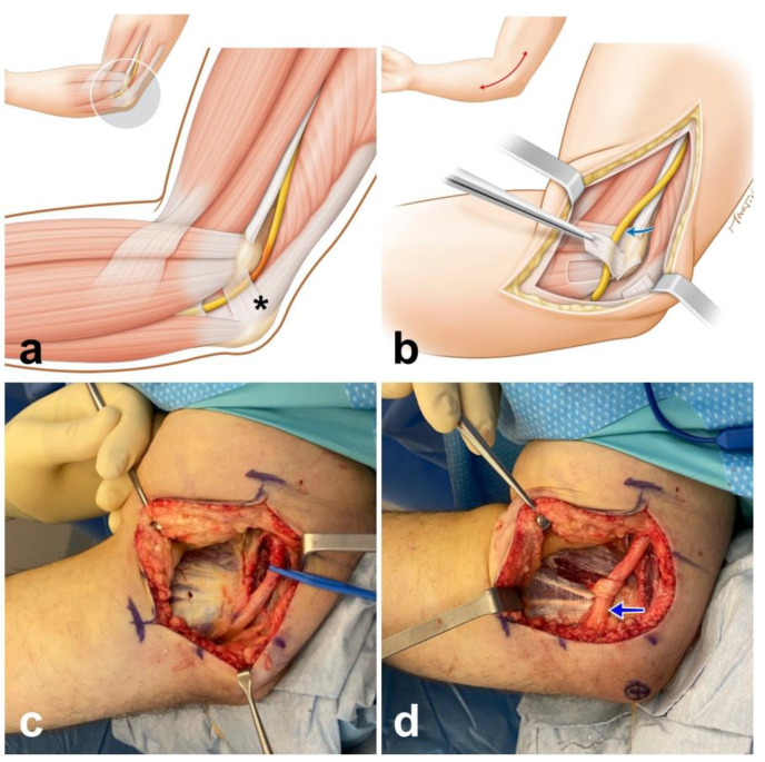
Background: Ulnar nerve compression at the elbow level is the second-most common entrapment neuropathy. The aim of this study was to use shear-wave elastography for the quantification of ulnar nerve elasticity in patients after ulnar nerve decompression with anterior transposition and in the contralateral non-operative side.

Method: Eleven patients with confirmed diagnosis and ulnar nerve decompression with anterior transposition were included and examinations were performed on an AixplorerTM ultrasound system (Supersonic Imagine, Aix-en-Provence, France).

Results: We observed significant differences at 0-degree (p < 0.001), 45-degree (p < 0.05), 90-degree (p < 0.01) and 120-degree (p < 0.001) elbow flexion in the shear elastic modulus of the ulnar nerve in the operative and non-operative sides. There were no statistically significant differences between the elasticity values of the ulnar nerve after transposition at 0-degree elbow flexion and in the non-operative side at 120-degree elbow flexion (p = 0.39), or in the ulnar nerve after transposition at 120-degree elbow flexion and in the non-operative side at 0-degree elbow flexion (p = 0.09).

Conclusion: Shear-wave elastography has the potential to be used postoperatively as a method for assessing nerve tension noninvasively by the estimation of mechanical properties, such as the shear elastic modulus.

Keywords: elastography; nerve compression; nerve transposition; ulnar nerve.


Conflict of interest statement

The authors declare no conflict of interest.

Figures

Figure 1
Figure 1
Sketches detailing ulnar nerve (yellow) entrapment in the cubital tunnel (a), which is where Obsorne’s ligament is located (asterix). The ulnar nerve is transposed anteriorly (blue arrow) and stabilized with a fascial sling (b). Intraoperative photograph after complete release of all points of ulnar nerve with elevation of the soft tissue above the flexor-pronator muscle mass (c); the ulnar nerve is transposed subcutaneously and stabilized anteriorly with a fascial sling (d).
Figure 2
Figure 2
Examples of shear elastic modulus measurement of the ulnar nerve at 0- (image at top), 45-, 90- and 120-degree flexion of the elbow in an Elbo™ brace (A). The colored region represents the shear elasticity map in a healthy ulnar nerve (B) and an ulnar nerve after anterior transposition (C) with a color scale (see bottom right), with blue representing lesser stiffness and red greater stiffness. The software allowed us to measure the mean shear elasticity value over a 2 mm2 Q-Box focus area.
Figure 3
Figure 3
Box plot of the shear elastic modulus of the ulnar nerve (kPa) at 0, 45, 90 and 120 degrees of elbow flexion in operative side (red) and non-operative side (blue). Box (interquartile range (IQR); 75th–25th percentile), central line (median; 50th percentile), lower whisker (5th percentile), upper whisker (95th percentile).

References

    1. Rayegani S.M., Raeissadat S.A., Kargozar E., Rahimi-Dehgolan S., Loni E. Diagnostic value of ultrasonography versus electrodiagnosis in ulnar neuropathy. Med. Devices (Auckl.) 2019;12:81–88. doi: 10.2147/MDER.S196106. - DOI - PMC - PubMed
    1. Kim J.H., Won S.J., Rhee W.I., Park H.J., Hong H.M. Diagnostic cutoff value for ultrasonography in the ulnar neuropathy at the elbow. Ann. Rehabil. Med. 2015;39:170–175. doi: 10.5535/arm.2015.39.2.170. - DOI - PMC - PubMed
    1. Paluch Ł., Noszczyk B., Nitek Ż., Walecki J., Osiak K., Pietruski P. Shear-wave elastography: A new potential method to diagnose ulnar neuropathy at the elbow. Eur. Radiol. 2018;28:4932–4939. doi: 10.1007/s00330-018-5517-9. - DOI - PMC - PubMed
    1. Nakashian M.N., Ireland D., Kane P.M. Cubital Tunnel Syndrome: Current Concepts. Curr. Rev. Musculoskelet. Med. 2020;13:520–524. doi: 10.1007/s12178-020-09650-y. - DOI - PMC - PubMed
    1. Wiesler E.R., Chloros G.D., Cartwright M.S., Shin H.W., Walker F.O. Ultrasound in the diagnosis of ulnar neuropathy at the cubital tunnel. J. Hand Surg. Am. 2006;31:1088–1093. doi: 10.1016/j.jhsa.2006.06.007. - DOI - PubMed