Review
. 2021 Sep 5;4(3):62.
doi: 10.3390/mps4030062.

What's Wrong in a Jump? Prediction and Validation of Splice Site Variants

Affiliations
Review

What's Wrong in a Jump? Prediction and Validation of Splice Site Variants

Giulia Riolo et al. Methods Protoc. .

Abstract

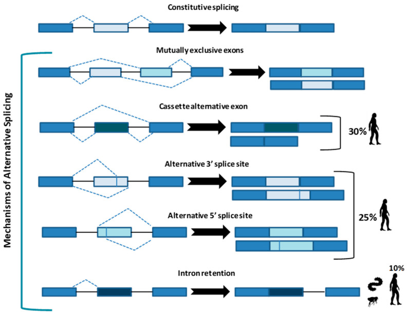
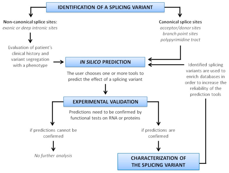
Alternative splicing (AS) is a crucial process to enhance gene expression driving organism development. Interestingly, more than 95% of human genes undergo AS, producing multiple protein isoforms from the same transcript. Any alteration (e.g., nucleotide substitutions, insertions, and deletions) involving consensus splicing regulatory sequences in a specific gene may result in the production of aberrant and not properly working proteins. In this review, we introduce the key steps of splicing mechanism and describe all different types of genomic variants affecting this process (splicing variants in acceptor/donor sites or branch point or polypyrimidine tract, exonic, and deep intronic changes). Then, we provide an updated approach to improve splice variants detection. First, we review the main computational tools, including the recent Machine Learning-based algorithms, for the prediction of splice site variants, in order to characterize how a genomic variant interferes with splicing process. Next, we report the experimental methods to validate the predictive analyses are defined, distinguishing between methods testing RNA (transcriptomics analysis) or proteins (proteomics experiments). For both prediction and validation steps, benefits and weaknesses of each tool/procedure are accurately reported, as well as suggestions on which approaches are more suitable in diagnostic rather than in clinical research.

Keywords: alternative splicing; experimental validation; machine learning; prediction tools; splice variant; splicing sites; variant classification.


Conflict of interest statement

The authors declare no conflict of interest.

Figures

Figure 1
Figure 1
Constitutive splicing and the five main types of alternative splicing. Cassette alternative exon and the alternative 3’ or 5’ splice site are the most common in humans (30% and 25%, respectively), while intron retention is typical of metazoans and less present in humans (10%). Arrows indicate the resulted sequence after intron/exon removal.
Figure 2
Figure 2
(a) U1 binds to exon 1 and U2 binds to exon 2 in order to define 5’ ends of the intron before removal. Addition of tri-snRNP U4/U6.U5 determines the full spliceosome assembly in humans. (b) Role of cis- and trans-regulatory sequences during alternative splicing. Cis-regulatory elements are located in the alternatively spliced exon or in its flanking introns. Cis-factors positively modulate intronic/exonic splicing enhancers (ISE/ESE) and negatively regulate intronic/exonic splice silencer (ISS/ESS). Cis-sequences are bound by trans-factors such as serine/arginine (SR) proteins or the heterogeneous nuclear ribonucleoprotein (hnRNP).
Figure 3
Figure 3
Microarray technology. RNA of two samples (normal/reference RNA and patient-isolated RNA) are differently labeled, mixed, and then spotted on the same microchip. After hybridization, the chip is scanned at two wavelengths to capture signals of the two different dyes. Scanner of the array generates an image for interpretation of the results. Green spots indicate expression in normal cells, while red spots indicate only expression in affected cells. Yellow signal means co-expression (not significant result).
Figure 4
Figure 4
RNA-sequencing workflow. RNA-seq is a three-step method: (1) library construction; (2) sequencing; (3) bioinformatic analysis. The RNA species of interest is selected and converted to complementary DNA, which is then amplified by PCR in order to prepare a sequencing library. Sequencing results in the generation of short sequences (reads) that need to be aligned to a reference genome. Then, different approaches can be used for transcript assembly to detect quantitative gene expression.
Figure 5
Figure 5
Overview of the protein truncation test. DNA or cDNA obtained from RNA by retrotrascription can be used as a template to perform PCR. During amplification process, an RNA polymerase promoter and a translation initiation sequence (ATG) are added to products, together with a consensus Kozak sequence to improve the process. Then, the RNA polymerase promoter initiates transcription and the ATG sequence is used to start translation of RNA into protein. PCR fragments are then separated on basis of their size by agarose gel-electrophoresis, and mutations affecting splicing can be revealed. In the radioactive PTT, addition of radiolabelled amino acids in nascent proteins requires blotting after SDS-PAGE and then exposition to X-ray to analyze results (not shown). Finally, only DNA sequencing can confirm if the production of truncated proteins is due to aberrant splicing.
Figure 6
Figure 6
Strategy for splice variant characterization.

References

    1. Gilbert W. Why genes in pieces? Nat. Cell Biol. 1978;271:501. doi: 10.1038/271501a0. - DOI - PubMed
    1. Nilsen T.W., Graveley B.R. Expansion of the eukaryotic proteome by alternative splicing. Nat. Cell Biol. 2010;463:457–463. doi: 10.1038/nature08909. - DOI - PMC - PubMed
    1. Wang Y., Liu J., Huang B.O., Xu Y.-M., Li J., Zhang J., Min Q.-H., Yang W.-M., Wang X.-Z. Mechanism of alternative splicing and its regulation. Biomed. Rep. 2015;3:152–158. doi: 10.3892/br.2014.407. - DOI - PMC - PubMed
    1. Richards S., Aziz N., Bale S., Bick D., Das S., Gastier-Foster J., Grody W.W., Hegde M., Lyon E., Spector E., et al. Standards and guidelines for the interpretation of sequence variants: A joint consensus recommendation of the American College of Medical Genetics and Genomics and the Association for Molecular Pathology. Genet. Med. 2015;17:405–423. doi: 10.1038/gim.2015.30. - DOI - PMC - PubMed
    1. Pohl M., Bortfeldt R.H., Grützmann K., Schuster S. Alternative splicing of mutually exclusive exons—A review. Biosystems. 2013;114:31–38. doi: 10.1016/j.biosystems.2013.07.003. - DOI - PubMed