Review
. 2021 Sep 16;7(3):57.
doi: 10.3390/ncrna7030057.

Modulation of MicroRNA Processing by Dicer via Its Associated dsRNA Binding Proteins

Affiliations
Review

Modulation of MicroRNA Processing by Dicer via Its Associated dsRNA Binding Proteins

Toyotaka Yoshida et al. Noncoding RNA. .

Abstract

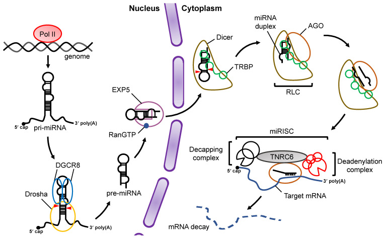
MicroRNAs (miRNAs) are small non-coding RNAs that are about 22 nucleotides in length. They regulate gene expression post-transcriptionally by guiding the effector protein Argonaute to its target mRNA in a sequence-dependent manner, causing the translational repression and destabilization of the target mRNAs. Both Drosha and Dicer, members of the RNase III family proteins, are essential components in the canonical miRNA biogenesis pathway. miRNA is transcribed into primary-miRNA (pri-miRNA) from genomic DNA. Drosha then cleaves the flanking regions of pri-miRNA into precursor-miRNA (pre-miRNA), while Dicer cleaves the loop region of the pre-miRNA to form a miRNA duplex. Although the role of Drosha and Dicer in miRNA maturation is well known, the modulation processes that are important for regulating the downstream gene network are not fully understood. In this review, we summarized and discussed current reports on miRNA biogenesis caused by Drosha and Dicer. We also discussed the modulation mechanisms regulated by double-stranded RNA binding proteins (dsRBPs) and the function and substrate specificity of dsRBPs, including the TAR RNA binding protein (TRBP) and the adenosine deaminase acting on RNA (ADAR).

Keywords: ADAR; Dicer-associated proteins; LGP2; PACT; TRBP; dsRBP; miRNA–mRNA network; microRNA biogenesis.


Conflict of interest statement

The authors declare no conflict of interest.

Figures

Figure 1
Figure 1
Overview of miRNA biogenesis and the RNA silencing pathway. Pri-miRNAs are transcribed from the genome by Pol II. In the nucleus, pri-miRNA is cleaved by a microprocessor complex consisting of Drosha and DGCR8 to produce pre-miRNA, which is then transported from the nucleus to the cytoplasm by EXP5 coupled with GTP-bound Ran (RanGTP). Cleavage of pre-miRNA is performed by Dicer and its cofactor, TRBP, in a canonical miRNA biogenesis pathway. After pre-miRNA cleavage, the miRNA duplex is loaded onto AGO proteins through formation of the RLC complex. The mature miRNA guides the RISC complex to target mRNAs that are complementary to the seed region of miRNA. Recruitment of the TNRC6 protein induces the destabilization and translational repression of the target mRNA.
Figure 2
Figure 2
Domain structure of proteins involved in miRNA biogenesis and structural features of processing complexes of pri-miRNA and pre-miRNA. (a) The domain structures of Drosha, DGCR8, Dicer, TRBP, PACT, ADAR1p150, and ADAR1p110. CED indicates central domain; RIIIDa, RNase III domain a; RIIIDb, RNase III domain b; dsRBD, double-stranded RNA binding domain; HBR, heme-binding region; DExD/H-box, DExD/H-box helicase; CTT, C-terminal tail; DUF283, domain of unknown function; PAZ, Piwi–Argonaute–Zwille; Zα and Zβ, Z-DNA binding domain α and β, respectively; NES, nuclear export signal; NLS, nuclear localization signal. (b) Pri-miRNA has a terminal loop, stem region, and flanking region as common structural features, and some pri-miRNAs have conserved sequence motifs. UG is recognized by Drosha, UGU is recognized by DGCR8, GHG is recognized by dsRBD of Drosha, and CNNC is recognized by Ser/Arg-rich splicing factor 3 (SRSF3). (c) Pre-miRNA is structurally characterized by a terminal loop, a stem region, and a 3′ end overhang.
Figure 3
Figure 3
The structure of human Drosha dsRBD interacting with pri-miR-16-2. The dsRBD binds to the stem region of pri-miRNA via three regions (Region 1: violet, Region 2: sky-blue, Region 3: orange). A pri-miR-16-2 RNA strand is colored in blue, and the other RNA strand is colored in cyan. Red lines indicate the interaction sites between Drosha dsRBD and pri-miR-16-2. This figure is modified from the report produced by Partin et al. [52] (Protein Data Bank ID: 6V5B) using PyMOL (The PyMOL Molecular Graphics System, Version 2.0 Schrödinger, LLC).
Figure 4
Figure 4
The pre-miRNA secondary structures preferentially bound by TRBP, ADAR1p110, or ADAR2. Secondary structures of pre-miRNAs, preferentially bound by TRBP, ADAR1p110, or ADAR2 as predicted by BPP. The pre-miRNAs bound to each dsRBP was determined by RIP-sequencing and secondary structures of pre-miRNAs are shown here schematically, modified from ref [93,97]. BPP is calculated for each base of pre-miRNAs and is a value in the range of 0 to 1; 0 indicates the low base-pairing probability and 1 indicates the high probability. The horizontal lines between 5′ strand and 3′ strand represent base-pairing, and the solid lines indicate the nucleotide positions with significantly high BPPs relative to the control and the dotted lines indicate the positions with significantly low BPPs. The BPP of the terminal loop of pre-miRNA was not calculated and shown in dotted line.
Figure 5
Figure 5
A schematic model of the miRNA–mRNA gene expression network through the modulation of Dicer-mediated processing by its associated proteins. Pre-miRNAs are transported from the nucleus to the cytoplasm by Exp5, which couples with RanGTP. Each dsRBP (TRBP, PACT, or ADAR1) binds to the pre-miRNAs based on their own substrate specificity and recruits them to Dicer to facilitate their maturation. The miRNAs shown in the figure represent the typical miRNAs that are specifically processed by Dicer–TRBP or Dicer–ADAR1 interaction and regulate the downstream gene expression networks. To date, the miRNAs specifically processed by Dicer–PACT interaction have not yet been identified. Note that the pathways shown here were reported by different studies. The numbers in the square brackets indicate the reference numbers.

References

    1. Hammond S.M., Bernstein E., Beach D., Hannon G.J. An RNA-directed nuclease mediates post-transcriptional gene silencing in Drosophila cells. Nature. 2000;404:293–296. doi: 10.1038/35005107. - DOI - PubMed
    1. Grimson A., Farh K.K.-H., Johnston W.K., Garrett-Engele P., Lim L.P., Bartel D.P. MicroRNA targeting specificity in mammals: Determinants beyond seed pairing. Mol. Cell. 2007;27:91–105. doi: 10.1016/j.molcel.2007.06.017. - DOI - PMC - PubMed
    1. Pasquinelli A.E., Reinhart B.J., Slack F., Martindale M.Q., Kuroda M.I., Maller B., Hayward D.C., Ball E.E., Degnan B., Müller P., et al. Conservation of the sequence and temporal expression of let-7 heterochronic regulatory RNA. Nature. 2000;408:86–89. doi: 10.1038/35040556. - DOI - PubMed
    1. Lagos-Quintana M., Rauhut R., Lendeckel W., Tuschl T. Identification of novel genes coding for small expressed RNAs. Science. 2001;294:853–858. doi: 10.1126/science.1064921. - DOI - PubMed
    1. Lau N.C., Lim L.P., Weinstein E.G., Bartel D.P. An abundant class of tiny RNAs with probable regulatory roles in Caenorhabditis elegans. Science. 2001;294:858–862. doi: 10.1126/science.1065062. - DOI - PubMed

LinkOut - more resources