. 2021 Aug 25;11(9):572.
doi: 10.3390/metabo11090572.

Can the Mitochondrial Metabolic Theory Explain Better the Origin and Management of Cancer than Can the Somatic Mutation Theory?

Affiliations

Can the Mitochondrial Metabolic Theory Explain Better the Origin and Management of Cancer than Can the Somatic Mutation Theory?

Thomas N Seyfried et al. Metabolites. .

Abstract

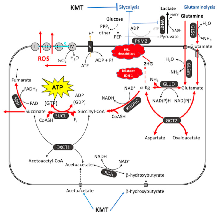
A theory that can best explain the facts of a phenomenon is more likely to advance knowledge than a theory that is less able to explain the facts. Cancer is generally considered a genetic disease based on the somatic mutation theory (SMT) where mutations in proto-oncogenes and tumor suppressor genes cause dysregulated cell growth. Evidence is reviewed showing that the mitochondrial metabolic theory (MMT) can better account for the hallmarks of cancer than can the SMT. Proliferating cancer cells cannot survive or grow without carbons and nitrogen for the synthesis of metabolites and ATP (Adenosine Triphosphate). Glucose carbons are essential for metabolite synthesis through the glycolysis and pentose phosphate pathways while glutamine nitrogen and carbons are essential for the synthesis of nitrogen-containing metabolites and ATP through the glutaminolysis pathway. Glutamine-dependent mitochondrial substrate level phosphorylation becomes essential for ATP synthesis in cancer cells that over-express the glycolytic pyruvate kinase M2 isoform (PKM2), that have deficient OxPhos, and that can grow in either hypoxia (0.1% oxygen) or in cyanide. The simultaneous targeting of glucose and glutamine, while elevating levels of non-fermentable ketone bodies, offers a simple and parsimonious therapeutic strategy for managing most cancers.

Keywords: IDH1; chimpanzees; evolution; fermentation; glutaminolysis; glycolysis; ketogenic metabolic therapy; metastasis; mitochondrial substrate level phosphorylation; mutations; oncogenes; respiration.


Conflict of interest statement

The authors declare no conflict of interest.

Figures

Figure 1
Figure 1
Role of the nucleus and mitochondria in the origin of tumors. Normal cells are shown in green with nuclear and mitochondrial morphology indicative of normal gene expression and OxPhos function, respectively. Tumor cells are shown in red with abnormal nuclear and mitochondrial morphology indicative of genomic instability and abnormal OxPhos function, respectively. “(1) Normal cells beget normal cells with regulated growth. (2) Tumor cells beget tumor cells with dysregulated growth. (3) Transfer of a tumor cell nucleus into a normal cytoplasm begets normal cells that have regulated growth, despite the presence of the tumor-associated genomic abnormalities. (4) Transfer of a normal cell nucleus into a tumor cell cytoplasm begets dead cells or tumor cells with dysregulated growth”. The general reproducibility of the findings across a broad range of tumor types, animal species, and experimental techniques is notable in light of major concerns regarding the irreproducibility of scientific results published in prestigious journals [40,48,49]. “The results of these experiments are profound in showing that nuclear genomic defects alone cannot account for the origin of tumors and that normal-functioning mitochondria can suppress tumorigenesis”. Original diagram from Jeffrey Ling and Thomas N. Seyfried with permission [41].
Figure 2
Figure 2
Cancer as a Mitochondrial Metabolic Disease. Cancer can arise from any number of unspecific influences (risk factors) that would alter the number, structure, and function of mitochondria thus affecting energy production through OxPhos. Unspecific cancer risk factors can include, age, viral infections, the Ras oncogene, rare inherited mutations, chronic inflammation, intermittent hypoxia, radiation exposure, chemical carcinogens etc. [2,65,66,67,68]. Any of these risk factors could cause chronic damage to OxPhos thus increasing the production of reactive oxygen species (ROS), which would ultimately link to the six major hallmarks of cancer [2,12,68]. The process by which each of these unspecific risk factors can chronically damage OxPhos was described previously in detail [16,66,69,70]. Excessive ROS, mostly generated from OxPhos dysfunction, are carcinogenic and mutagenic and would cause significant damage to lipids, proteins, and nucleic acids in both the mitochondria and the in the nucleus [71]. Nuclear genomic instability, including the vast array of somatic mutations and aneuploidy, would arise because of ROS damage together with extracellular acidification and inflammation through a bidirectional interaction between the provocative agent and cells within a tissue [1,2,57,72,73]. Indeed, mutations in the p53 tumor suppressor gene and genomic instability have been linked directly to OxPhos deficiency and mitochondrial ROS production in cancer stem cells [55,74]. Fermentation metabolism and ROS formation underlie the hyperproliferation of tumor cells. A gradual reduction in OxPhos efficiency would elicit a mitochondrial stress response through retrograde (RTG) signaling [69,75,76,77]. RTG activation would cause persistent expression of various oncogenes, e.g., Hif-1a and c-Myc, that upregulate receptors and enzymes in both the glycolysis and the glutaminolysis pathways [75,78,79,80,81,82]. Oncogenes become facilitators of fermentation metabolism. ATP synthesis through mSLP (Q effect) will compensate for lost ATP synthesis through OxPhos or from PKM2 expression in glycolysis [83,84]. The path to carcinogenesis will occur only in those cells capable of sustaining energy production through substrate level phosphorylation, (SLP). Cells unable to replace OxPhos with SLP, e.g., CNS neurons or cardiomyocytes, would die and rarely become tumorigenic. Despite the shift from respiration to SLP, the ΔG’ATP hydrolysis remains fairly constant at approximately −56 kJ, indicating that the energy from SLP compensates for the reduced energy from OxPhos. When respiration becomes unable to maintain energy homeostasis, the RTG will initiate oncogene up-regulation and tumor suppressor gene inactivation. Protracted RTG activation becomes necessary to maintain the viability of incipient cancer cells. Genomic instability will arise as a secondary consequence of protracted mitochondrial stress from disturbances in the intracellular and extracellular environments. Metastasis arises from respiratory damage in cells of myeloid/macrophage origin either directly or after fusion hybridization with epithelial-derived tumor cells [4,85]. Tumor progression and degree of malignancy is linked directly to ultrastructure abnormalities (mitochondrial cristolysis) and to the energy transition from OxPhos to substrate level phosphorylation (Warburg effect and Q effect) [83]. The T signifies an arbitrary threshold when the shift from OxPhos to SLP becomes irreversible. This scenario links all major cancer hallmarks to an extrachromosomal and epigenetic respiratory dysfunction and can explain the oncogenic paradox [70]. Reprinted with modifications from [68,83].
Figure 3
Figure 3
Fusion-hybrid hypothesis for cancer cell metastasis. According to the fusion hybrid hypothesis, metastatic cancer cells can arise following fusion-hybridization between neoplastic epithelial cells and myeloid cells (macrophages). The fusion hybrid hypothesis originated with the work of Aichel in 1911 and was expanded by the Pawelek and the Munzarova groups [105,128,129,130,131,132]. Macrophages are known to invade in situ carcinoma as if it were an unhealed wound [95,109,133]. This creates a protracted inflammatory microenvironment leading to fusion hybridization between the neoplastic epithelial cell and the mesenchymal macrophage. Mitochondrial damage becomes the driver for the neoplastic transformation of the epithelial cell and of the fusion hybrids. Inflammation damages mitochondria leading to enhanced fermentation and acidification of the microenvironment. The gradual replacement of normal macrophage mitochondria with dysfunctional mitochondria in the hybrid cell cytoplasm leads to rogue behavior in cells that naturally possess the capability to, (1) move through tissues, (2) suppress the immune system, (3) enter (intravasate), and to exit (extravasate) the circulation. In addition to explaining the “seed-soil” hypothesis of metastasis, the fusion hybrid hypothesis can also explain how metastatic cells can re-capitulate the epithelial characteristics of the primary tumor at secondary micro-metastatic growth sites [4,85]. Furthermore, this hypothesis can explain the phenomenon of mesenchymal epithelial transition without invoking a mutation suppression mechanism. See text for more details. Modified from [85,134].
Figure 4
Figure 4
Glutamine-driven mSLP as a major source of ATP synthesis in cancer cells and inhibition by the therapeutic IDH1(R132H) mutation. ATP synthesis through mSLP at the succinate-CoA ligase reaction (SUCL) in the glutaminolysis pathway (red) can compensate for inefficient ATP synthesis through OxPhos in tumor cells with mitochondrial abnormalities [83]. mSLP can also compensate for inefficient ATP synthesis through glycolysis in cancer cells that express the cytoplasmic PKM2 isoform, which produces less ATP than the PKM1 isoform [84]. These bioenergetic compensations will hold the ΔG’ATP hydrolysis at −56 kJ/mole, thus maintaining cancer cell viability in either the presence or the absence or oxygen. Oxygen consumption in cancer cells is used more for production of ROS, which are carcinogenic and mutagenic, than for ATP synthesis. Moreover, mSLP will maintain the forward operation of the adenine nucleotide translocase thus preventing depletion of cytosolic ATP reserves from the reverse operation of the F0-F1 ATP synthase [84,182]. Release of succinate to the cytoplasm can help stabilize Hif-1α, thus linking lactic acid fermentation through glycolysis to glutamine fermentation through glutaminolysis. Ketogenic metabolic therapy (KMT) will reduce availability of glucose to the glycolytic and the PPP pathways while diverting CoA from succinate to acetoacetate under the metabolism of ketone bodies (β-hydroxybutyrate and acetoacetate) thus indirectly reducing ATP synthesis through the SUCL reaction. The therapeutic IDH1 mutation will further reduce ATP synthesis through mSLP by increasing synthesis of 2-hydroxyglutarate (2HG) from α-ketoglutarate and thus reduce the succinyl CoA substrate for the SUCL reaction [83,215]. Besides its potential effect in reducing glutaminolysis, 2-hydroxyglutarate can also target multiple HIF1α-responsive genes and enzymes in the glycolysis pathway thus limiting synthesis of metabolites and one-carbon metabolism needed for rapid tumor growth [83,182,216,217]. The down regulation of Hif1-α-regulated lactate dehydrogenase A (LDHA), through the action of both KMT and the IDH1 mutation, would reduce extracellular lactate levels thus reducing microenvironment inflammation and tumor cell invasion. The simultaneous restriction of glucose and glutamine, while elevating circulating ketone bodies, will stress the majority of signaling pathways necessary for maintaining tumor cell viability [123,182]. See text for additional details. KMT = ketogenic metabolic therapy; 2HG = 2-hydroxyglutarate; BDH  =  β-hydroxybutyrate dehydrogenase; FAD  =  flavin adenine dinucleotide; GLSc  =  glutaminase, cytosolic; GLSm  =  glutaminase, mitochondrial; GLUD  =  glutamate dehydrogenase; GOT2  =  aspartate aminotransferase; KGDHC  =  α-ketoglutarate dehydrogenase complex; LDH: lactate dehydrogenase; NME  =  nucleoside diphosphate kinase; OXCT1  =  succinyl-CoA:3-ketoacid coenzyme A transferase 1; PC  =  pyruvate carboxylase; PDH  =  pyruvate dehydrogenase; PEP  =  phosphoenolpyruvate; PKM2  =  pyruvate kinase M2; SDH: succinate dehydrogenase; SUCL  =  succinate-CoA ligase. Reprinted with modifications from [83,213].

References

    1. Seyfried T.N., Yu G., Maroon J.C., D’Agostino D.P. Press-pulse: A novel therapeutic strategy for the metabolic management of cancer. Nutr. Metab. 2017;14:19. doi: 10.1186/s12986-017-0178-2. - DOI - PMC - PubMed
    1. Seyfried T.N., Flores R.E., Poff A.M., D’Agostino D.P. Cancer as a metabolic disease: Implications for novel therapeutics. Carcinogenesis. 2014;35:515–527. doi: 10.1093/carcin/bgt480. - DOI - PMC - PubMed
    1. Sonnenschein C., Soto A.M. Somatic mutation theory of carcinogenesis: Why it should be dropped and replaced. Mol. Carcinog. 2000;29:205–211. doi: 10.1002/1098-2744(200012)29:4<205::AID-MC1002>3.0.CO;2-W. - DOI - PubMed
    1. Seyfried T.N., Huysentruyt L.C. On the origin of cancer metastasis. Crit. Rev. Oncog. 2013;18:43–73. doi: 10.1615/CritRevOncog.v18.i1-2.40. - DOI - PMC - PubMed
    1. Fidler I.J. The pathogenesis of cancer metastasis: The ‘seed and soil’ hypothesis revisited. Nat. Rev. Cancer. 2003;3:453–458. doi: 10.1038/nrc1098. - DOI - PubMed

LinkOut - more resources