. 2021 Sep 15;11(9):627.
doi: 10.3390/metabo11090627.

Comparative Untargeted Metabolomic Profiling of Induced Mitochondrial Fusion in Pancreatic Cancer

Affiliations

Comparative Untargeted Metabolomic Profiling of Induced Mitochondrial Fusion in Pancreatic Cancer

Nicholas D Nguyen et al. Metabolites. .

Abstract

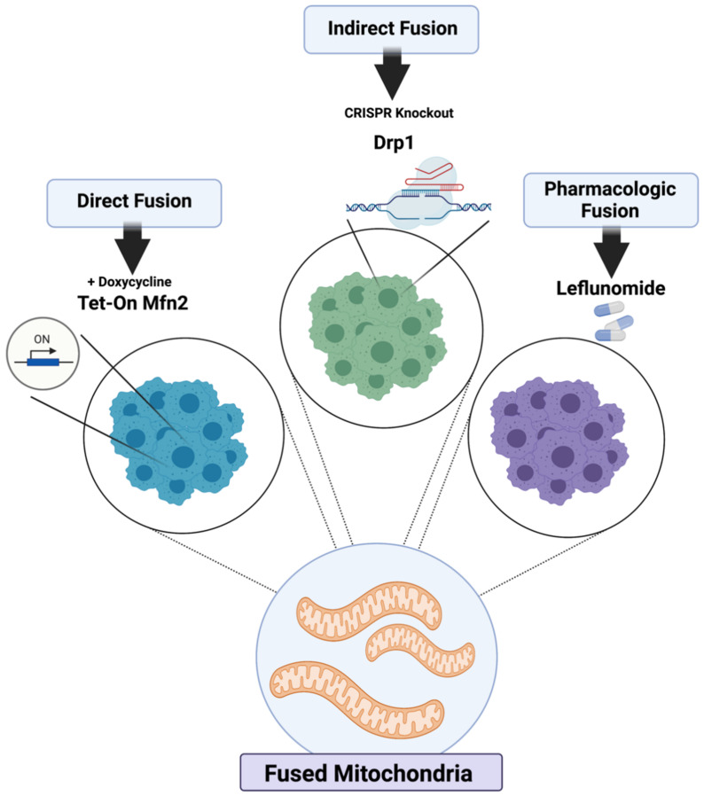
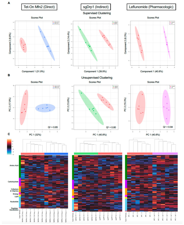
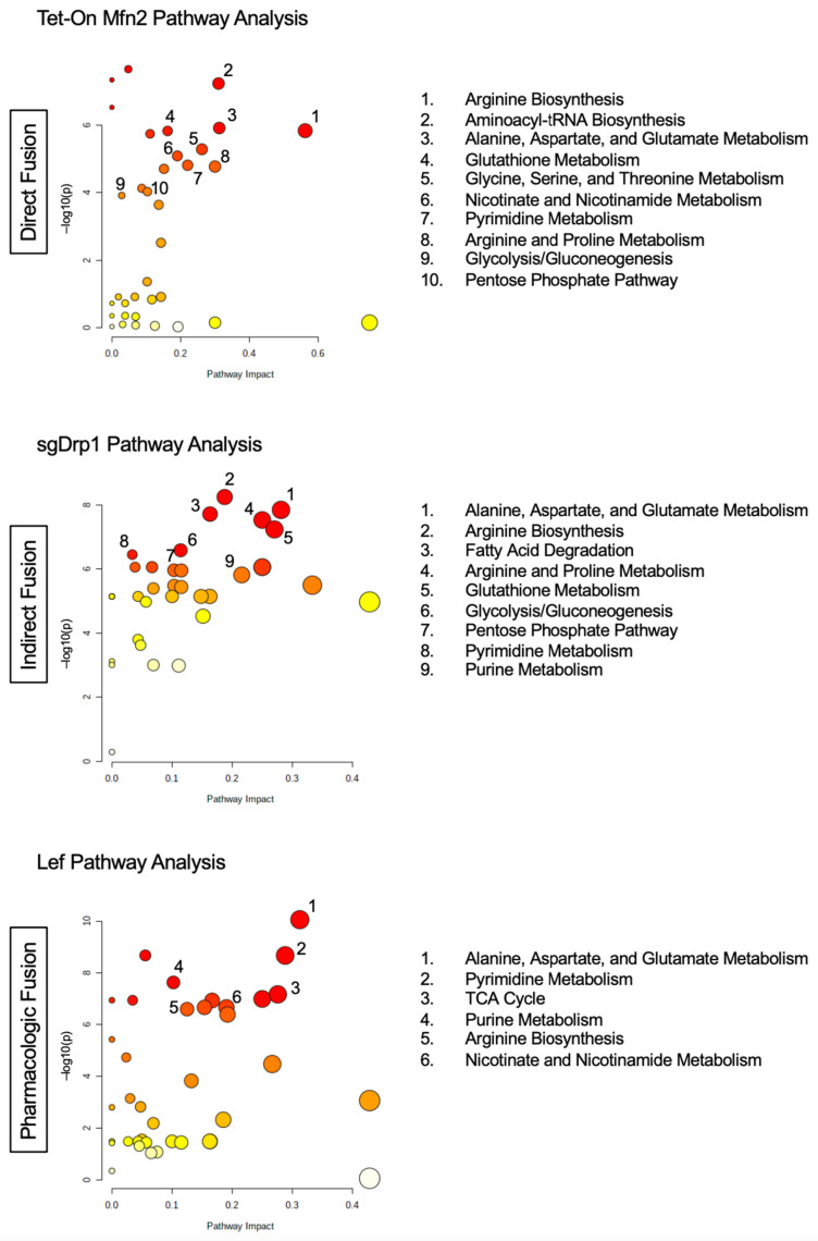
Mitochondria are dynamic organelles that constantly alter their shape through the recruitment of specialized proteins, like mitofusin-2 (Mfn2) and dynamin-related protein 1 (Drp1). Mfn2 induces the fusion of nearby mitochondria, while Drp1 mediates mitochondrial fission. We previously found that the genetic or pharmacological activation of mitochondrial fusion was tumor suppressive against pancreatic ductal adenocarcinoma (PDAC) in several model systems. The mechanisms of how these different inducers of mitochondrial fusion reduce pancreatic cancer growth are still unknown. Here, we characterized and compared the metabolic reprogramming of these three independent methods of inducing mitochondrial fusion in KPC cells: overexpression of Mfn2, genetic editing of Drp1, or treatment with leflunomide. We identified significantly altered metabolites via robust, orthogonal statistical analyses and found that mitochondrial fusion consistently produces alterations in the metabolism of amino acids. Our unbiased methodology revealed that metabolic perturbations were similar across all these methods of inducing mitochondrial fusion, proposing a common pathway for metabolic targeting with other drugs.

Keywords: fission; fusion; leflunomide; metabolomic reprogramming; metabolomics; mitochondrial morphology; mitofusin-2; pancreatic cancer.


Conflict of interest statement

C.M.T. was on the clinical advisory board of Accuray during this study, has a patent for oral amifostine as a radioprotectant of the upper GI tract issues that is licensed with royalties paid from Xerient Pharmaceuticals, and PHD inhibitors as a radioprotectant of the GI tract pending, and was the lead principal investigator of a multicenter trial testing the effects of high-dose SBRT with the radiomodulator, GC4419. All other authors declare no conflict of interest.

Figures

Figure 1
Figure 1
Models of mitochondrial fusion induction. KPC cells were genetically modified to directly overexpress Mfn2 in a tetracycline-inducible manner, indirectly fuse through CIRSPR knockout of Drp1, and pharmacologically fuse after treatment with Leflunomide.
Figure 2
Figure 2
Confocal microscopy reveals changes in mitochondrial morphology in KPC cells after multiple independent fusion induction modalities. (A) Doxycycline-induced overexpression of Mfn2 (blue triangles) significantly increased tubular morphology while significantly decreasing fragmented mitochondria when compared to its Tet-Off control (red circles). (B) Indirect fusion through abrogation of Drp1 (green squares) significantly increased tubular morphology while significantly decreasing intermediate and fragmented mitochondria when compared to sgGFP controls (red diamonds). (C) Pharmacologic induction of fusion with Lef (purple hexagons) significantly increased tubular and intermediate mitochondrial morphology while decreasing fragmented morphology when compared to vehicle controls (red triangles). Red fluorescence represents mitochondria; blue fluorescence represents DAPI-labeled nuclei. Mitochondrial morphology quantified using n = 100–200 cells. Statistical analysis by Student’s t-test, **** p < 0.0001, ** p < 0.01. Original magnification, ×60. Scale bar = 10 µm. Data presented as mean ± SEM.
Figure 3
Figure 3
Multivariate clustering reveals distinct separation after inducing mitochondrial fusion when compared to controls. (A) Supervised PLS-DA and (B) unsupervised PCA score plots of Tet-On Mfn2 (blue), sgDrp1 (green), and Leflunomide (purple) treated KPC cells with respect to their corresponding controls (red). (C) Heatmap with unsupervised hierarchical clustering of affected super pathways across Tet-On Mfn2 (blue), sgDrp1 (green), and Leflunomide (purple). Both unsupervised and supervised clustering methods revealed a distinct separation between each method of fusion induction and its respective control. Predictive power of PLS-DA in component 1 represented by Q2 = 0.85 for Tet-On Mfn2, Q2 = 0.80 for sgDrp1, and Q2 = 0.94 for Leflunomide.
Figure 4
Figure 4
Total pathway analysis of filtered metabolites reveals similar impact of Amino Acid, Nucleotide, and Carbohydrate metabolism pathways as a function of mitochondrial fusion: Tet-On Mfn2 (direct), sgDrp1 (indirect), and leflunomide treatment (pharmacologic).
Figure 5
Figure 5
Statistical methods to identify differentially expressed metabolites after inducing mitochondrial fusion. (A) univariate Student’s t-test, FDR-adjusted p-value < 0.05, (B) SAM, FDR < 0.05, (C) PLS-DA, VIP score < 1.0, (D) RF model, Mean Decrease Accuracy > 0.
Figure 6
Figure 6
Pathway analysis of overlapped discriminant metabolites from direct fusion in Tet-On Mfn2, indirect fusion in sgDrp1, and pharmacologic fusion in leflunomide-treated KPC cells.

References

    1. Hollinshead K.E.R., Parker S.J., Eapen V.V., Encarnacion-Rosado J., Sohn A., Oncu T., Cammer M., Mancias J.D., Kimmelman A.C. Respiratory Supercomplexes Promote Mitochondrial Efficiency and Growth in Severely Hypoxic Pancreatic Cancer. Cell Rep. 2020;33:108231. doi: 10.1016/j.celrep.2020.108231. - DOI - PMC - PubMed
    1. Zhao J., Zhang J., Yu M., Xie Y., Huang Y., Wolff D.W., Abel P.W., Tu Y. Mitochondrial Dynamics Regulates Migration and Invasion of Breast Cancer Cells. Oncogene. 2013;32:4814–4824. doi: 10.1038/onc.2012.494. - DOI - PMC - PubMed
    1. Yu M., Nguyen N.D., Huang Y., Lin D., Fujimoto T.N., Molkentine J.M., Deorukhkar A., Kang Y., San Lucas F.A., Fernandes C.J., et al. Mitochondrial Fusion Exploits a Therapeutic Vulnerability of Pancreatic Cancer. JCI Insight. 2019;4:e126915. doi: 10.1172/jci.insight.126915. - DOI - PMC - PubMed
    1. Kashatus J.A., Nascimento A., Myers L.J., Sher A., Byrne F.L., Hoehn K.L., Counter C.M., Kashatus D.F. Erk2 Phosphorylation of Drp1 Promotes Mitochondrial Fission and MAPK-Driven Tumor Growth. Mol. Cell. 2015;57:537–551. doi: 10.1016/j.molcel.2015.01.002. - DOI - PMC - PubMed
    1. Senft D., Ronai Z.A. Regulators of Mitochondrial Dynamics in Cancer. Curr. Opin. Cell Biol. 2016;39:43–52. doi: 10.1016/j.ceb.2016.02.001. - DOI - PMC - PubMed