. 2021 Aug 30;11(9):667.
doi: 10.3390/membranes11090667.

Highly Segregated Biocomposite Membrane as a Functionally Graded Template for Periodontal Tissue Regeneration

Affiliations

Highly Segregated Biocomposite Membrane as a Functionally Graded Template for Periodontal Tissue Regeneration

Syed Saad B Qasim et al. Membranes (Basel). .

Abstract

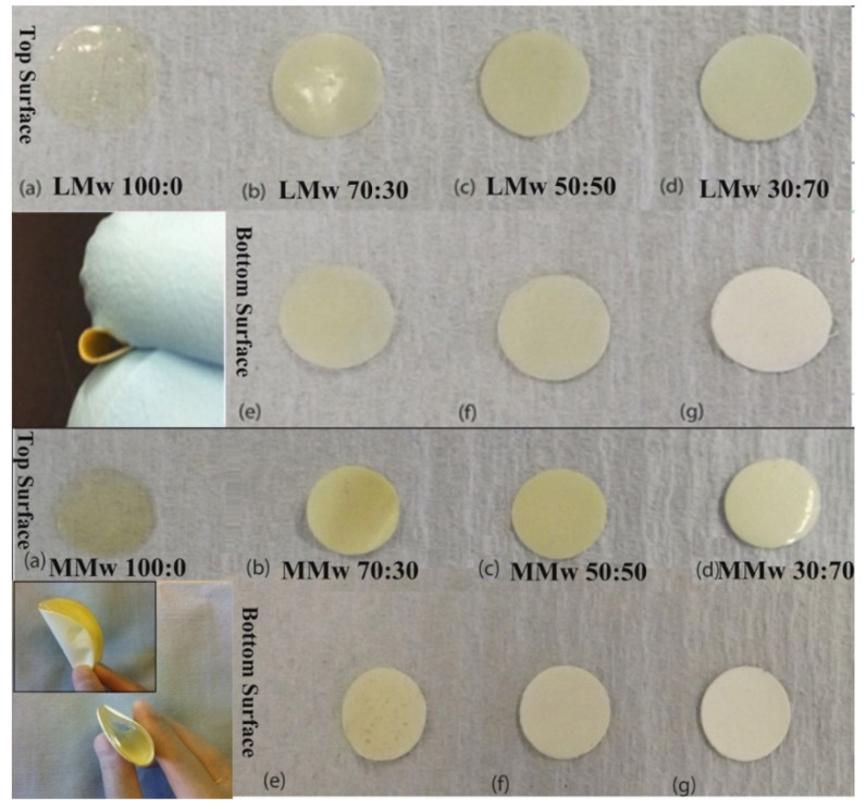
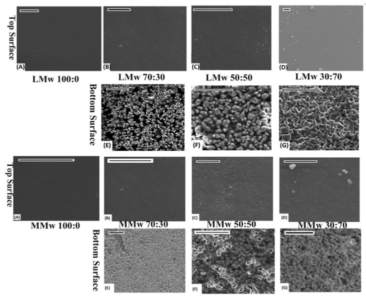
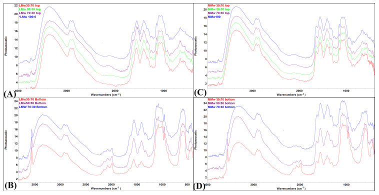
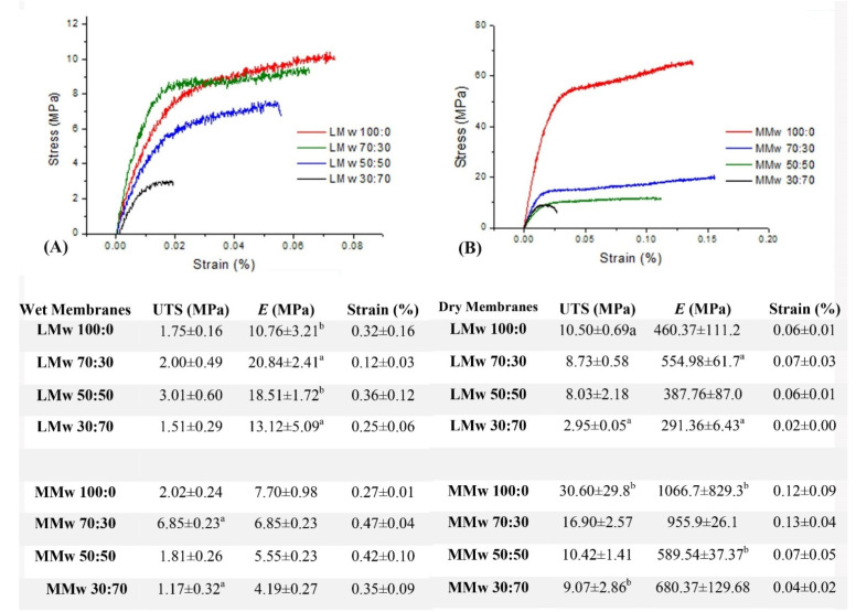
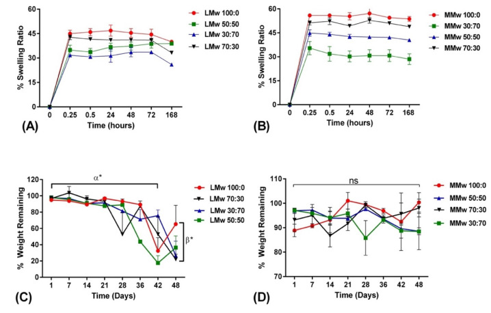
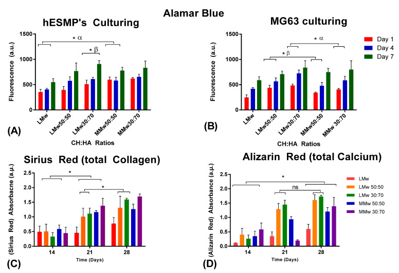
Guided tissue regeneration (GTR) membranes are used for treating chronic periodontal lesions with the aim of regenerating lost periodontal attachment. Spatially designed functionally graded bioactive membranes with surface core layers have been proposed as the next generation of GTR membranes. Composite formulations of biopolymer and bioceramic have the potential to meet these criteria. Chitosan has emerged as a well-known biopolymer for use in tissue engineering applications due to its properties of degradation, cytotoxicity and antimicrobial nature. Hydroxyapatite is an essential component of the mineral phase of bone. This study developed a GTR membrane with an ideal chitosan to hydroxyapatite ratio with adequate molecular weight. Membranes were fabricated using solvent casting with low and medium molecular weights of chitosan. They were rigorously characterised with scanning electron microscopy, Fourier transform infrared spectroscopy in conjunction with photoacoustic sampling accessory (FTIR-PAS), swelling ratio, degradation profile, mechanical tensile testing and cytotoxicity using human osteosarcoma and mesenchymal progenitor cells. Scanning electron microscopy showed two different features with 70% HA at the bottom surface packed tightly together, with high distinction of CH from HA. FTIR showed distinct chitosan dominance on top and hydroxyapatite on the bottom surface. Membranes with medium molecular weight showed higher swelling and longer degradation profile as compared to low molecular weight. Cytotoxicity results indicated that the low molecular weight membrane with 30% chitosan and 70% hydroxyapatite showed higher viability with time. Results suggest that this highly segregated bilayer membrane shows promising potential to be adapted as a surface layer whilst constructing a functionally graded GTR membrane on its own and for other biomedical applications.

Keywords: chitosan; guided tissue regeneration; hydroxyapatite; periodontal engineering.


Conflict of interest statement

The authors declare no conflict of interest.

Figures

Figure 1
Figure 1
Optical images of LMw and MMwCH:HA membranes in different ratios of CH to HA with clear distinction between top and bottom surfaces of both molecular weight variants. Images taken with a DSLR S5600. The smaller inset image depicts the change in colour in between the top and bottom surface and their ability of the membranes to be bent, which is useful when using as a GTR in periodontal defect side. All (ad) are top surfaces of LMw and MMw neat and composite membranes. All (eg) are bottom surfaces of LMw and MMw composite membranes.
Figure 2
Figure 2
SEM performed on LMW and MMwCH:HA membranes. Top and bottom surface of 100:0, 70:30, 50:50 and 30:70 ratios of CH:HA. All images scaled at 20 µm. (AD) are top surfaces of LMw and MMw Neat and composite membranes. (EG) are bottom surfaces of composite LMw and MMw membranes.
Figure 3
Figure 3
FTIR-PAS spectral profile of (A) low molecular weight (LMw) 100:0, 70:30, 50:50 and 30:70 obtained from the top surface; (B) LMw 70:30, 50:50 and 30:70 obtained from the bottom surface; (C) medium molecular weight (MMw) 100:0, 70:30, 50:50 and 30:70 obtained from the top surface; (D) MMw 70:30, 50:50 and 30:70 obtained from the bottom surface.
Figure 4
Figure 4
The correlation of stress to strain. (A) Representation of the LMw composite membranes in wet conditions, (B) representation of the MMw composite membranes in dry conditions. Results of the tensile test are shown in the table, values are a mean ± SD (n = 6). Ultimate tensile strength (MPa), Young’s Modulus (E) or Elastic Modulus (MPa) and Strain (%). Similar superscripted letters in the same column indicate statistical significance (p < 0.05).
Figure 5
Figure 5
Swelling percentage of (A) LMw and (B) MMw CH and composite membranes performed over a period of 168 h. Weight remaining percentage of (C) LMw and (D) MMw composite membranes, performed over an experimental time period of 48 days. Shown values are mean ± SD (n = 3).
Figure 6
Figure 6
Water contact angle analysis of (A) LMw and (B) MMw neat and composite membranes 70:30. 50:50 and 30:70. The graphs show mean ± SD (n = 3), (*) indicates statistically significant difference. LMw 100:0, 78° ± 6; LMw 70:30, 50°± 1.6; MMw 90° ± 3; MMw 30:70, 55° ± 1.6; MMw 50:50, 45° ± 1.
Figure 7
Figure 7
Cytotoxicity conducted via culturing (A) hES-MP and (B) MG63s. Cellular viability analysed with Alamar blue staining until day 7. Values shown are taken from mean ± SD where n = 3. (C) Collagen and (D) calcium accumulated by hES-MPs at day 14, 21 and 28. (ns, no statistically significant difference, α and β represent statistically significant difference in between the observed groups). (*) indicates statistically significant difference.

References

    1. Foey A.D., Habil N., Al-Shaghdali K., Crean S.J. Porphyromonas gingivalis-stimulated macrophage subsets exhibit differential induction and responsiveness to interleukin-10. Arch. Oral Biol. 2017;73:282–288. doi: 10.1016/j.archoralbio.2016.10.029. - DOI - PubMed
    1. Sculean A., Nikolidakis D., Nikou G., Ivanovic A., Chapple I.L.C., Stavropoulos A. Biomaterials for promoting periodontal regeneration in human intrabony defects: A systematic review. Periodontol. 2000. 2015;68:182–216. doi: 10.1111/prd.12086. - DOI - PubMed
    1. Susin C., Wikesjö U.M.E. Regenerative periodontal therapy: 30 years of lessons learned and unlearned. Periodontol. 2000. 2013;62:232–242. doi: 10.1111/prd.12003. - DOI - PubMed
    1. Chen F.-M.M., Jin Y. Periodontal tissue engineering and regeneration: Current approaches and expanding opportunities. Tissue Eng. Part B-Rev. 2010;16:219–255. doi: 10.1089/ten.teb.2009.0562. - DOI - PubMed
    1. Menicanin D., Hynes K., Han J., Gronthos S., Bartold P.M. Cementum and periodontal ligament regeneration. Adv. Exp. Med. Biol. 2015;881:207–236. doi: 10.1007/978-3-319-22345-2_12. - DOI - PubMed

LinkOut - more resources