. 2021 Aug 24;8(9):170.
doi: 10.3390/vetsci8090170.

Establishment of a Newborn Lamb Gut-Loop Model to Evaluate New Methods of Enteric Disease Control and Reduce Experimental Animal Use

Affiliations

Establishment of a Newborn Lamb Gut-Loop Model to Evaluate New Methods of Enteric Disease Control and Reduce Experimental Animal Use

Ambre Baillou et al. Vet Sci. .

Abstract

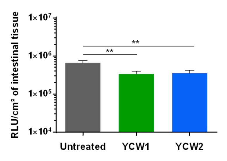
Enteric infectious diseases are not all well controlled, which leads to animal suffering and sometimes death in the most severe cases, in addition to economic losses for farmers. Typical symptoms of enteric infections include watery diarrhea, stomach cramps or pain, dehydration, nausea, vomiting, fever and weight loss. Evaluation of new control methods against enteric infections requires the use of many animals. We aimed to develop a new method for an initial in vivo screen of promising compounds against neonatal diseases such as cryptosporidiosis while limiting experimental animal use. We therefore adapted an in vivo method of multiple consecutive but independent intestinal loops to newborn lambs delivered by cesarean section, in which endotoxin responsiveness is retained. This new method allowed for the screening of natural yeast fractions for their ability to stimulate immune responses and to limit early Cryptosporidium parvum development. This model may also be used to investigate host-pathogen interactions and immune responses in a neonatal controlled environment.

Keywords: 3R; Cryptosporidium parvum; multiple intestinal loop model; newborn lamb; yeast cell wall fractions.


Conflict of interest statement

The authors declared no potential conflict of interest with respect to the research, authorship, and/or publication of this article.

Figures

Figure 1
Figure 1
Intestinal loops surgery on the ileum of cesarean-section born lamb. (a) Picture of lamb that was anesthetized before surgery, intubated with endotracheal tube and put on assisted ventilation; (b) Schematic representation of the intestinal loop generation in the ileum of caesarean-born lamb. Series of triplicates of loops (L1, L2, L3) were generated between the two areas (I and II) indicated by arrows. Interloop region separates series of triplicates and are indicated in shaded areas. The first ligature of the triplicates harbors longer ends, so do the one of each triplicate to facilitate the identification; (c) Schematic representation of end-to-end anastomosis is visualized (I/II) allowing intestinal transit restauration and the separation of the intestinal segment containing the isolated loops; (d) Picture of a loop creation within intestinal segment during surgery; (e) Food intake represented by the volume of milk drunk per ml and per kg of lamb body weight per 24 h for the gut-loop lamb and its littermate. Each point shape corresponds to an independent experimentation (n = 4 for each group); (f) Body temperature (°C) (mean ± SEM) was monitored from birth to 20-h post-surgery (ps) for the gut-loop lamb and its littermate. The grey box corresponds to the surgery intervention (from start to end).
Figure 2
Figure 2
Intestinal responsiveness to LPS stimulation within the gut loop generated with a cesarean-born neonatal lamb. Intestinal tissues from the ileal gut loops and the ileum were recovered from the same neonatal lamb 24 h post-surgery. Small pieces of intestine, designed as “explants”, were placed in a CO2 incubator at 37 °C in culture medium alone (untreated) or with LPS at a concentration of 10 µg/mL for 4 h (n = 3 explants per condition). Explants were processed for RNA extraction and cxcl1 (a), cxcl2 (b), cxcl8 (c) and tnfα (d) gene expressions were quantified by RT-qPCR. Results are expressed as 2e−ΔCt (mean ± SEM), following normalization with the expression of three reference genes (hprt; gapdh; actb).
Figure 3
Figure 3
Innate ileal immune response to TLR-ligands and yeast cell wall fractions. Lamb’s gut-loops were stimulated in vivo immediately after the surgery procedure by in situ injection of LPS (10 µg/mL), R848 (10 µg/mL), yeast cell wall fraction 1 (YCW1) or 2 (YCW2) (5 mg/mL) into distinct intestinal loops. Twenty-four hours later, intestinal loops were recovered and samples processed for RNA extraction. cxcl8 (a), il1α (b), il1β (c) and mx1 (d) gene expressions were quantified by RT-qPCR. Results are expressed as 2e−ΔCt following normalization with the expression of three reference genes (gapdh; hprt; actb).
Figure 4
Figure 4
Establishment of Cryptosporidium parvum experimental infection in the gut-loop model. Data are cumulative results from three independent experimentations each performed with one newborn lamb. For each experimental condition, three loops were used; (a) Parasite load were determined by measuring luminescence activity in the ileal tissue from the loop 24 h after infection with oocysts of C. parvum nluc-INRAE transgenic strain (n = 9). Results are expressed as RLU per cm2 of intestinal tissue (mean ± SEM); (b) Immunofluorescence microscopy of ileal tissue section from a C. parvum infected gut loop (1.5 × 10⁵ oocysts) 24 h after infection. Parasites were stained in red with anti-C. parvum antibodies and nuclei in blue with DAPI. “GC” corresponds to the germinal centers of the ileal Peyer’s patch. White arrows indicate parasites developing into intestinal epithelial cells lining the ileal villi; (c) Quantification of Cp18S gene expression in the intestinal loops was performed on the same samples as above. RNA was extracted from a piece of each intestinal loop after 24 h of infection with C. parvum nluc-INRAE transgenic strain, reverse transcribed and amplified by quantitative PCR. Results are expressed as 2e−ΔCt (mean ± SEM) following normalization with the expression of three reference genes (gapdh; hprt; actb); (d) Correlation between parasite load evaluated by luminescence and Cp18S gene expression was determined by linear regression analysis. Statistical analyses in (a,b) were realized with the Kruskal–Wallis non-parametric test and the Dunn’s multiple comparison test, and significative difference was determined by a p-value < 0.05 (** p < 0.01, *** p < 0.001).
Figure 5
Figure 5
Evaluation of yeast cell wall fractions on early Cryptosporidium parvum invasion and development. Data are cumulative results from three independent experimentations each with one newborn lamb. For each experimental condition, three loops were used. Loops were infected by in situ injection of 1.5 × 10⁴ oocysts of C. parvum nluc-INRAE transgenic strain alone (untreated), or associated with YCW1 or YCW2 at a concentration of 5 mg/mL. Parasite loads were evaluated by quantifying luminescence in intestinal tissues after 24 h of infection. Results are expressed as RLU per cm2 of intestinal tissue with a logarithmic scale (mean ± SEM). Statistical analyses were performed with the Kruskal–Wallis non-parametric test followed by the Dunn’s multiple comparison test (** p < 0.01).

Similar articles

Cited by

References

    1. Mayr L.M., Bojanic D. Novel trends in high-throughput screening. Curr. Opin. Pharmacol. 2009;9:580–588. doi: 10.1016/j.coph.2009.08.004. - DOI - PubMed
    1. Brodin P., Christophe T. High-content screening in infectious diseases. Curr. Opin. Chem. Biol. 2011;15:534–539. doi: 10.1016/j.cbpa.2011.05.023. - DOI - PubMed
    1. Li V.S.W. Modelling intestinal inflammation and infection using ‘mini-gut’ organoids. Nat. Rev. Gastroenterol. Hepatol. 2021;18:89–90. doi: 10.1038/s41575-020-00391-4. - DOI - PMC - PubMed
    1. Beaumont M., Blanc F., Cherbuy C., Egidy G., Giuffra E., Lacroix-Lamande S., Wiedemann A. Intestinal organoids in farm animals. Vet. Res. 2021;52:33. doi: 10.1186/s13567-021-00909-x. - DOI - PMC - PubMed
    1. Richmond J. The 3Rs—Past, present and future. Scand. J. Lab. Anim. Sci. 2000;27:84–92.

LinkOut - more resources