Comparative Study
. 2021 Aug 24;13(9):592.
doi: 10.3390/toxins13090592.

Helicobacter pylori CagA EPIYA Motif Variations Affect Metabolic Activity in B Cells

Affiliations
Comparative Study

Helicobacter pylori CagA EPIYA Motif Variations Affect Metabolic Activity in B Cells

Sebastian Diechler et al. Toxins (Basel). .

Abstract

Background: Helicobacter pylori (Hp) colonizes the human stomach and can induce gastric cancer and mucosa-associated lymphoid tissue (MALT) lymphoma. Clinical observations suggest a role for the Hp virulence factor cytotoxin-associated gene A (CagA) in pathogenesis. The pathogenic activity of CagA is partly regulated by tyrosine phosphorylation of C-terminal Glu-Pro-Ile-Tyr-Ala (EPIYA) motifs in host cells. However, CagA differs considerably in EPIYA motifs, whose functions have been well characterized in epithelial cells. Since CagA is fragmented in immune cells, different CagA variants may exhibit undetected functions in B cells.

Methods: B cells were infected with Hp isolates and isogenic mutants expressing different CagA EPIYA variants. CagA translocation and tyrosine phosphorylation were investigated by Western blotting. Apoptosis was analyzed by flow cytometry and metabolic activity was detected by an MTT assay.

Results: Isogenic CagA EPIYA variants are equally well translocated into B cells, followed by tyrosine phosphorylation and cleavage. B cell apoptosis was induced in a CagA-independent manner. However, variants containing at least one EPIYA-C motif affected metabolic activity independently of phosphorylation or multiplication of EPIYA-C motifs.

Conclusions: The diverse structure of CagA regulates B cell physiology, whereas B cell survival is independent of CagA.

Keywords: B cells; CagA; EPIYA motifs; Helicobacter pylori.


Conflict of interest statement

The authors declare no conflict of interest.

Figures

Figure 1
Figure 1
CagA expression in Hp isolates. (A,B) Variable 3‣ terminal regions of cagA genes from Hp isolates NCTC 11638 (AB), Hp 26695 (ABC), P12 (ABCC), NCTC 11637 (ABCCC), HPAG1 (AABC), J99 (BC), 42GX (ABD) and 48GX (AABD) were sequenced to verify EPIYA-A, -B and -C/D motifs. (C) CagA expression was confirmed by Western blot analysis of Hp lysates. Full-length CagA (CagAFL) was detected using an antibody directed against CagA N-terminus (α-CagANT). Bacterial HtrA was detected as a loading control.
Figure 2
Figure 2
Hp isolates translocate CagA harboring different EPIYA motifs into B cells and gastric epithelial cells. (A) CagA translocation and phosphorylation in B cell line MEC1 (left panel) and gastric epithelial AGS cells (right panel) were investigated in infection experiments using the indicated Hp strains. Cells were infected at an MOI of 100 for 4 h or remained uninfected (mock). Phosphorylated full-length CagA (pCagAFL) and C-terminal CagA (pCagACT) were analyzed by Western blotting of whole cell lysates using an anti-phosphotyrosine-specific antibody (α-pTyr). Full-length CagA (CagAFL) was detected by α-CagANT. GAPDH was detected as a loading control. (B) CagA cleavage was analyzed after infection of MEC1 (left panel) and AGS cells (right panel). CagA was detected using an antibody (α-CagACT) recognizing C-terminal CagA (CagACT) and CagAFL. Equal sample loading was confirmed by detection of GAPDH. (C,D) pCagAFL (white bars) and pCagACT (grey bars) were quantified in MEC1 (C) and AGS cells (D) by blot densitometry, normalized to the loading control GAPDH and expressed as arbitrary units (* p < 0.05, ** p < 0.01, *** p < 0.001, **** p < 0.0001).
Figure 3
Figure 3
Infections with isogenic Hp mutant strains reveal that the presence of an EPIYA-C motif enhances CagA phosphorylation. (A) The variable 3´ terminal regions of cagA genes containing EPIYA-C motifs (AB, ABC, ABCC, ABCCC) or phosphorylation-deficient EPIFA motifs (ABF, ABFF, ABFFF) of isogenic Hp P12 mutants and P12 wild types (wt) were verified by sequencing. (B) Expression of CagA variants in bacterial lysates was analyzed by Western blotting. Full-length CagA (CagAFL) was detected using a CagA N-terminal antibody (α-CagANT). Detection of HtrA served as a loading control. (C) MEC1 cells were infected at an MOI of 100 for 4 h with the indicated Hp mutants, P12 wt, or remained uninfected (mock). CagA cleavage in MEC1 cells was analyzed using an anti-CagA antibody (α-CagACT) recognizing the C-terminal region of CagA (CagACT) and CagAFL. GAPDH was detected as a loading control. (D) Phosphorylation of full-length CagA (pCagAFL) and C-terminal CagA fragments (pCagACT) was analyzed using antibodies against phosphorylated tyrosine (α-p-Tyr) and CagAFL was detected by α-CagANT. (E) pCagAFL (white bars) and pCagACT (grey bars) were quantified by blot densitometry, normalized to GAPDH and expressed as arbitrary units (* p < 0.05, **** p < 0.0001, ns: not significant).
Figure 4
Figure 4
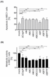
Isogenic Hp mutants induce apoptosis in B cells independently of CagA, but decrease metabolic activity dependently on EPIYA-C motifs. (A) MEC1 cells were infected with P12 mutants at an MOI of 50 as indicated. Apoptosis was measured after 24 h by flow cytometry. Results represent the means of three independent experiments (* p < 0.05, ** p < 0.01, *** p < 0.001, ns: not significant). (B) MEC1 cells were infected with Hp strains at an MOI of 200. After 24 h, metabolic activity was measured using an MTT assay. The data represent the results from three independent experiments, each performed in triplicate. The metabolic activities of Hp-infected cells are shown in relation to the mock control (** p < 0.01, ns: not significant).

References

    1. Amieva M., Peek R.M., Jr. Pathobiology of Helicobacter pylori-Induced Gastric Cancer. Gastroenterology. 2016;150:64–78. doi: 10.1053/j.gastro.2015.09.004. - DOI - PMC - PubMed
    1. Tegtmeyer N., Neddermann M., Asche C.I., Backert S. Subversion of host kinases: A key network in cellular signaling hijacked by Helicobacter pylori CagA. Mol. Microbiol. 2017;105:358–372. doi: 10.1111/mmi.13707. - DOI - PubMed
    1. Eck M., Schmausser B., Haas R., Greiner A., Czub S., Müller-Hermelink H.K. MALT-type lymphoma of the stomach is associated with Helicobacter pylori strains expressing the CagA protein. Gastroenterology. 1997;112:1482–1486. doi: 10.1016/S0016-5085(97)70028-3. - DOI - PubMed
    1. Tsai H.F., Hsu P.N. Interplay between Helicobacter pylori and immune cells in immune pathogenesis of gastric inflammation and mucosal pathology. Cell Mol. Immunol. 2010;7:255–259. doi: 10.1038/cmi.2010.2. - DOI - PMC - PubMed
    1. Hoy B., Löwer M., Weydig C., Carra G., Tegtmeyer N., Geppert T., Schröder P., Sewald N., Backert S., Schneider G., et al. Helicobacter pylori HtrA is a new secreted virulence factor that cleaves E-cadherin to disrupt intercellular adhesion. EMBO Rep. 2010;11:798–804. doi: 10.1038/embor.2010.114. - DOI - PMC - PubMed

Publication types

MeSH terms