. 2021 Sep 25;12(10):875.
doi: 10.1038/s41419-021-04154-0.

Combination of tyrosine kinase inhibitors and the MCL1 inhibitor S63845 exerts synergistic antitumorigenic effects on CML cells

Affiliations

Combination of tyrosine kinase inhibitors and the MCL1 inhibitor S63845 exerts synergistic antitumorigenic effects on CML cells

Alena Malyukova et al. Cell Death Dis. .

Abstract

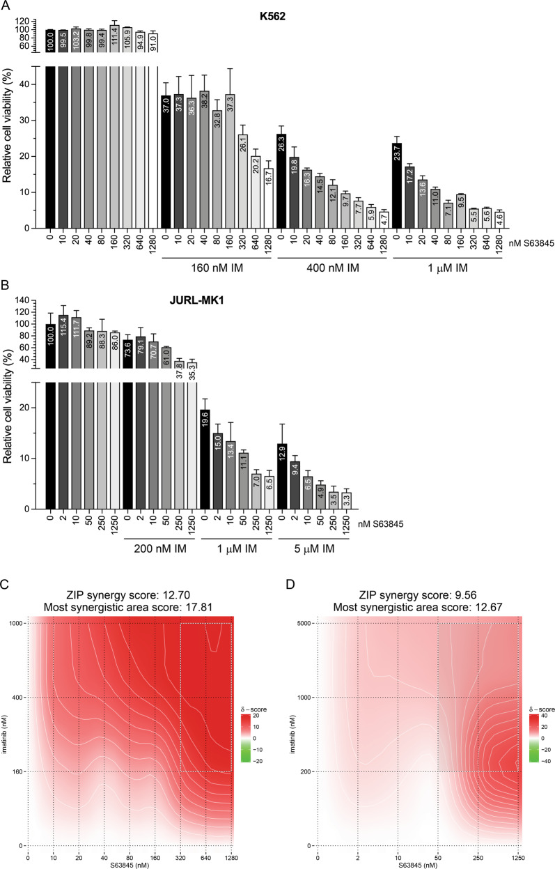
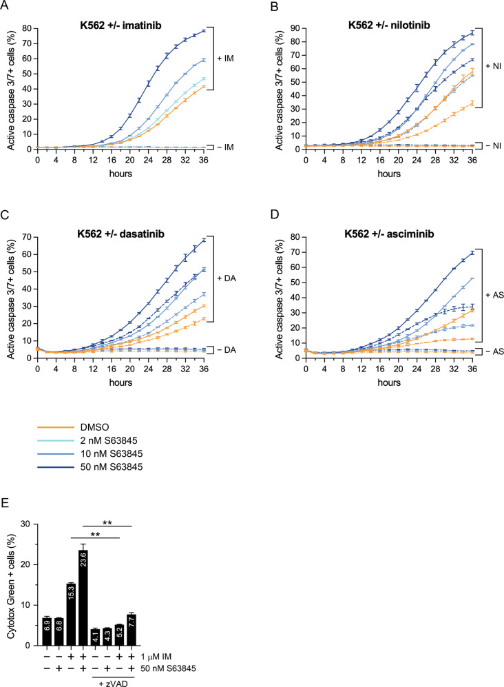
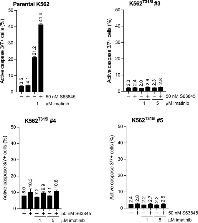
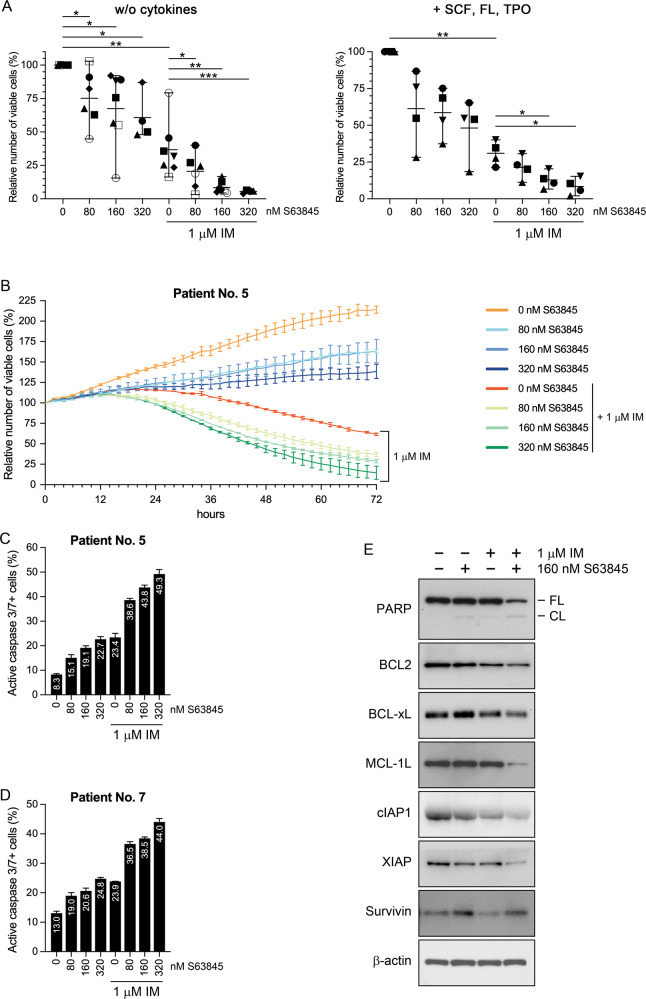
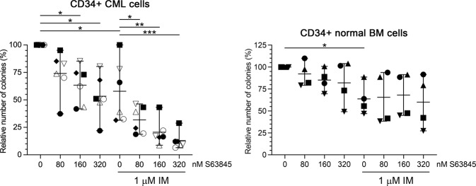
Tyrosine kinase inhibitor (TKI) treatment has dramatically improved the survival of chronic myeloid leukemia (CML) patients, but measurable residual disease typically persists. To more effectively eradicate leukemia cells, simultaneous targeting of BCR-ABL1 and additional CML-related survival proteins has been proposed. Notably, several highly specific myeloid cell leukemia 1 (MCL1) inhibitors have recently entered clinical trials for various hematologic malignancies, although not for CML, reflecting the insensitivity of CML cell lines to single MCL1 inhibition. Here, we show that combining TKI (imatinib, nilotinib, dasatinib, or asciminib) treatment with the small-molecule MCL1 inhibitor S63845 exerted strong synergistic antiviability and proapoptotic effects on CML lines and CD34+ stem/progenitor cells isolated from untreated CML patients in chronic phase. Using wild-type BCR-ABL1-harboring CML lines and their T315I-mutated sublines (generated by CRISPR/Cas9-mediated homologous recombination), we prove that the synergistic proapoptotic effect of the drug combination depended on TKI-mediated BCR-ABL1 inhibition, but not on TKI-related off-target mechanisms. Moreover, we demonstrate that colony formation of CML but not normal hematopoietic stem/progenitor cells became markedly reduced upon combination treatment compared to imatinib monotherapy. Our results suggest that dual targeting of MCL1 and BCR-ABL1 activity may efficiently eradicate residual CML cells without affecting normal hematopoietic stem/progenitors.


Conflict of interest statement

The authors declare no competing interests.

Figures

Fig. 1
Fig. 1. Effects of S63845 and imatinib administered as single drug or in combination on the viability of K562 and JURL-MK1 cells.
Relative viability (A, B) and synergy maps (C, D) of K562 (A, C) and JURL-MK1 (B, D) cells left untreated or treated for 48 h (A, C) or 32 h (B, D) with the indicated concentrations of imatinib (IM) and/or S63845, analyzed by alamarBlue (A, C) or SYTOX Green (B, D) assays. Data represent mean with standard deviation derived from three technical replicates. Experiments were performed at least twice with similar results.
Fig. 2
Fig. 2. Effects of S63845 and imatinib administered as single drug or in combination on the expression of apoptotic and pyroptotic markers and key antiapoptotic members of the BCL2 and BIRC gene families in K562 and TCCS cells.
Immunoblot analysis of PARP, caspase-3, caspase-7, GSDME, GSDMD, BCL-xL, MCL-1L (the longest isoform of MCL1), cIAP1, XIAP, survivin and β-actin protein expression in total cell extracts of K562 (A) and TCCS (B) cells left untreated (−) or treated (+) for 24 h (A) or 16 h (B) with the indicated concentrations of imatinib (IM) and/or S63845. Cleaved caspase-3 could not be detected in any lysates of TCCS, while cleaved caspase-7 was not detectable in any lysates of K562 cells (not shown). BCL2 and cIAP2 could not be detected in any lysates of either of the cell lines (not shown). Experiments were performed at least twice with similar results. Representative blots are shown. FL full length, CL cleaved (89 kDa C-terminal cleavage product of PARP or large subunits of cleaved caspase-3/7 or N-terminal cleavage product of GSDME), p43 p43 GSDMD fragment, p30 p30 GSDMD fragment.
Fig. 3
Fig. 3. Combination treatment with S63845 and a TKI synergistically induces caspase-3/7 activation and caspase-dependent death in K562 cells.
Proportion of active caspase-3/7 positive K562 cells left untreated or treated for the indicated hours with the indicated concentrations of S63845, in the absence (dotted lines) or presence of 1 μM (solid lines) imatinib (IM) (A), 20 nM (dashed lines) or 100 nM (solid lines) nilotinib (NI) (B), 1 nM (dashed lines) or 5 nM (solid lines) dasatinib (DA) (C), or 4 nM (dashed lines) or 20 nM (solid lines) asciminib (AS) (D) analyzed by fluorescence live cell microscopy. Data represent mean with standard deviation derived from three technical replicates. Experiments were performed at least twice with similar results. E Proportion of Cytotox Green positive K562 cells left untreated (−) or treated (+) for 36 h with 1 μM imatinib (IM) and/or 50 nM S63845, in the absence (−) or presence (+) of 40 μM Z-VAD-FMK (zVAD), analyzed by fluorescence live cell microscopy. Data represent mean with standard deviation derived from three independent experiments. **P < 0.01.
Fig. 4
Fig. 4. Effects of S63845 and TKIs administered as single drug or in combination on the morphology of K562 and TCCS cells.
Representative fluorescence live cell microscopy images of K562 (A) and TCCS (B, C) cells left untreated or treated with 50 nM S63845 and/or 100 nM nilotinib (NI) for 24 h (A) or 50 nM S63845 and/or 200 nM imatinib (IM) for 16 h (B) or 24 h (C) in the presence of IncuCyte Caspase-3/7 Green Dye for Apoptosis. Experiments were performed at least twice with similar results.
Fig. 5
Fig. 5. Effects of S63845 and TKIs administered as single drug or in combination on the apoptotic rate of the parental TCCS line and its imatinib resistant sublines harboring the T315I mutation in BCR-ABL1.
Proportion of active caspase-3/7 positive cells in the parental TCCS line and its T315I mutation harboring sublines left untreated or treated for the indicated hours with the indicated concentrations of S63845 in the absence (dotted lines) or presence of 200 nM (dashed lines) or 1 μM (solid lines) imatinib (IM) (left panels), or in the absence (dotted lines) or presence of 50 nM (solid lines) asciminib (AS) (right panels), analyzed by fluorescence live cell microscopy. Data represent mean with standard deviation derived from three technical replicates. Experiments were performed at least twice with similar results.
Fig. 6
Fig. 6. Effects of S63845 and imatinib administered as single drug or in combination on the apoptotic rate of the parental K562 line and its imatinib resistant sublines harboring the T315I mutation in BCR-ABL1.
Proportion of active caspase-3/7 positive cells in the parental K562 line and its T315I mutation harboring sublines left untreated or treated for 60 h with 50 nM S63845 in the absence or presence of the indicated concentrations of imatinib, analyzed by fluorescence live cell microscopy. Data represent mean with standard deviation derived from three technical replicates. Experiments were performed at least twice with similar results.
Fig. 7
Fig. 7. Effects of S63845 and imatinib administered as single drug or in combination on the viability, apoptotic rate and antiapoptotic protein expression of primary human CD34+ stem/progenitor cells obtained from the peripheral blood of untreated CML patients in chronic phase.
A Relative numbers of viable CD34+ stem/progenitor cells obtained from the peripheral blood of untreated CML patients in chronic phase and left untreated or treated ex vivo for 96 h with 1 μM imatinib (IM) and/or the indicated concentrations of S63845 in unsupplemented (left panel; n = 7 for 0 and 160 nM, n = 6 for 80 nM, and n = 4 for 320 nM S63845) or SCF, FL and TPO (0.5 ng/ml each) supplemented (right panel; n = 4 for all S63845 concentrations) StemSpan SFEM Medium. Data represent mean with range derived from four to seven independent experiments. *P < 0.05; **P < 0.01; ***P < 0.001. Patient no. 1: ⚫; patient no. 2: ◼; patient no. 3: ▲; patient no. 4: ▼; patient no. 5: ◆; patient no. 6: ○; patient no. 7: ⬜. B Relative numbers of viable (SYTOX Green negative) CD34+ stem/progenitor cells obtained from the peripheral blood of CML patient No. 5 and left untreated or treated ex vivo for the indicated hours with 1 μM imatinib (IM) and/or the indicated concentrations of S63845 (represented by different colors) in StemSpan SFEM Medium supplemented with SCF, FL and TPO (0.5 ng/ml each), analyzed by fluorescence live cell microscopy. Proportion of active caspase-3/7 positive CD34+ stem/progenitor cells obtained from the peripheral blood of CML patient Nos. 5 (C) and 7 (D), and left untreated or treated ex vivo for 24 h with 1 μM imatinib (IM) and/or the indicated concentrations of S63845 in StemSpan SFEM Medium supplemented with SCF, FL and TPO (0.5 ng/ml each), analyzed by fluorescence live cell microscopy. Data shown in BD represent mean with standard deviation derived from two (B) or three (C, D) technical replicates. E Immunoblot analysis of PARP, BCL2, BCL-xL, MCL-1L, cIAP1, XIAP, survivin and β-actin protein expression in total cell extracts of CD34+ stem/progenitor cells obtained from the peripheral blood of CML patient No. 2, and left untreated or treated ex vivo for 24 h with 1 μM imatinib (IM) and/or 160 nM S63845 in StemSpan SFEM Medium supplemented with SCF, FL and TPO (0.5 ng/ml each). cIAP2, the p30 GSDMD fragment, and the full length and cleaved GSDME proteins were not detectable in any of the lysates (not shown). FL full length, CL 89 kDa C-terminal cleavage product of PARP.
Fig. 8
Fig. 8. Effects of S63845 and imatinib administered as single drug or in combination on the colony forming capacity of primary human CD34+ stem/progenitor cells obtained from the peripheral blood of untreated CML patients in chronic phase or from the bone marrow of healthy donors.
Relative colony forming capacity of CD34+ stem/progenitor cells obtained from the peripheral blood of untreated CML patients in chronic phase (left panel; n = 6 for all S63845 concentrations) or from the bone marrow (BM) of healthy donors (right panel; n = 4 for all S63845 concentrations) in the absence or presence of 1 μM imatinib (IM) and/or the indicated concentrations of S63845. Data represent mean with range derived from four (CD34+ normal BM cells) to six (CD34+ CML cells) independent experiments. *P < 0.05; **P < 0.01; ***P < 0.001. Patient no. 1: ⚫; patient no. 2: ◼; patient no. 5: ◆; patient no. 6: ○; patient no. 8: △; patient no. 9: ▽; healthy donor no. 1 ⚫; donor no. 2: ◼; donor no. 3: ▲; donor no. 4: ▼.

References

    1. O’Hare T, Zabriskie MS, Eiring AM, Deininger MW. Pushing the limits of targeted therapy in chronic myeloid leukaemia. Nat Rev Cancer. 2012;12:513–26. doi: 10.1038/nrc3317. - DOI - PubMed
    1. Braun TP, Eide CA, Druker BJ. Response and resistance to BCR-ABL1-targeted therapies. Cancer Cell. 2020;37:530–42. doi: 10.1016/j.ccell.2020.03.006. - DOI - PMC - PubMed
    1. Hochhaus A, Larson RA, Guilhot F, Radich JP, Branford S, Hughes TP,, et al. Long-term outcomes of imatinib treatment for chronic myeloid leukemia. N Engl J Med. 2017;376:917–27. doi: 10.1056/NEJMoa1609324. - DOI - PMC - PubMed
    1. Saussele S, Richter J, Guilhot J, Gruber FX, Hjorth-Hansen H, Almeida A, et al. Discontinuation of tyrosine kinase inhibitor therapy in chronic myeloid leukaemia (EURO-SKI): a prespecified interim analysis of a prospective, multicentre, non-randomised, trial. Lancet Oncol. 2018;19:747–57. doi: 10.1016/S1470-2045(18)30192-X. - DOI - PubMed
    1. Massimino M, Stella S, Tirrò E, Romano C, Pennisi MS, Puma A, et al. Non ABL-directed inhibitors as alternative treatment strategies for chronic myeloid leukemia. Mol Cancer. 2018;17:56. doi: 10.1186/s12943-018-0805-1. - DOI - PMC - PubMed

Publication types

MeSH terms