. 2021 Sep 26;28(1):65.
doi: 10.1186/s12929-021-00763-1.

Clinical and functional characterization of a novel STUB1 frameshift mutation in autosomal dominant spinocerebellar ataxia type 48 (SCA48)

Affiliations

Clinical and functional characterization of a novel STUB1 frameshift mutation in autosomal dominant spinocerebellar ataxia type 48 (SCA48)

Huan-Yun Chen et al. J Biomed Sci. .

Abstract

Background: Heterozygous pathogenic variants in STUB1 are implicated in autosomal dominant spinocerebellar ataxia type 48 (SCA48), which is a rare familial ataxia disorder. We investigated the clinical, genetic and functional characteristics of STUB1 mutations identified from a Taiwanese ataxia cohort.

Methods: We performed whole genome sequencing in a genetically undiagnosed family with an autosomal dominant ataxia syndrome. Further Sanger sequencing of all exons and intron-exon boundary junctions of STUB1 in 249 unrelated patients with cerebellar ataxia was performed. The pathogenicity of the identified novel STUB1 variant was investigated.

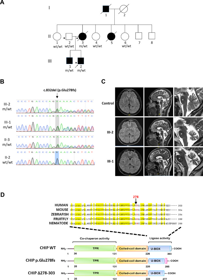
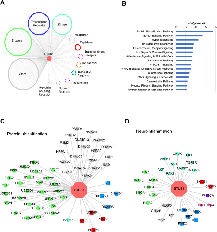
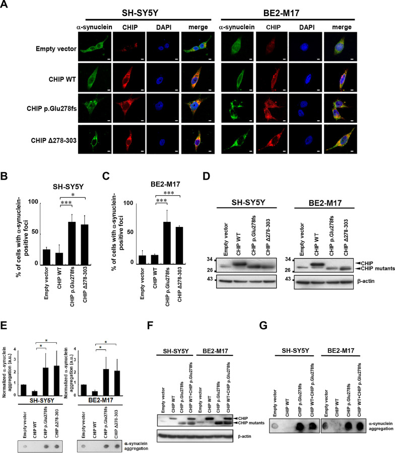
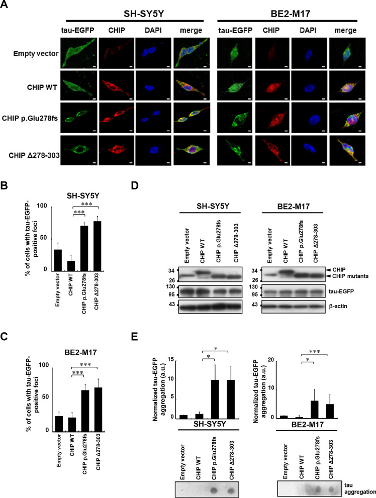
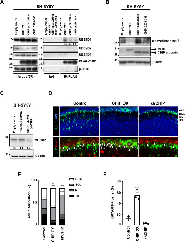
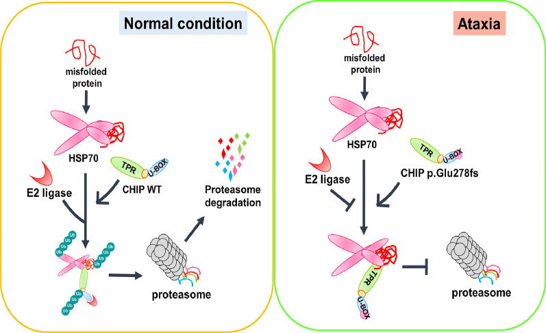
Results: We identified a novel heterozygous frameshift variant, c.832del (p.Glu278fs), in STUB1 in two patients from the same family. This rare mutation is located in the U-box of the carboxyl terminus of the Hsc70-interacting protein (CHIP) protein, which is encoded by STUB1. Further in vitro experiments demonstrated that this novel heterozygous STUB1 frameshift variant impairs the CHIP protein's activity and its interaction with the E2 ubiquitin ligase, UbE2D1, leading to neuronal accumulation of tau and α-synuclein, caspase-3 activation, and promoting cellular apoptosis through a dominant-negative pathogenic effect. The in vivo study revealed the influence of the CHIP expression level on the differentiation and migration of cerebellar granule neuron progenitors during cerebellar development.

Conclusions: Our findings provide clinical, genetic, and a mechanistic insight linking the novel heterozygous STUB1 frameshift mutation at the highly conserved U-box domain of CHIP as the cause of autosomal dominant SCA48. Our results further stress the importance of CHIP activity in neuronal protein homeostasis and cerebellar functions.

Keywords: Ataxia; CHIP; STUB1; Spinocerebellar ataxia type 48; Tau; α-Synuclein.


Conflict of interest statement

All authors report no competing interests.

Figures

Fig. 1
Fig. 1
Pedigree, genetic and brain MRI features of the index SCA48 family with the novel STUB1 frameshift mutation. A Pedigree of the index family with the heterozygous rare frameshift variant, c.832del (p.Glu278fs), in the STUB1 gene. m/wt = heterozygous carriers of the STUB1 mutation; wt/wt = non-carriers; open symbols = unaffected; filled symbols = affected; symbol with a diagonal line = deceased; arrow = proband. Asterisks indicate family members whose whole genomes were sequenced. B Sanger sequencing traces confirming the c.832del (p.Glu278fs) variant in STUB1 identified in the proband and affected members of the index family. C Brain MRI scans show cerebellar atrophy (arrows) with preserved pons (asterisk) in patients III:1 and III:2 of the index family, and an age- and gender-matched healthy control participant. D CHIP comprises three functional domains: TPR, coiled-coil, and U-Box. The protein structure shows that the p.Glu278fs variant amino acid residue (red) is located in the U-Box domain of CHIP. The CHIP Δ278–303 mutation results in a truncated protein without the C-terminal 22 aa of the U-box domain (278–303). A sequence alignment (top) demonstrates the evolutionary conservation of E278 in the U-Box domain of CHIP proteins across the indicated species. Identical residues are labeled in yellow
Fig. 2
Fig. 2
STUB1 interactome analysis. A STUB1-interacting proteins retrieved from the IPA database. Node shape and color denote the different molecular types. B Top 15 significant enriched pathways of STUB1-interacting proteins. C and D Subnetworks of STUB1-interacting proteins involved in protein ubiquitination and neuroinflammation pathways
Fig. 3
Fig. 3
The CHIP mutations promote α-synuclein aggregation in SH-SY5Y and BE2-M17 cells. SH-SY5Y and BE2-M17 cells were transfected for 48 h with WT or mutant CHIP. A Confocal Images of SH-SY5Y and BE2-M17 cells following CHIP WT, p.Glu278fs, or CHIP Δ278-303 ectopic expression were captured. Scale bar, 10 μm. B, C Quantified results in A are shown as the percentage of cells with α-synuclein aggregated foci. D Ectopic expression of CHIP WT, p.Glu278fs, or Δ278–303 in A was examined using Western blot analysis. E CHIP mutants increase SDS-insoluble aggregation of α-synuclein in SH-SY5Y and BE2-M17 cells. α-synuclein aggregation was detected by the filter-trap assay in cells transfected with the CHIP WT, p.Glu278fs, or Δ278–303 plasmid. The lysate was diluted in SDS and filtered through nitrocellulose membranes. α-synuclein immunostaining was detected using α-synuclein antibody. A representative image and the densitometry data are shown (a.u., arbitrary unit). The values of α-synuclein aggregation were normalized to the amount of aggregation in the empty vector control (one-way ANOVA, *p < 0.05, ***p < 0.001). F, G The α-synuclein aggregations detected by a filter trap assay in F SH-SY5Y and BE2-M17 cells overexpressing both wild-type and mutant CHIP at the same time G were comparable between cells expressing CHIP p.Glu278fs mutant alone and those co-expressing both CHIP WT and p.Glu278fs mutant
Fig. 4
Fig. 4
The CHIP mutations cause tau aggregation. A After 48-h transfection, SH-SY5Y and BE2-M17 cells were paraformaldehyde-fixed and DAPI (blue) was used to stain the nuclear DNA. The images (× 630) were acquired using a Zeiss LSM780 laser scanning fluorescence confocal microscope. Scale bar, 10 μm. B, C Quantified results in A are shown as the percentage of tau aggregated foci in SH-SY5Y and BE2-M17 cells. D Ectopic expression of CHIP WT, p.Glu278fs, or Δ278–303 in A was examined using Western blot analysis. E CHIP mutants increase SDS-insoluble aggregation of tau in SH-SY5Y and BE2-M17 cells. Tau aggregation was detected by the filter-trap assay in cells transfected with the CHIP WT, p.Glu278fs, or Δ278–303 plasmid. The lysate was diluted in SDS and filtered through nitrocellulose membranes. Tau immunostaining was detected using the tau antibody. A representative image and the densitometry data are shown (a.u., arbitrary unit). The values of tau aggregation were normalized to the amount of aggregation in the empty vector control (one-way ANOVA, *p < 0.05, ***p < 0.001)
Fig. 5
Fig. 5
The CHIP mutations abolish the interaction between E2 ubiquitin ligase and CHIP, enhance caspase-3 cleavage and alter cerebellum development. A SH-SY5Y cells were transfected with the CHIP WT, p.Glu278fs, or Δ278–303 plasmid for 48 h. Immunoprecipitations were performed using an anti-FLAG antibody. Immunoprecipitates were sequentially probed with anti-UBE2D1, anti-UBE2D2, anti-UBE2D3, and anti-FLAG antibodies. Five percent of lysates used for immunoprecipitation were loaded as the inputs and probed with anti-UBE2D1, anti-UBE2D2, anti-UBE2D3, and anti-FLAG antibodies. IgG served as an IP negative control, and β-actin as a loading control. B SH-SY5Y cells were transfected with the CHIP WT, p.Glu278fs, or Δ278–303 plasmid for 48 h and subjected to Western blot analysis. Cleaved caspase-3 was detected. β-actin served as a loading control. C SH-SY5Y cells were transfected with the plko.1-puro empty vector, plko.1-puro scramble shRNA and plko.1-puro STUB1 shRNA plasmid for 48 h and subjected to Western blot analysis. CHIP was detected to examine the knockdown efficiency of plko.1-puro STUB1 shRNA. β-actin served as a loading control. (D) Mouse cerebellum was electroporated with CHIP cDNA or shCHIP along with GFP at P6 and dissected 2 days later. The upper panel shows the distribution of electroporated GFP + GNPs (green). The lower panel shows the staining of the cell cycle marker Ki67 (red). Brain slices were stained with the DNA dye, DAPI (blue). Arrows: GFP + , Ki67 + cells. E Bar graph showing the distribution of GFP + cells in different layers. In the control cerebellum, about half the GNPs migrated from the EGL to the ML and IGL. Overexpression of CHIP arrested cells mostly in the oEGL, while knockdown of CHIP arrested cells mostly in the iEGL. F Bar graph showing the percentage of Ki67 + cells among all GFP + cells. CHIP overexpression leads to a dramatic increase in the percentage of Ki67 + cells. (***p < 0.001, **p < 0.002, n = 3 animals, one-way ANOVA.)
Fig. 6
Fig. 6
The CHIP p.Glu278fs mutation may impair α-synuclein and tau degradation by disrupting the E2-U-box interaction, thereby promoting ataxia progression. CHIP targets misfolded proteins for proteasome degradation. Under normal conditions, CHIP binds to E2 ligase to form an HSP70 chaperone complex and initiate ubiquitination of the misfolded protein. Under the ataxia condition, the interaction between CHIP and its specific E2 ligase is inhibited, which disrupts CHIP’s E3 ligase activity

Similar articles

Cited by

References

    1. Connell P, Ballinger CA, Jiang J, Wu Y, Thompson LJ, Hohfeld J, et al. The co-chaperone CHIP regulates protein triage decisions mediated by heat-shock proteins. Nat Cell Biol. 2001;3(1):93–96. doi: 10.1038/35050618. - DOI - PubMed
    1. Ballinger CA, Connell P, Wu Y, Hu Z, Thompson LJ, Yin LY, et al. Identification of CHIP, a novel tetratricopeptide repeat-containing protein that interacts with heat shock proteins and negatively regulates chaperone functions. Mol Cell Biol. 1999;19(6):4535–4545. doi: 10.1128/MCB.19.6.4535. - DOI - PMC - PubMed
    1. Tawo R, Pokrzywa W, Kevei É, Akyuz ME, Balaji V, Adrian S, et al. The ubiquitin ligase CHIP integrates proteostasis and aging by regulation of insulin receptor turnover. Cell. 2017;169(3):470–482. doi: 10.1016/j.cell.2017.04.003. - DOI - PMC - PubMed
    1. Seo J, Lee EW, Sung H, Seong D, Dondelinger Y, Shin J, et al. CHIP controls necroptosis through ubiquitylation- and lysosome-dependent degradation of RIPK3. Nat Cell Biol. 2016;18(3):291–302. doi: 10.1038/ncb3314. - DOI - PubMed
    1. Sha Y, Rao L, Settembre C, Ballabio A, Eissa NT. STUB1 regulates TFEB-induced autophagy-lysosome pathway. EMBO J. 2017;36(17):2544–2552. doi: 10.15252/embj.201796699. - DOI - PMC - PubMed

Substances