

An experimental approach to analyze aerosol and splatter formations due to a dental procedure
- PMID: 34566249
- PMCID: PMC8449526
- DOI: 10.1007/s00348-021-03289-2
An experimental approach to analyze aerosol and splatter formations due to a dental procedure
Abstract
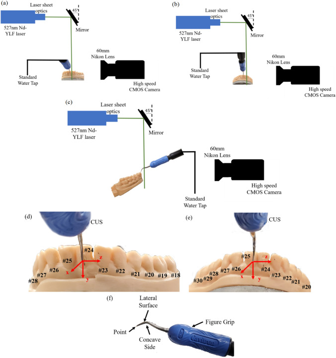
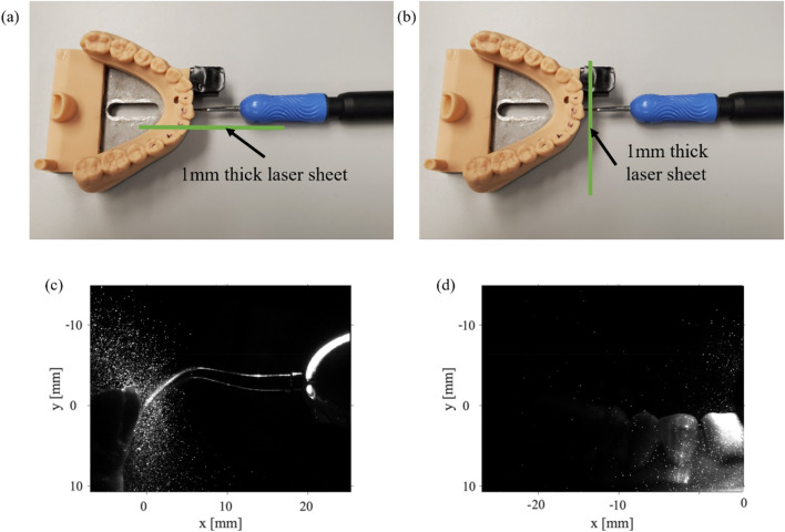
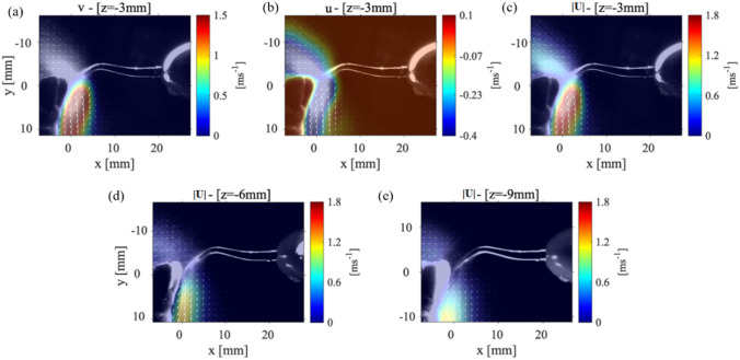
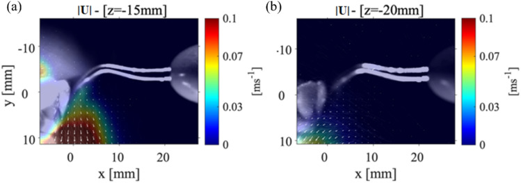
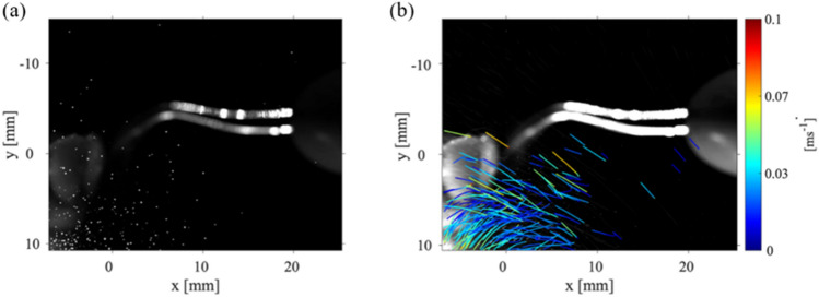
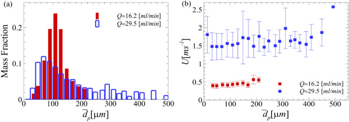
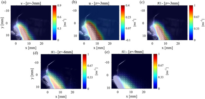
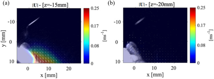
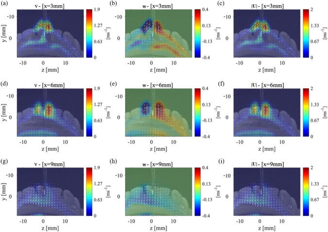
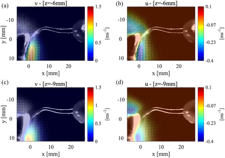
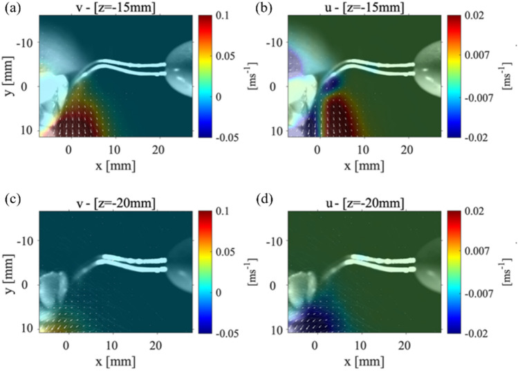
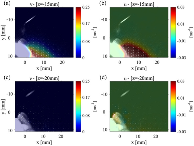
Throughout 2020 and beyond, the entire world has observed a continuous increase in the infectious spread of the novel coronavirus (SARS-CoV-2) otherwise known as COVID-19. The high transmission of this airborne virus has raised countless concerns regarding safety measures employed in the working conditions for medical professionals. Specifically, those who perform treatment procedures on patients which intrinsically create mists of fine airborne droplets, i.e., perfect vectors for this and other viruses to spread. The present study focuses on understanding the splatter produced due to a common dentistry technique to remove plaque buildup on teeth. This technique uses a high-speed dentistry instrument, e.g., a Cavitron ultrasonic scaler, to scrape along the surface of a patient's teeth. This detailed understanding of the velocity and the trajectory of the droplets generated by the splatter will aid in the development of hygiene mechanisms to guarantee the safety of those performing these procedures and people in clinics or hospitals. Optical flow tracking velocimetry (OFTV) method was employed to obtain droplet velocity and trajectory in a two-dimensional plane. Multiple data collection planes were taken in different orientations around a model of adult mandibular teeth. This technique provided pseudo-three-dimensional velocity information for the droplets within the splatter developed from this high-speed dental instrument. These results indicated that within the three-dimensional splatter produced there were high velocities (1-2 m/s) observed directly below the intersection point between the front teeth and the scaler. The splatter formed a cone-shape structure that propagated 10-15 mm away from the location of the scaler tip. From the droplet trajectories, it was observed that high velocity isolated droplets propagate away from the bulk of the splatter. It is these droplets which are concerning for health safety to those performing the medical procedures. Using a shadowgraphy technique, we further characterize the individual droplets' size and their individual velocity. We then compare these results to previously published distributions. The obtained data can be used as a first step to further examine flow and transport of droplets in clinics/dental offices.
© This is a U.S. government work and not under copyright protection in the U.S.; foreign copyright protection may apply 2021.
Figures

Similar articles
-
Aerosol formation due to a dental procedure: insights leading to the transmission of diseases to the environment.J R Soc Interface. 2021 Mar;18(176):20200967. doi: 10.1098/rsif.2020.0967. Epub 2021 Mar 24. J R Soc Interface. 2021. PMID: 33757291 Free PMC article.
-
Simulated and clinical aerosol spread in common periodontal aerosol-generating procedures.Clin Oral Investig. 2022 Sep;26(9):5751-5762. doi: 10.1007/s00784-022-04532-8. Epub 2022 May 17. Clin Oral Investig. 2022. PMID: 35581347 Free PMC article.
-
Dissemination of Aerosol and Splatter in Clinical Environment during Cavity Preparation: An In Vitro Study.Int J Environ Res Public Health. 2021 Apr 4;18(7):3773. doi: 10.3390/ijerph18073773. Int J Environ Res Public Health. 2021. PMID: 33916609 Free PMC article.
-
Dental bioaerosol as an occupational hazard in a dentist's workplace.Ann Agric Environ Med. 2007;14(2):203-7. Ann Agric Environ Med. 2007. PMID: 18247451 Review.
-
Interventions to reduce contaminated aerosols produced during dental procedures for preventing infectious diseases.Cochrane Database Syst Rev. 2020 Oct 12;10(10):CD013686. doi: 10.1002/14651858.CD013686.pub2. Cochrane Database Syst Rev. 2020. PMID: 33047816 Free PMC article.
Cited by
-
Precise control of digital dental unit to reduce aerosol and splatter production: new challenges for future epidemics.BMC Oral Health. 2024 Feb 10;24(1):213. doi: 10.1186/s12903-024-03980-w. BMC Oral Health. 2024. PMID: 38341576 Free PMC article.
-
Novel use of riboflavin as a fluorescent tracer in the dissemination of aerosol and splatter in an open operatory dental clinic.Clin Exp Dent Res. 2023 Jun;9(3):500-508. doi: 10.1002/cre2.727. Epub 2023 Mar 31. Clin Exp Dent Res. 2023. PMID: 37000173 Free PMC article.
-
Quantifying strategies to minimize aerosol dispersion in dental clinics.Exp Comput Multiph Flow. 2023;5(3):290-303. doi: 10.1007/s42757-022-0157-3. Epub 2023 Mar 28. Exp Comput Multiph Flow. 2023. PMID: 37305074 Free PMC article.
References
-
- Beggs CB (2020) Is there an airborne component to the transmission of COVID-19?: a quantitative analysis study. medRxiv
-
- Bennett A, Fulford M, Walker J, Bradshaw D, Martin M, Marsh P (2000) Microbial aerosols in general dental practice. Br Dent J 189:664–667 - PubMed
-
- CDC Best Practices for Environmental Cleaning in Healthcare Facilities in Resource-Limited Settings. Atlanta, GA: US Department of Health and Human Services, ; Cape Town, South Africa: Infection Control Africa Network
LinkOut - more resources
Full Text Sources
Miscellaneous