

Visual Explanation for Identification of the Brain Bases for Developmental Dyslexia on fMRI Data
- PMID: 34566613
- PMCID: PMC8458961
- DOI: 10.3389/fncom.2021.594659
Visual Explanation for Identification of the Brain Bases for Developmental Dyslexia on fMRI Data
Abstract
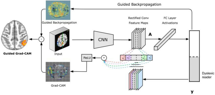
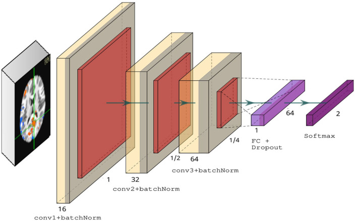
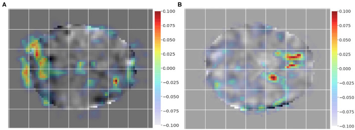
Problem: Brain imaging studies of mental health and neurodevelopmental disorders have recently included machine learning approaches to identify patients based solely on their brain activation. The goal is to identify brain-related features that generalize from smaller samples of data to larger ones; in the case of neurodevelopmental disorders, finding these patterns can help understand differences in brain function and development that underpin early signs of risk for developmental dyslexia. The success of machine learning classification algorithms on neurofunctional data has been limited to typically homogeneous data sets of few dozens of participants. More recently, larger brain imaging data sets have allowed for deep learning techniques to classify brain states and clinical groups solely from neurofunctional features. Indeed, deep learning techniques can provide helpful tools for classification in healthcare applications, including classification of structural 3D brain images. The adoption of deep learning approaches allows for incremental improvements in classification performance of larger functional brain imaging data sets, but still lacks diagnostic insights about the underlying brain mechanisms associated with disorders; moreover, a related challenge involves providing more clinically-relevant explanations from the neural features that inform classification. Methods: We target this challenge by leveraging two network visualization techniques in convolutional neural network layers responsible for learning high-level features. Using such techniques, we are able to provide meaningful images for expert-backed insights into the condition being classified. We address this challenge using a dataset that includes children diagnosed with developmental dyslexia, and typical reader children. Results: Our results show accurate classification of developmental dyslexia (94.8%) from the brain imaging alone, while providing automatic visualizations of the features involved that match contemporary neuroscientific knowledge (brain regions involved in the reading process for the dyslexic reader group and brain regions associated with strategic control and attention processes for the typical reader group). Conclusions: Our visual explanations of deep learning models turn the accurate yet opaque conclusions from the models into evidence to the condition being studied.
Keywords: deep learning; dyslexia; fMRI; neuroimaging; visual explanation.
Copyright © 2021 Tomaz Da Silva, Esper, Ruiz, Meneguzzi and Buchweitz.
Conflict of interest statement
The authors declare that the research was conducted in the absence of any commercial or financial relationships that could be construed as a potential conflict of interest.
Figures


References
-
- American Psychiatric Association (2013). Diagnostic and Statistical Manual of Mental disorders (DSM-5®). American Psychiatric Association.
-
- Buchweitz A., Costa A. C., Toazza R., de Moraes A. B., Cara V. M., Esper N. B., et al. (2019). Decoupling of the occipitotemporal cortex and the brain's default-mode network in dyslexia and a role for the cingulate cortex in good readers: a brain imaging study of brazilian children. Dev. Neuropsychol. 44, 146–157. 10.1080/87565641.2017.1292516 - DOI - PubMed
LinkOut - more resources
Full Text Sources