Review
. 2021 Sep 9:12:734215.
doi: 10.3389/fphys.2021.734215. eCollection 2021.

The Mechanobiology of Endothelial-to-Mesenchymal Transition in Cardiovascular Disease

Affiliations
Review

The Mechanobiology of Endothelial-to-Mesenchymal Transition in Cardiovascular Disease

Shahrin Islam et al. Front Physiol. .

Abstract

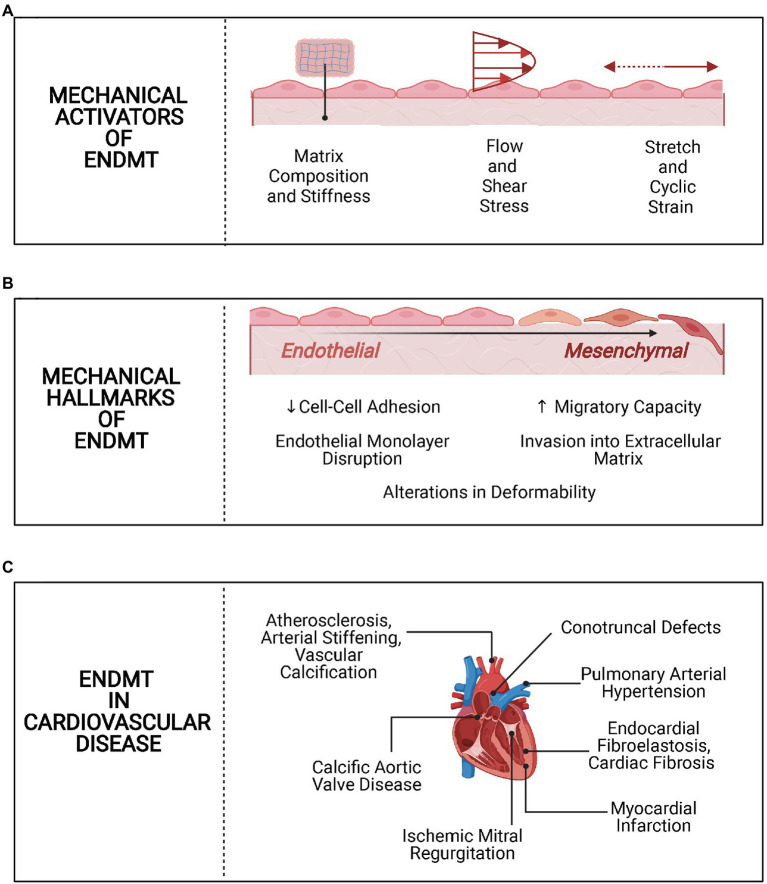
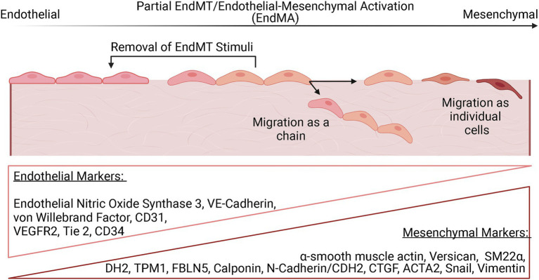
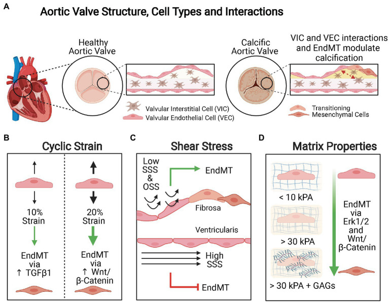
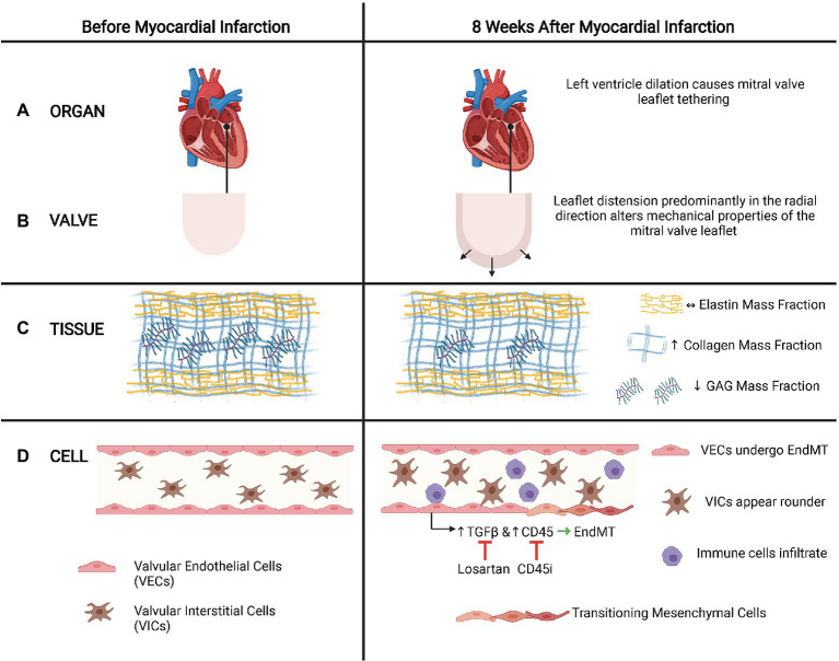
Endothelial cells (ECs) lining the cardiovascular system are subjected to a highly dynamic microenvironment resulting from pulsatile pressure and circulating blood flow. Endothelial cells are remarkably sensitive to these forces, which are transduced to activate signaling pathways to maintain endothelial homeostasis and respond to changes in the environment. Aberrations in these biomechanical stresses, however, can trigger changes in endothelial cell phenotype and function. One process involved in this cellular plasticity is endothelial-to-mesenchymal transition (EndMT). As a result of EndMT, ECs lose cell-cell adhesion, alter their cytoskeletal organization, and gain increased migratory and invasive capabilities. EndMT has long been known to occur during cardiovascular development, but there is now a growing body of evidence also implicating it in many cardiovascular diseases (CVD), often associated with alterations in the cellular mechanical environment. In this review, we highlight the emerging role of shear stress, cyclic strain, matrix stiffness, and composition associated with EndMT in CVD. We first provide an overview of EndMT and context for how ECs sense, transduce, and respond to certain mechanical stimuli. We then describe the biomechanical features of EndMT and the role of mechanically driven EndMT in CVD. Finally, we indicate areas of open investigation to further elucidate the complexity of EndMT in the cardiovascular system. Understanding the mechanistic underpinnings of the mechanobiology of EndMT in CVD can provide insight into new opportunities for identification of novel diagnostic markers and therapeutic interventions.

Keywords: biomechanical; cardiovascular disease; endothelial; endothelial-to-mesenchymal transition; mechanobiology; mesenchymal.


Conflict of interest statement

The authors declare that the research was conducted in the absence of any commercial or financial relationships that could be construed as a potential conflict of interest.

Figures

Figure 1
Figure 1
Overview of mechanobiology of endothelial-to-mesenchymal transition (EndMT) in cardiovascular disease (CVD). (A) Mechanical activators of EndMT. Alterations in matrix stiffness and composition, shear stress, and cyclic strain activate EndMT in endothelial cells. (B) Mechanical hallmarks of EndMT. Following activation of EndMT, there are mechanical changes, such as loss of cell-cell adhesion and increased migration. (C) EndMT in CVD. EndMT is implicated in several CVD processes, with a potential role for biomechanical alterations in each.
Figure 2
Figure 2
The EndMT continuum. EndMT is thought to exist on a spectrum with evidence for partial activation of EndMT that may be reversed with removal of the EndMT stimuli. Endothelial cells that have undergone partial EndMT can migrate as a chain without loss of cell-cell adhesion. ECs that undergo EndMT can also migrate as individual cells. Complete EndMT is defined as the presence of an entirely mesenchymal state. The progression of EndMT is evaluated by the monitoring the expression of various endothelial and mesenchymal markers, with ECs that have undergone partial EndMT expressing some level of both types of markers.
Figure 3
Figure 3
Mechanical modulation of EndMT in valvular endothelial cells (VECs). (A) Aortic valve structure, cell types, and interactions. The aortic valve is a trilaminar structure with valvular interstitial cells (VICs) sandwiched in between two layers of VECs. Interactions between the VICs and VECs and EndMT are both important in valvular calcification. (B) Cyclic strain. Cyclic strain induces greater extent of EndMT when applied orthogonally to the alignment of the valve endothelium and the pathways activated are magnitude dependent. When VECs are subject to 10% cyclic strain, EndMT is activated via TGFβ1 signaling, whereas 20% cyclic strain promotes EndMT via Wnt/β-Catenin signaling. (C) Shear stress. Low steady shear stress (SSS) and oscillatory shear stress (OSS) promote EndMT, whereas high SSS is protective against EndMT. Calcification is more common on the fibrosa side where VECs are exposed to low SSS or OSS than on the ventricularis side where VECs are subject to high SSS. (D) Matrix composition and stiffness. Increasing matrix stiffness and increased glycosaminoglycans (GAGs) content promote EndMT.
Figure 4
Figure 4
Leaflet tethering and EndMT in ischemic mitral regurgitation (IMR). (A). Organ. After a myocardial infarction, the heart often dilates, which causes tethering of the mitral valves that can lead to IMR. (B) Valve. Tethering distends the leaflet predominantly in the radial direction. (C) Tissue. Leaflet tethering alters the mitral valve matrix composition by increasing the collagen mass fraction and decreasing the GAG mass fraction. However, the elastic mass fraction does not change. (D) Cell. These biomechanical alterations of the valve leaflet may trigger EndMT of the mitral valve endothelial cells via mechanisms that involve TGFβ signaling and CD45. Inhibiting TGFβ with losartan or CD45 with a CD45 inhibitor reduces EndMT and associated maladaptive responses observed in IMR. In addition to EndMT of VECs, VICs also appear rounder and immune cells may infiltrate. (Adapted with permission from Howsmon et al., 2020).
Figure 5
Figure 5
Disturbed flow (d-flow), EndMT, and plaque stability in atherosclerosis. (A) d-flow that occurs at the lesser curvature of the aortic arch is atherogenic, whereas stable flow typically presents in the descending aorta is atheroprotective (EndICLT). (B) The mechanosensor Piezo1 is crucial to the differential responses to these hemodynamic profiles. In response to d-flow, Piezo1 activates the NF-κB pathway, which is known to be important to inflammation-mediated EndMT. In addition to EndMT, d-flow also activates of endothelial-to-immune-cell-like transition. The mechanosensor Alk5 and associated protein Shc have been directly linked to activation of Smad-2 mediated EndMT pathways in response to d-flow in atherosclerosis. (C) EndMT can contribute to both plaque instability and plaque stability depending on whether EndMT results in transition of ECs to fibroblast-like cell types with increased matrix metalloproteinase expression or to myofibroblasts.

References

    1. Adjuto-Saccone M., Soubeyran P., Garcia J., Audebert S., Camoin L., Rubis M., et al. . (2021). TNF-α induces endothelial–mesenchymal transition promoting stromal development of pancreatic adenocarcinoma. Cell Death Dis. 12:649. 10.1038/s41419-021-03920-4, PMID: - DOI - PMC - PubMed
    1. Ahmmed S. M., Bithi S. S., Pore A. A., Mubtasim N., Schuster C., Gollahon L. S., et al. . (2018). Multi-sample deformability cytometry of cancer cells. APL Bioeng. 2:032002. 10.1063/1.5020992, PMID: - DOI - PMC - PubMed
    1. Ajami N. E., Gupta S., Maurya M. R., Nguyen P., Li J. Y.-S., Shyy J. Y.-J., et al. . (2017). Systems biology analysis of longitudinal functional response of endothelial cells to shear stress. Proc. Natl. Acad. Sci. U. S. A. 114, 10990–10995. 10.1073/pnas.1707517114, PMID: - DOI - PMC - PubMed
    1. Ali S. R., Ranjbarvaziri S., Talkhabi M., Zhao P., Subat A., Hojjat A., et al. . (2014). Developmental heterogeneity of cardiac fibroblasts does not predict pathological proliferation and activation. Circ. Res. 115, 625–635. 10.1161/CIRCRESAHA.115.303794, PMID: - DOI - PubMed
    1. Alonso-Herranz L., Sahún-Español Á., Paredes A., Gonzalo P., Gkontra P., Núñez V., et al. . (2020). Macrophages promote endothelial-to-mesenchymal transition via MT1-MMP/TGFβ1 after myocardial infarction. elife 9:e57920. 10.7554/eLife.57920, PMID: - DOI - PMC - PubMed