. 2021 Sep 8:12:722526.
doi: 10.3389/fneur.2021.722526. eCollection 2021.

Age-at-Injury Determines the Extent of Long-Term Neuropathology and Microgliosis After a Diffuse Brain Injury in Male Rats

Affiliations

Age-at-Injury Determines the Extent of Long-Term Neuropathology and Microgliosis After a Diffuse Brain Injury in Male Rats

Yasmine V Doust et al. Front Neurol. .

Abstract

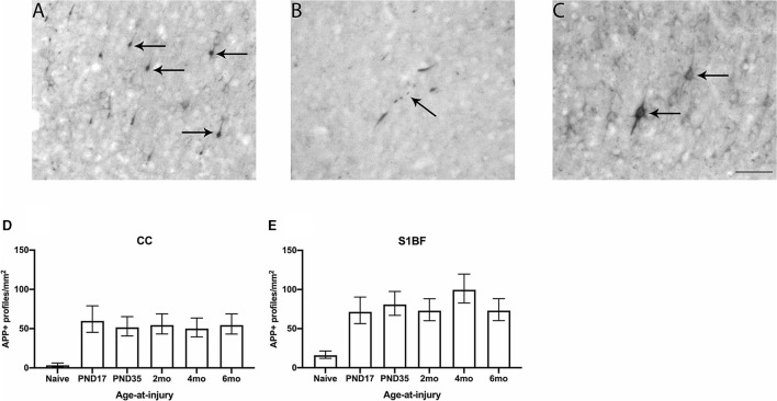
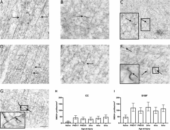
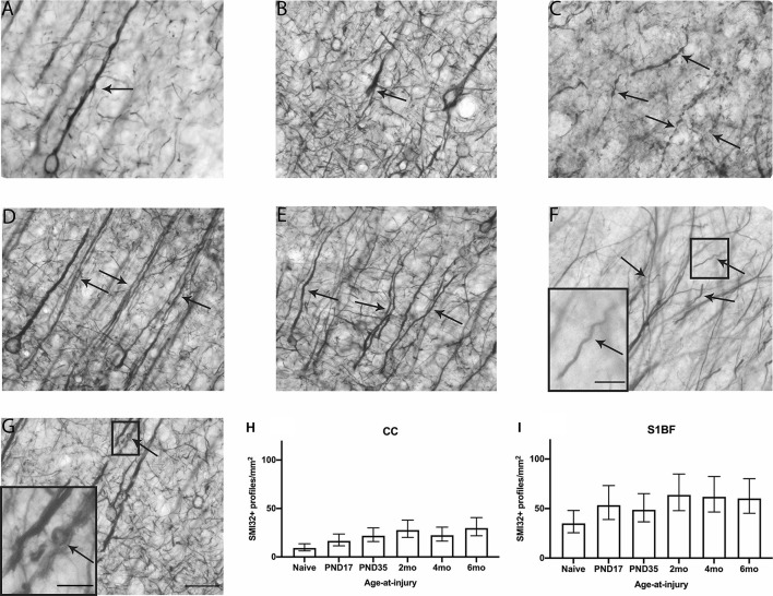
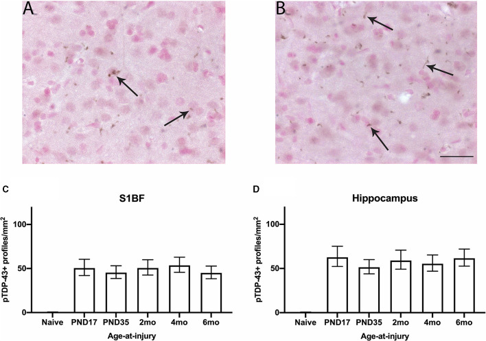
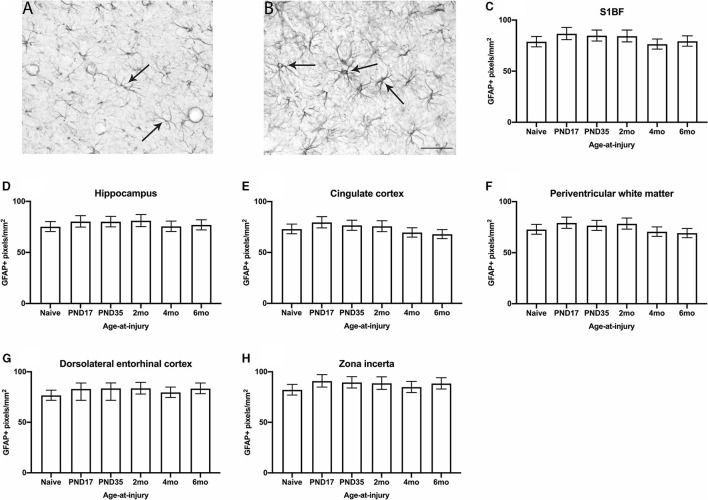
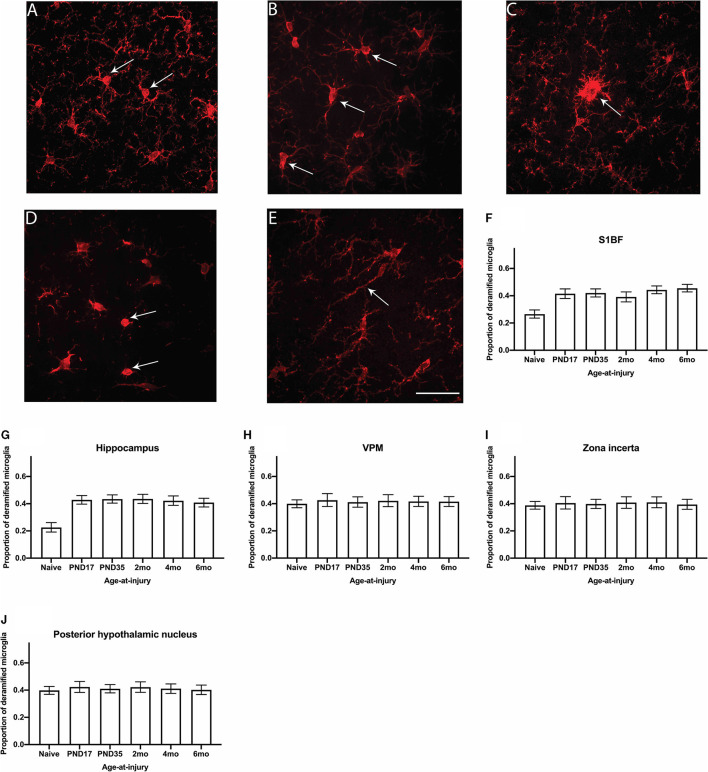
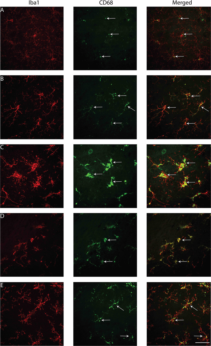
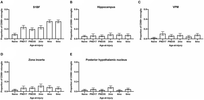
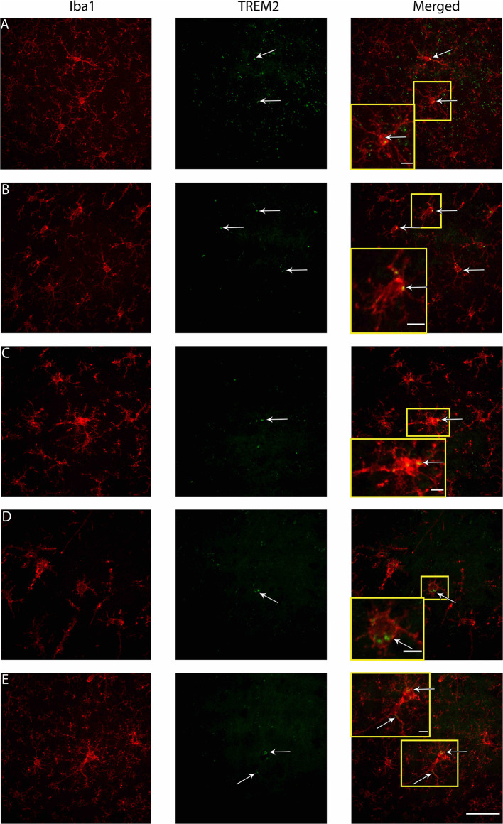
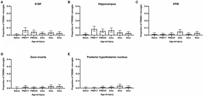
Traumatic brain injury (TBI) can occur at any age, from youth to the elderly, and its contribution to age-related neuropathology remains unknown. Few studies have investigated the relationship between age-at-injury and pathophysiology at a discrete biological age. In this study, we report the immunohistochemical analysis of naïve rat brains compared to those subjected to diffuse TBI by midline fluid percussion injury (mFPI) at post-natal day (PND) 17, PND35, 2-, 4-, or 6-months of age. All brains were collected when rats were 10-months of age (n = 6-7/group). Generalized linear mixed models were fitted to analyze binomial proportion and count data with R Studio. Amyloid precursor protein (APP) and neurofilament (SMI34, SMI32) neuronal pathology were counted in the corpus callosum (CC) and primary sensory barrel field (S1BF). Phosphorylated TAR DNA-binding protein 43 (pTDP-43) neuropathology was counted in the S1BF and hippocampus. There was a significantly greater extent of APP and SMI34 axonal pathology and pTDP-43 neuropathology following a TBI compared with naïves regardless of brain region or age-at-injury. However, age-at-injury did determine the extent of dendritic neurofilament (SMI32) pathology in the CC and S1BF where all brain-injured rats exhibited a greater extent of pathology compared with naïve. No significant differences were detected in the extent of astrocyte activation between brain-injured and naïve rats. Microglia counts were conducted in the S1BF, hippocampus, ventral posteromedial (VPM) nucleus, zona incerta, and posterior hypothalamic nucleus. There was a significantly greater proportion of deramified microglia, regardless of whether the TBI was recent or remote, but this only occurred in the S1BF and hippocampus. The proportion of microglia with colocalized CD68 and TREM2 in the S1BF was greater in all brain-injured rats compared with naïve, regardless of whether the TBI was recent or remote. Only rats with recent TBI exhibited a greater proportion of CD68-positive microglia compared with naive in the hippocampus and posterior hypothalamic nucleus. Whilst, only rats with a remote brain-injury displayed a greater proportion of microglia colocalized with TREM2 in the hippocampus. Thus, chronic alterations in neuronal and microglial characteristics are evident in the injured brain despite the recency of a diffuse brain injury.

Keywords: TBI; age-at-injury; aging; concussion; juvenile; pathology; puberty; traumatic brain injury.


Conflict of interest statement

The authors declare that the research was conducted in the absence of any commercial or financial relationships that could be construed as a potential conflict of interest.

Figures

Figure 1
Figure 1
APP neuronal profiles were quantified in the CC and S1BF. Indicative images of neuronal pathology visualized by APP immunohistochemistry were taken in the primary sensory barrel field (S1BF) of rats brain-injured at 6-months of age. Quantitative analysis included bulbs (A), blebbing (B), and cell bodies (C) indicated by the black arrows (scale bar = 50 μm). Counts of neuronal pathology are expressed as the number of APP-positive (APP+) profiles/mm2 (mean ± 95%CI) in the corpus callosum (CC) and S1BF. Significantly more APP+ profiles were observed following mFPI, regardless of age-at-injury, compared to naïves in both the CC (D) and the S1BF (E).
Figure 2
Figure 2
SMI34 axonal profiles were quantified in the CC and S1BF. Indicative images of healthy axons (A) and axonal pathology visualized by SMI34 immunohistochemistry were taken in the primary sensory barrel field (S1BF) of rats brain-injured at 6-months of age. Analysis quantified bulbs (B), blebbing (C), thickened axons (white arrows; D) or undulated axons (black arrows; D), thickened and undulated axons (E), corkscrews (F), and rings (G) indicated by the arrows (scale bar = 50 μm, inset = 10 μm). Counts of axonal pathology are expressed as the number of SMI34+ profiles/mm2 (mean ± 95%CI) in the corpus callosum (CC) and S1BF. There were significantly more SMI34-positive (SMI34+) profiles observed post-injury, regardless of age-at-injury, compared to naïves in both the CC (H) and the S1BF (I).
Figure 3
Figure 3
SMI32 dendritic profiles were quantified in the CC and S1BF. Indicative images of healthy dendrites (A) and dendritic pathology visualized by SMI32 immunohistochemistry were taken in the primary sensory barrel field (S1BF) of rats brain-injured at 6-months of age. Quantitative analysis included bulbs (B), blebbing (C), thickened axons (white arrows; D), or undulated axons (black arrows; D), thickened and undulated axons (E), corkscrews (F), and rings (G) indicated by the arrows (scale bar = 50 μm, inset = 10 μm). Dendritic pathology was expressed as the number of SMI32+ profiles/mm2 (mean ± 95%CI) in the corpus callosum (CC) and S1BF. Significantly more SMI32+ profiles were observed following mFPI compared to naïves in the CC (H), regardless of whether the TBI was recent or remote. However, a greater extent of SMI32 pathology in the S1BF (I) compared with naïves was only evident with a more recent TBI (see Table 3).
Figure 4
Figure 4
pTDP-43 neuronal profiles were quantified in the S1BF and hippocampus. Indicative images of neuronal pathology visualized by pTDP-43 immunohistochemistry were taken in the primary sensory barrel field (S1BF) of rats injured at 6-months of age. Quantitative analysis included nuclei inclusions (A) and cytoplasmic neurites (B) indicated by the black arrows (scale bar = 50 μm). Counts of neuronal pathology are expressed as the number of pTDP-43+ profiles/mm2 (mean ± 95%CI) in the S1BF and hippocampus. Significantly more pTDP-43+ profiles were observed following mFPI, regardless of age-at-injury, compared to naïves in both the S1BF (C) and the hippocampus (D).
Figure 5
Figure 5
GFAP immunoreactivity was quantified in the S1BF, hippocampus, cingulate cortex, periventricular white matter, dorsolateral entorhinal cortex, and zona incerta. Representative images of ramified (A) and hypertrophic (B) astrocytes, indicated with black arrows (scale bar = 50 μm), visualized by GFAP immunohistochemistry were taken in the primary sensory barrel field (S1BF) of rats injured at 6-months of age. The expression of GFAP increases when astrocytes become hypertrophic and, thus, activated. Therefore, the extent of GFAP immunoreactivity is used as a measure of astrocyte activation and is expressed as the number of GFAP-positive pixels/mm2 (mean ± 95%CI) in the S1BF, hippocampus, cingulate cortex, lateral ventricle, dorsolateral entorhinal cortex, and zona incerta. There were no significant differences in the percentage of GFAP+ profiles observed following mFPI, regardless of age-at-injury, compared to naïves in the S1BF (C), hippocampus (D), cingulate cortex (E), periventricular white matter (F), dorsolateral entorhinal cortex (G), or the zona incerta (H).
Figure 6
Figure 6
The proportion of deramified microglia were quantified in the S1BF, hippocampus, VPM, zona incerta, and posterior hypothalamic nucleus. Representative images of ramified (A) and deramified (B–E) microglia, indicated by the black arrows (scale bar = 50 μm), visualized by Iba1 immunohistochemistry were taken in the primary sensory barrel field (S1BF) of rats injured at 6-months of age. The proportion of deramified microglia (mean ± 95%CI) was determined in the S1BF, hippocampus, ventral posteromedial nucleus (VPM), zona incerta, and posterior hypothalamic nucleus. There was a significantly greater proportion of deramified microglia observed after brain injury, regardless of age-at-injury, compared to naïves in the S1BF (F), hippocampus (G) but not in the zona incerta (H), VPM (I), or posterior hypothalamic nucleus (J).
Figure 7
Figure 7
Examples of microglial morphologies colocalized with CD68 in the S1BF of rats brain-injured at 6-months of age. Representative images of microglial morphologies were taken in the primary sensory barrel field (S1BF) of rats injured at 6-months of age; ramified (A), hyper-ramified (B), hypertrophic (C), amoeboid (D), and rod (E), and their colocalization with CD68 indicated with the white arrows (scale bar = 50 μm).
Figure 8
Figure 8
The proportion of microglia colocalized with CD68 were quantified in the S1BF, hippocampus, VPM, zona incerta, and posterior hypothalamic nucleus. Microglial colocalization with CD68 is expressed as the proportion of CD68+ microglia (mean ± 95%CI) in the primary sensory barrel field (S1BF), hippocampus, ventral posteromedial nucleus (VPM), zona incerta, and posterior hypothalamic nucleus. Significantly more CD68+ microglia were observed following mFPI compared to naïves in the S1BF (A), zona incerta (C), and VPM (D) regardless of whether the TBI was recent or remote. However, in the hippocampus (B) and posterior hypothalamic nucleus (E), a greater proportion of microglia colocalized with CD68 was only evident compared with naïves with a more recent TBI (see Table 5).
Figure 9
Figure 9
Examples of microglial morphologies in colocalization with TREM2 in the S1BF of rats brain-injured at 6-months of age. Representative images of microglial morphologies were taken in the primary sensory barrel field (S1BF) of rats injured at 6-months of age; ramified (A), hyper-ramified (B), deramified (C), amoeboid (D), and rod (E), and their colocalization with TREM2 indicated with the white arrows (scale bar = 50 μm, inset = 10 μm).
Figure 10
Figure 10
The proportion of microglia colocalized with TREM2 were quantified in the S1BF, hippocampus, VPM, zona incerta, and posterior hypothalamic nucleus. Microglial colocalization with TREM2 is expressed as the proportion of TREM2+ microglia (mean ± 95%CI) in the primary sensory barrel field (S1BF), hippocampus, ventral posteromedial nucleus (VPM), zona incerta, and posterior hypothalamic nucleus. Significantly more TREM2+ microglia were observed following mFPI compared to naïves in the S1BF (A), regardless of whether the TBI was recent or remote. However, in the hippocampus (B), a greater proportion of microglia colocalized with TREM2 was only evident compared with naïves with a more remote TBI (see Table 14). This trend is the opposite in the zona incerta (C), VPM (D), and posterior hypothalamic nucleus (E), where a greater proportion of microglia colocalized with TREM2 was only evident compared with naïves with a more recent TBI (see Table 5).

Similar articles

Cited by

References

    1. Dhandapani S, Manju D, Sharma B, Mahapatra A. Prognostic significance of age in traumatic brain injury. J Neurosci Rural Pract. (2012) 3:131–5. 10.4103/0976-3147.98208 - DOI - PMC - PubMed
    1. Prins M, Greco T, Alexander D, Giza CC. The pathophysiology of traumatic brain injury at a glance. Dis Model Mech. (2013) 6:1307–15. 10.1242/dmm,0.011585 - DOI - PMC - PubMed
    1. Cao T, Thomas TC, Ziebell JM, Pauly JR, Lifshitz J. Morphological and genetic activation of microglia after diffuse traumatic brain injury in the rat. Neuroscience. (2012) 225:65–75. 10.1016/j.neuroscience.2012.08.058 - DOI - PMC - PubMed
    1. Johnson VE, Stewart JE, Begbie FD, Trojanowski JQ, Smith DH, Stewart W. Inflammation and white matter degeneration persist for years after a single traumatic brain injury. Brain. (2013) 136:28–42. 10.1093/brain/aws322 - DOI - PMC - PubMed
    1. Johnson VE, Stewart W, Smith DH. Axonal pathology in traumatic brain injury. Exp Neurol. (2013) 246:35–43. 10.1016/j.expneurol.2012.01.013 - DOI - PMC - PubMed

LinkOut - more resources