. 2021 Sep 9:12:723010.
doi: 10.3389/fimmu.2021.723010. eCollection 2021.

Immunomonitoring of Human Breast Milk Cells During HCMV-Reactivation

Affiliations

Immunomonitoring of Human Breast Milk Cells During HCMV-Reactivation

Katrin Lazar et al. Front Immunol. .

Abstract

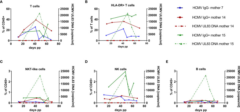
Background: Breast milk leukocytes may play a role in protecting the infant from pathogens. The dynamics and the role of lymphocytes in human cytomegalovirus (HCMV)-seropositive mothers shedding HCMV into breast milk during the first months postpartum (p.p.) are mostly unclear.

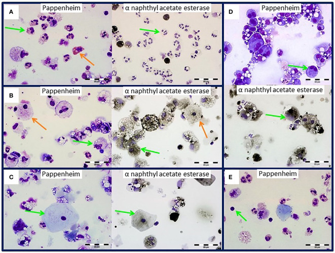
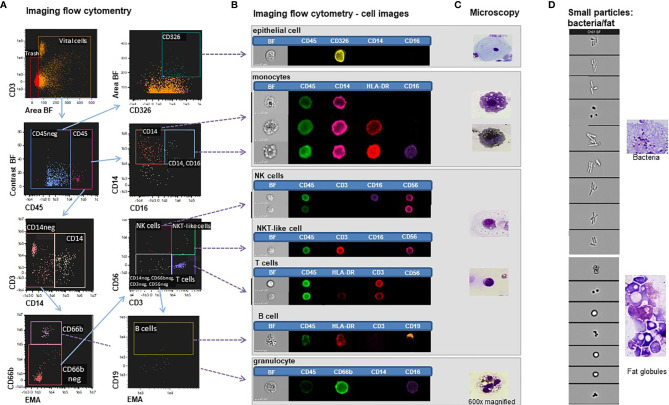
Methods: Breast milk cells were analyzed by Pappenheim panoptic and alpha-naphthyl acetate esterase staining as well as by imaging and polychromatic flow cytometry to simultaneously establish their morphological and phenotypic properties. The latter were characterized in HCMV-seropositive and seronegative mothers´ breast milk cells at different time points p.p.

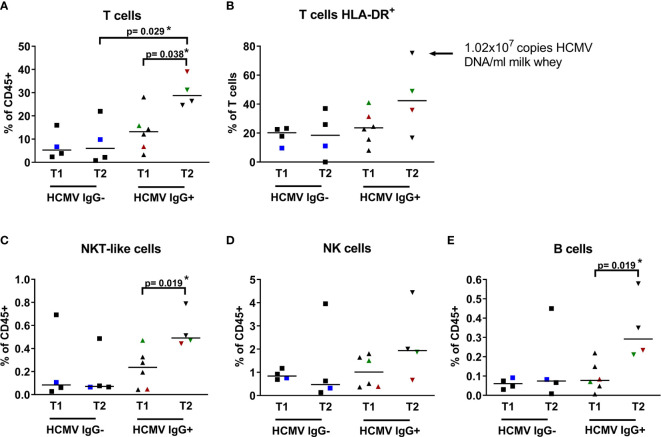
Results: Panoptic staining of breast milk cells revealed the presence of monocytes/macrophages, granulocytes and lymphocytes. Imaging flow cytometry data combining phenotypic and morphological analysis identified NKT-like cells, NK cells, epithelial cells, T cells and monocytes/macrophages. HCMV-seropositive but not -seronegative mothers had significantly higher T cell frequencies in mature milk.

Conclusions: The presence of lymphocyte subsets in breast milk may be more influenced by the HCMV-seropositivity of the mother than previously recognized.

Keywords: (imaging) flow cytometry; B cells; T cells; breastfeeding; human cytomegalovirus (HCMV); lactation; phenotyping.


Conflict of interest statement

The authors declare that the research was conducted in the absence of any commercial or financial relationships that could be construed as a potential conflict of interest.

Figures

Figure 1
Figure 1
Breast milk cells of an HCMV-seropositive mother. Green arrows indicate (A) granulocytes, (B) monocytes, (C) epithelial cells, (D) monocytes with two nuclei and (E) lymphocytes by panoptic Pappenheim (left) and α-naphthyl acetate esterase (right) staining. Orange arrows show eosinophils (A) or lactocytes (B). Images at 600x magnification.
Figure 2
Figure 2
Imaging flow cytometry of breast milk cells. (A) Gating strategy in IDEAS® of breast milk cells stained with EMA, CD45-BV510, CD3-APC, CD56-APC-H7, CD14-PE-Cy7, CD16-Pacific blue, CD66b-FITC, CD326-PE and HLADR-PerCPCy5.5. (B) Bright field (BF) and fluorescent images of epithelial cells, monocytes, NK cells, NKT-like cells, and T cells, B -cells and granulocytes. (C) Microscopy images (600x magnification) of the corresponding panoptic staining of cytospin preparations. (D) Small particles excluded as trash in the imaging flow cytometry experiments (left panel) showing morphologic characteristics of bacteria and fat globules compared to panoptically stained images (right panel).
Figure 3
Figure 3
Breast milk lymphocyte subpopulations of 15 HCMV seropositive and seronegative mothers. Frequencies within CD45+ leukocytes of (A) T cells (CD45+ CD3+ CD56-), (B) activated T cells (CD45+ CD3+ HLA-DRdim), (C) NKT-like cells (CD45+ CD3+ CD56+), (D) NK cells (CD45+CD3-CD56dim), (E) B cells (CD45+ CD3- CD19+) of HCMV-IgG- and IgG+ mothers at two time points (T1: 6-22, T2 39-108 days p.p.). Mothers with consecutive samples are color coded (blue: mother 7, red: mother 14 and green: mother 15). Statistical analysis was performed by Mann-Whitney U-test. The arrow in (B) highlights very high HLA-DR expression on CD3 T cells of one mother.
Figure 4
Figure 4
Longitudinal case studies of 3 mothers. (A) Breast milk T cell (CD45+ CD3+ CD56-), (B) HLA-DR-positive T cell (CD45+ CD3+ HLA-DRdim), (C) NKT-like cell (CD45+ CD3+ CD56+), (D) NK cell (CD45+CD3-CD56dim) and (E) B cell (CD45+ CD3- CD19+) frequencies of one HCMV-seronegative (mother 7, blue), and two HCMV-seropositive mothers (mother 15, green, with HCMV-DNA in breast milk and mother 14, red, *without HCMV reactivation in breast milk). Dotted lines present HCMV UL83 DNA viral load in milk whey.

References

    1. Andreas NJ, Kampmann B, Le-Doare KM. Human Breast Milk: A Review on its Composition and Bioactivity. Early Hum Dev (2015) 91(11):629–35. doi: 10.1016/j.earlhumdev.2015.08.013 - DOI - PubMed
    1. Boix-Amorós A, Collado MC, Van’t Land B, Calvert A, Le Doare K, Garssen J, et al. . Reviewing the Evidence on Breast Milk Composition and Immunological Outcomes. Nutr Rev (2019) 77:541–56. doi: 10.1093/nutrit/nuz019 - DOI - PubMed
    1. Gao X, McMahon RJ, Woo JG, Davidson BS, Morrow AL, Zhang Q. Temporal Changes in Milk Proteomes Reveal Developing Milk Functions. J Proteome Res (2012) 11(7):3897–907. doi: 10.1021/pr3004002 - DOI - PubMed
    1. Molinari CE, Casadio YS, Hartmann BT, Livk A, Bringans S, Arthur PG, et al. . Proteome Mapping of Human Skim Milk Proteins in Term and Preterm Milk. J Proteome Res (2012) 11(3):1696–714. doi: 10.1021/pr2008797 - DOI - PubMed
    1. Hurley WL, Theil PK. Perspectives on Immunoglobulins in Colostrum and Milk. Nutrients (2011) 3(4):442–74. doi: 10.3390/nu3040442 - DOI - PMC - PubMed

Publication types

Substances