

Congenital hepatic cyst: Prenatal and postnatal imaging findings
- PMID: 34567232
- PMCID: PMC8366221
- DOI: 10.1177/1742271X20970601
Congenital hepatic cyst: Prenatal and postnatal imaging findings
Abstract
Introduction: Congenital hepatic cyst is a rare hepatobiliary malformation that can present as an asymptomatic, unilocular, upper abdominal cystic mass in the fetus.
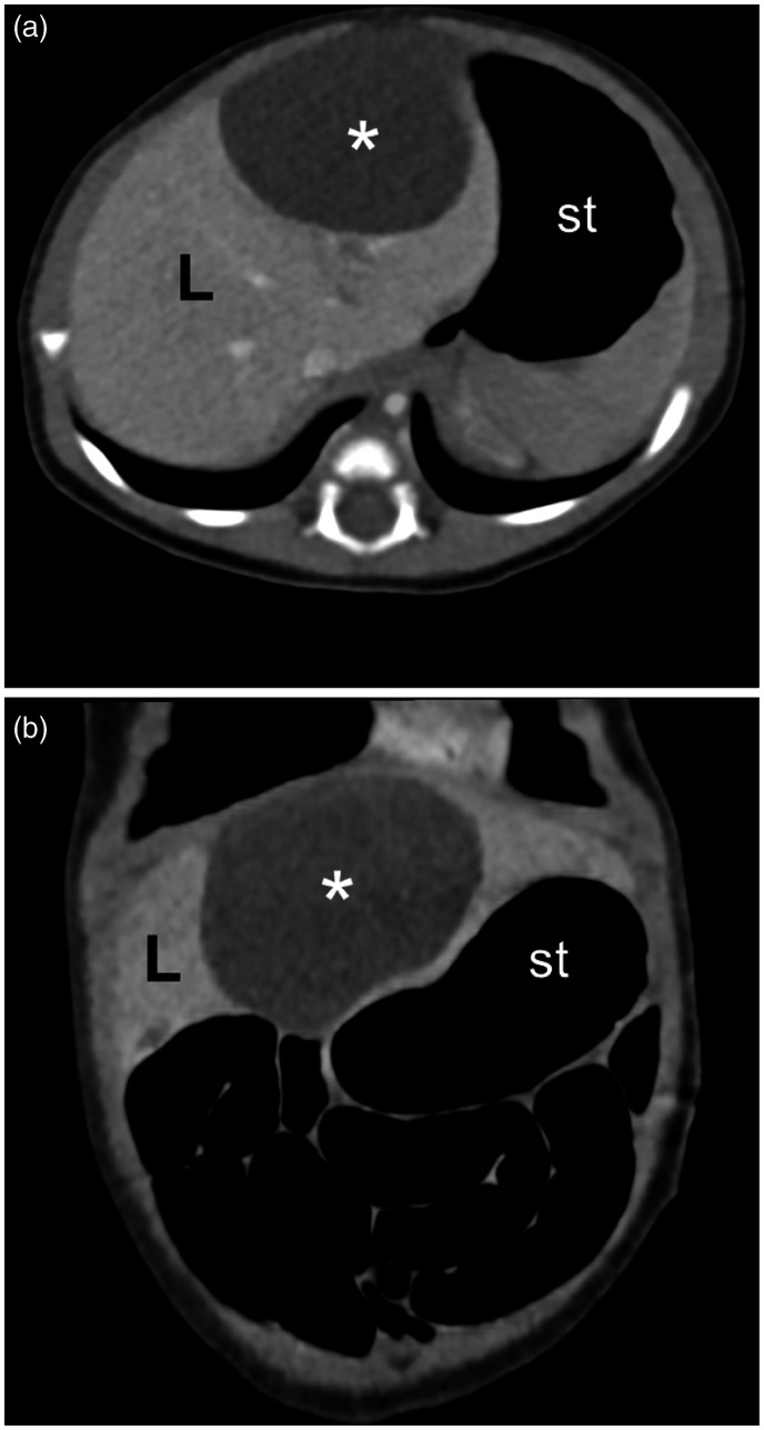
Cases: We report two cases of congenital hepatic cyst in which the diagnosis was made by prenatal ultrasound at 25 and 33 weeks' gestation. The diagnosis was confirmed postnatally by abdominal ultrasound and radiologic imaging studies. Although the infants remained asymptomatic, laparoscopic excision was performed due to the increasing size of the cyst in both cases. Pathological examination of the resected specimens confirmed a simple cyst in one case and an epidermoid cyst in the other.
Conclusions: Our cases and those described in the literature demonstrate the usefulness of incidental prenatal detection of congenital hepatic cyst, especially during late pregnancy. Such a diagnosis can allow for proper perinatal surveillance, selection of the route of delivery, and timely postnatal surgical intervention if required.
Keywords: Congenital anomalies; fetal ultrasound; hepatic cyst; prenatal diagnosis.
© The Author(s) 2020.
Conflict of interest statement
Declaration of Conflicting Interests: The author(s) declared no potential conflicts of interest with respect to the research, authorship, and/or publication of this article.
Figures


References
-
- Allan M, Davenport M.Congenital hepatic cysts. In: Lima M, Reinberg O. (eds) Neonatal surgery. Contemporary strategies from fetal life to the first year of age. Cham: Springer Nature Switzerland AG, 2019, pp. 401− 408.
-
- McEwing R, Hayward C, Furness M.Foetal cystic abdominal masses. Australas Radiol 2003; 47: 101−110. - PubMed
-
- Ficara A, Syngelaki A, Hammami A, et al.. Value of routine ultrasound examination at 35–37 weeks’ gestation in diagnosis of fetal abnormalities. Ultrasound Obstet Gynecol 2020; 55: 75−80. - PubMed
-
- Macken MB, Wright JR, Lau H, et al.. Prenatal sonographic detection of congenital hepatic cyst in third trimester after normal second-trimester sonographic examination. J Clin Ultrasound 2000; 28: 307−310. - PubMed
Publication types
LinkOut - more resources
Full Text Sources