Review
. 2020 Oct 5:6:76.
doi: 10.1038/s41378-020-00185-3. eCollection 2020.

Microtechnology-based methods for organoid models

Affiliations
Review

Microtechnology-based methods for organoid models

Vanessa Velasco et al. Microsyst Nanoeng. .

Abstract

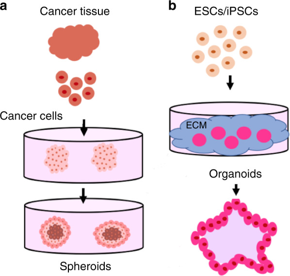
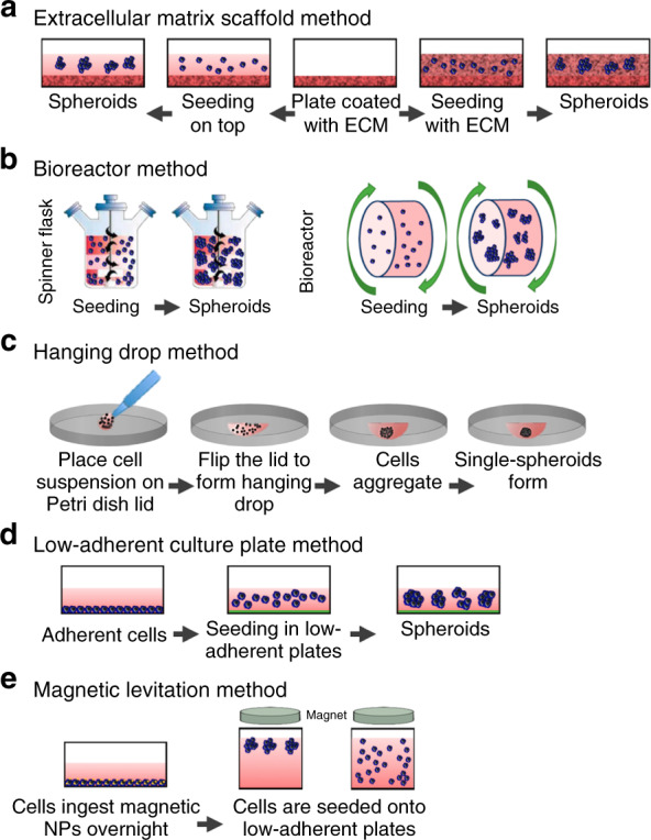
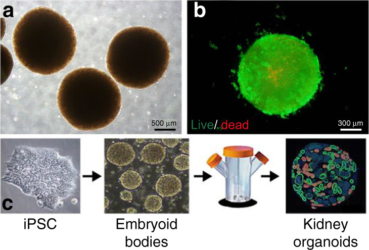
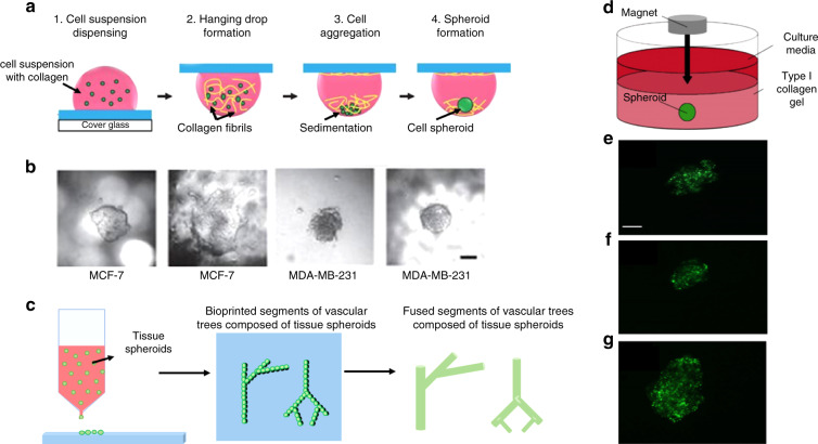
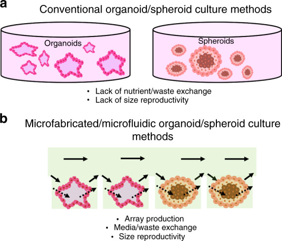
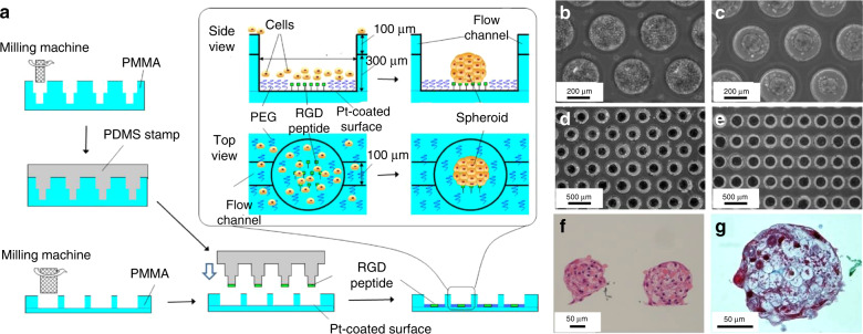
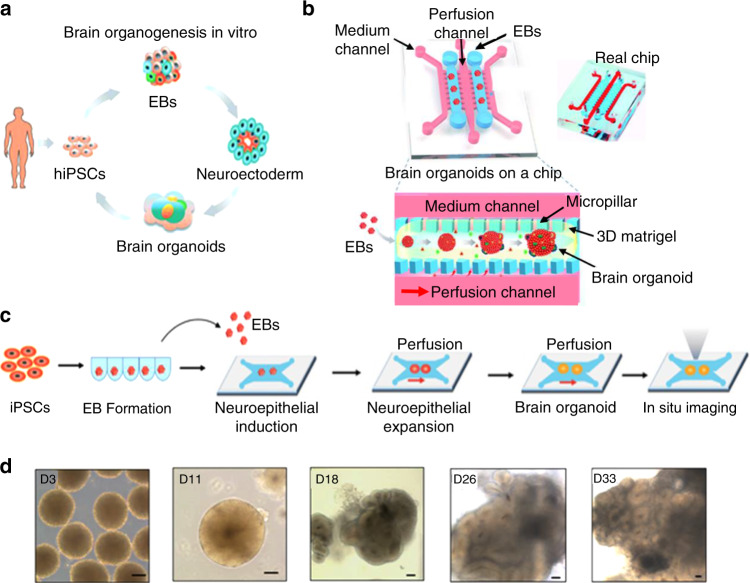
Innovations in biomaterials and stem cell technology have allowed for the emergence of novel three-dimensional (3D) tissue-like structures known as organoids and spheroids. As a result, compared to conventional 2D cell culture and animal models, these complex 3D structures have improved the accuracy and facilitated in vitro investigations of human diseases, human development, and personalized medical treatment. Due to the rapid progress of this field, numerous spheroid and organoid production methodologies have been published. However, many of the current spheroid and organoid production techniques are limited by complexity, throughput, and reproducibility. Microfabricated and microscale platforms (e.g., microfluidics and microprinting) have shown promise to address some of the current limitations in both organoid and spheroid generation. Microfabricated and microfluidic devices have been shown to improve nutrient delivery and exchange and have allowed for the arrayed production of size-controlled culture areas that yield more uniform organoids and spheroids for a higher throughput at a lower cost. In this review, we discuss the most recent production methods, challenges currently faced in organoid and spheroid production, and microfabricated and microfluidic applications for improving spheroid and organoid generation. Specifically, we focus on how microfabrication methods and devices such as lithography, microcontact printing, and microfluidic delivery systems can advance organoid and spheroid applications in medicine.

Keywords: Bionanoelectronics; Electrical and electronic engineering.


Conflict of interest statement

Conflict of interestThe authors declare that they have no conflict of interest.

Figures

Fig. 1
Fig. 1. Diagram of spheroid and organoid development.
a From human cancer tissue, tumor cells can be isolated and placed in culture to yield spheroids. b Embryonic stem cells (ESCs) and induced pluripotent stem cells (iPSCs) are two common stem cell types used as the cellular source for organoid production. Both ESCs and iPSCs can form a variety of organoid models when given the right signaling cues and ECM
Fig. 2
Fig. 2. Summary of conventional organoid/spheroid production methods.
A comprehensive schematic on the methods used for the generation of organoids or spheroids including (a) extracellular matrix scaffold, (b) spinning bioreactor, (c) hanging drop, (d) low-adherent cell culture plates, and (e) magnetic levitation method. Altered and reproduced with permission of MDPI
Fig. 3
Fig. 3. An example of liver organoids generated by the extracellular matrix scaffold method.
These liver organoids were achieved by seeding hepatocarcinoma, human mesenchymal, and endothelial cells in a liver-derived 3D ECM hydrogel termed LEMgel. a Phase contrast and (b) fluorescence images show live (green) and dead (red) cells within liver organoids. Reproduced with the permission of Wiley. c Schematic of the generation of kidney organoids using the spinning flask method. This particular example showed how embryoid bodies were formed from pluripotent stem cells and placed into the spinning flask to produce the kidney organoids. Reproduced with the permission of MDPI
Fig. 4
Fig. 4. Schematic of an example of the hanging drop method. In this version of the hanging drop method, 3D spheroid culture is achieved using a PDMS-based hanging drop array (PDMS-HDA).
a Hanging drop steps of 3D cell culture using the PDMS-HDA device. b Breast cancer spheroid models of MCF7 and MDA-MB-231 cells in 500 μg/ml (left) and 1 mg/ml (right) collagen-containing medium drops, respectively, were formed. Scale bar: 100 μm. Reproduced with the permission of Scientific Reports. c General schematic of the 3D bioprinting method. Tissue spheroids are dispensed by a bioprinter in vascular tree segments and are allowed to morphologically evolve into vascular tree geometries during tissue fusion. d Demonstration of magnetic levitation. Implementing magnetic levitation, mesenchymal stem cell (MSC) spheroids derived from cells seeded at different concentrations: (e) 6×10³, (f) 1×10⁴, and (g) 2×10⁴ cells/mL. Images show that there is a direct relationship between seeded cell concentration and spheroid size. Scale bar: 10 μm. Reproduced with permission from MDPI
Fig. 5
Fig. 5. Comparison between conventional and microfluidic organoid/spheroid production methods.
Diagram demonstrating the current challenges in (a) conventional organoid and spheroid production techniques including lack of proper nutrient delivery and exchange, as well as lack of size reproducibility. b Microfabricated and microfluidic-based approaches allow for array production with improved media exchange, as well as improved size control due to a defined culture area
Fig. 6
Fig. 6. Schematic of an example of a micropatterning technique to generate arrays of spheroids.
a A microcontact stamp was fabricated by milling a PMMA plate to form wells with 100 μm head diameter. This milled plate was ultimately used as a stamp mold onto which PDMS was casted to produce the microcontact PDMS stamp. A second PMMA plate was also milled to form wells (300 μm in diameter and 400 μm in height) that connect flow channels (100 μm wide and deep). The entire assembly (also termed the spheroid microarray chip) was then coated with a thin film of platinum, and the PDMS stamp was used to create cell attachment areas by printing 1 mM RGD peptide onto the bottom of the wells. The device was then dipped in 5 mM PEG-SH in ethanol to eliminate nonspecific cell binding around printed RGD peptide areas, resulting in spheroid production. Images show the spheroid microarray chip with (b) primary hepatocytes. Within 2 days of culture, (c) hepatocyte spheroids are observed that exhibit uniform diameter. d An image of the array hepatocyte spheroids within wells and those within (e) flow-type chips after 7 days of culture. Cross sections of hepatocyte spheroids generated with the spheroid microarray chip stained with (f) hematoxylin and eosin (g) and Masson trichrome after 3 days of culture. Reproduced with the permission of AIP publishing
Fig. 7
Fig. 7. Example of a microfluidic device for brain organoid production.
a Diagram of the in vitro brain organoid generation process from hiPSCs. b Schematic of the microfluidic chip used as an organ-on-a-chip device for the derivation of brain organoids where embryoid bodies result from hiPSCs and encapsulated with Matrigel. Adjacent perfusion channels were used to infuse the mixtures; these conditions allowed for the differentiation and organization of embryoid bodies into brain organoids. c Depiction of the process of brain organoid culture and differentiation within the microfluidic chip. d Images of cell organoids acquired on days 3, 11, 18, 26, and 33 of culture. Scale bars: 100 µm. Reproduced with the permission of the Royal Chemistry Society

References

    1. Jackson EL, Lu H. Three-dimensional models for studying development and disease: moving on from organisms to organs-on-a-chip and organoids. Integr. Biol. (Camb.) 2016;8:672–683. - PMC - PubMed
    1. Fang Y, Eglen RM. Three-dimensional cell cultures in drug discovery and development. SLAS Disco. 2017;22:456–472. - PMC - PubMed
    1. Breslin S, O’Driscoll L. Three-dimensional cell culture: the missing link in drug discovery. Drug Discov. Today. 2013;18:240–249. - PubMed
    1. Clevers H. Modeling development and disease with organoids. Cell. 2016;165:1586–1597. - PubMed
    1. Lazzari G, Couvreur P, Mura S. Multicellular tumor spheroids: a relevant 3D model for the: In vitro preclinical investigation of polymer nanomedicines. Polym. Chem. 2017;8:4947–4969.