. 2020 Oct 5:6:100.
doi: 10.1038/s41378-020-00208-z. eCollection 2020.

3D-Printed electrochemical sensor-integrated transwell systems

Affiliations

3D-Printed electrochemical sensor-integrated transwell systems

Pradeep Ramiah Rajasekaran et al. Microsyst Nanoeng. .

Abstract

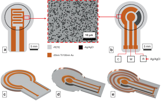
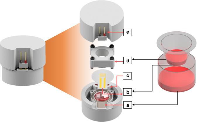
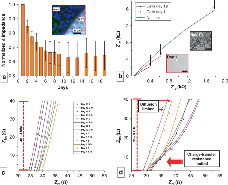
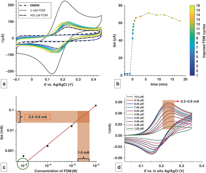
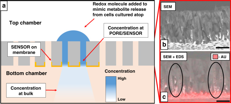
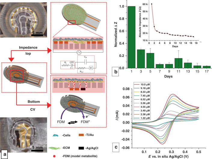
This work presents a 3D-printed, modular, electrochemical sensor-integrated transwell system for monitoring cellular and molecular events in situ without sample extraction or microfluidics-assisted downstream omics. Simple additive manufacturing techniques such as 3D printing, shadow masking, and molding are used to fabricate this modular system, which is autoclavable, biocompatible, and designed to operate following standard operating protocols (SOPs) of cellular biology. Integral to the platform is a flexible porous membrane, which is used as a cell culture substrate similarly to a commercial transwell insert. Multimodal electrochemical sensors fabricated on the membrane allow direct access to cells and their products. A pair of gold electrodes on the top side of the membrane measures impedance over the course of cell attachment and growth, characterized by an exponential decrease (~160% at 10 Hz) due to an increase in the double layer capacitance from secreted extracellular matrix (ECM) proteins. Cyclic voltammetry (CV) sensor electrodes, fabricated on the bottom side of the membrane, enable sensing of molecular release at the site of cell culture without the need for downstream fluidics. Real-time detection of ferrocene dimethanol injection across the membrane showed a three order-of-magnitude higher signal at the membrane than in the bulk media after reaching equilibrium. This modular sensor-integrated transwell system allows unprecedented direct, real-time, and noninvasive access to physical and biochemical information, which cannot be obtained in a conventional transwell system.

Keywords: Chemistry; Electrical and electronic engineering.


Conflict of interest statement

Conflict of interestThe authors declare that they do not have any conflicts of interest.

Figures

Fig. 1
Fig. 1. Electrode-integrated membrane designs.
Case I: a, b Schematics of a impedance interdigitated electrodes (IDEs) and b circular CV electrodes fabricated separately on two different PETE membranes. The image at the center shows an SEM image of the Au-coated membrane, showing that the track-etched pores are open even after Au deposition. The pore diameter is 1µm, and the porosity is 2×10⁷ pores/cm2. C, W and R represent the counter, working, and reference electrodes, respectively. Case II: ce Schematics of the c concentric impedance electrodes on the top side of the membrane, d CV electrodes on the bottom side, and e integrated arrangement of both impedance and CV electrodes on one membrane. The rectangular trace in all electrodes represents the contact pads that protrude outside the sensing area of the platform
Fig. 2
Fig. 2. 3D-printed transwell platform design.
A side-by-side comparison of our sensor-integrated 3D-printed transwell (left) and a commercial transwell (right). (Left) Illustration showing the exploded view of the 3D-printed sensor-integrated cell culture platform, with labeled components: a 3D-printed bottom chamber filled with cell media (red) and in situ PDMS gaskets for membrane attachment, b electrode-integrated PETE membrane with contact pads, c access port to provide fluid to the bottom chamber and serving as an accessibility point for TEER electrodes, (see also Supplementary Fig. S1), d top chamber, and e electrodes to connect to the sensor contact pads. Analogous parts are labeled in the transwell on the right
Fig. 3
Fig. 3. Impedimetric analysis of cell culture on IDEs.
a Normalized change in impedance values measured at 10 Hz over the course of 19 days (n = 3 measurements from separate devices). The inset shows the endpoint (day 19) fluorescence micrograph of the cell-covered sensor surface superimposed on the brightfield image of the sensor. The dark area denotes the area covered by the Au electrode, and the bright area corresponds to PETE. b Nyquist plot showing the impedance at 3 stages of cell culture. The black arrows indicate impedance at 10 Hz. Insets show the phase contrast image of the triculture on day 1 and day 19 (Scale bar: 100 µm). c, d Zoomed in Nyquist plots for c control device with no cells and d device with cell culture. The latter indicates the transition of the system from a diffusion-limited state to a charge transfer-limited state as the culture progresses. (dots = raw data, line = fitting with model on Supplementary Fig. S2). c, d Share the same legend
Fig. 4
Fig. 4. Electrochemical sensing of a model redox molecule through flexible and porous CV electrodes.
a CV demonstrating the diffusion of FDM through the pores in real time, with peak height proportional to the local FDM concentration. Scan rate = 300 mV/s, 3.7s/cycle. b Evolution of anodic peak current (Ipa) vs time. c Calibration plot for FDM in DMEM + 10% FBS in the porous CV sensor. The concentration corresponding to the Ipa values measured from d is shown by the brown trace in c. The experimental bulk concentration in the bottom chamber is marked by the green circle in c. d CV following the addition of 10 aliquots of 100 μL of 10 mM FDM. The legend displays the final bulk concentration
Fig. 5
Fig. 5. CV detection scheme of molecular diffusion across an electrode-integrated porous membrane.
a Cross-sectional schematic of the porous sensor and diffusion of FDM. The blue semicircle represents the addition of FDM to the top chamber. The Au working electrode (sensor) fabricated on the bottom side of the porous membrane is shown as yellow caps. The gradient in blue illustrates the diffusion-assisted concentration gradient in the system. b The cross-sectional SEM of the membrane shows pore channels in all directions. c Cross-sectional SEM + EDS shows the distribution of e-beam-deposited Au (scale bar: 8 μm)
Fig. 6
Fig. 6. Multimodal sensor-integrated transwell system.
a Optical images and schematic illustrations of the multimodal sensors displaying both impedimetric (top view) and CV (bottom view) sensors. Electrodes in view are lustrous Au and black Ag/AgCl, while electrodes on the opposite side of the translucent membrane are gray due to the Ti adhesion layer. Membranes appear wrinkled due to thermal expansion during PDMS curing. b Normalized change in impedance values measured at 10 Hz over the course of 17 days. (n = 3, measurements from 3 separate devices). The inset shows the absolute change in impedance from one of the devices tested. c CV following the addition of 10 aliquots of 100 µL of 10 mM FDM on the apical side of the gut triculture. The legend displays the final experimental concentration

References

    1. Corning. Transwell Permeable Supports Selection and Use Guide. Corning Inc. 1–11 (2006).
    1. Hatherell K, Couraud PO, Romero IA, Weksler B, Pilkington GJ. Development of a three-dimensional, all-human in vitro model of the blood-brain barrier using mono-, co-, and tri-cultivation transwell models. J. Neurosci. Methods. 2011;199:223–229. doi: 10.1016/j.jneumeth.2011.05.012. - DOI - PubMed
    1. Gamboa JM, Leong KW. In vitro and in vivo models for the study of oral delivery of nanoparticles. Adv. Drug Deliv. Rev. 2013;65:800–810. doi: 10.1016/j.addr.2013.01.003. - DOI - PMC - PubMed
    1. Marshall J. Cell migration transwell assay. Methods. 2011;769:111–136.
    1. Chen YW, Chen JK, Wang JS. Exercise affects platelet-promoted tumor cell adhesion and invasion to endothelium. Eur. J. Appl. Physiol. 2009;105:45–50. doi: 10.1007/s00421-008-0916-2. - DOI - PubMed