

Low-Grade Fibromyxoid Sarcoma of the Back
- PMID: 34567863
- PMCID: PMC8451521
- DOI: 10.7759/cureus.17308
Low-Grade Fibromyxoid Sarcoma of the Back
Abstract
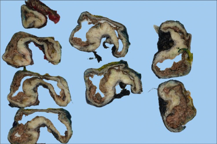
A 29-year-old male presented with a seven-year history of a slow-growing, painless, firm, mobile mass in the right upper back that was bothersome when supine or with direct pressure. On initial presentation, a clinical diagnosis of lipoma was given. The mass progressively increased in size over several years but remained painless. The mass measured 15 x 10 cm on examination. Excision of the lesion was performed, which revealed a white cut surface with cystic degenerative changes. Histologically, the lesion revealed spindle cell morphology with occasional mitosis. Diffuse immunohistochemical staining with MUC4 supports a diagnosis of low-grade fibromyxoid sarcoma (LGFMS). Tumor was present with focal extension into the deep margin. However, serial magnetic resonance imaging studies performed suggest no residual disease and negative regional lymph node involvement. This case demonstrates the growth pattern of LGFMS, but also denotes the importance of correlating radiological and pathological features to accurately diagnose and treat these tumors in a timely fashion.
Keywords: lipoma; low-grade fibromyxoid sarcoma; magnetic resonance imaging; metastasis; soft tissue tumours.
Copyright © 2021, Cantu et al.
Conflict of interest statement
The authors have declared that no competing interests exist.
Figures


References
-
- Low-grade fibromyxoid sarcoma. A report of two metastasizing neoplasms having a deceptively benign appearance. Evans HL. Am J Clin Pathol. 1987;88:615–619. - PubMed
-
- Low-grade fibromyxoid sarcoma: clinical, morphologic and genetic features. Mohamed M, Fisher C, Thway K. Ann Diagn Pathol. 2017;28:60–67. - PubMed
-
- Low-grade fibromyxoid sarcoma: a clinicopathologic study of 33 cases with long-term follow-up. Evans HL. Am J Surg Pathol. 2011;35:1450–1462. - PubMed
Publication types
LinkOut - more resources
Full Text Sources