Case Reports
. 2021 Jul 31;10(4):334-340.
doi: 10.1007/s13691-021-00505-y. eCollection 2021 Oct.

Successful treatment of cervical and upper thoracic esophageal adenocarcinoma using induction chemotherapy followed by surgery: a case report

Affiliations
Case Reports

Successful treatment of cervical and upper thoracic esophageal adenocarcinoma using induction chemotherapy followed by surgery: a case report

Takeharu Imai et al. Int Cancer Conf J. .

Abstract

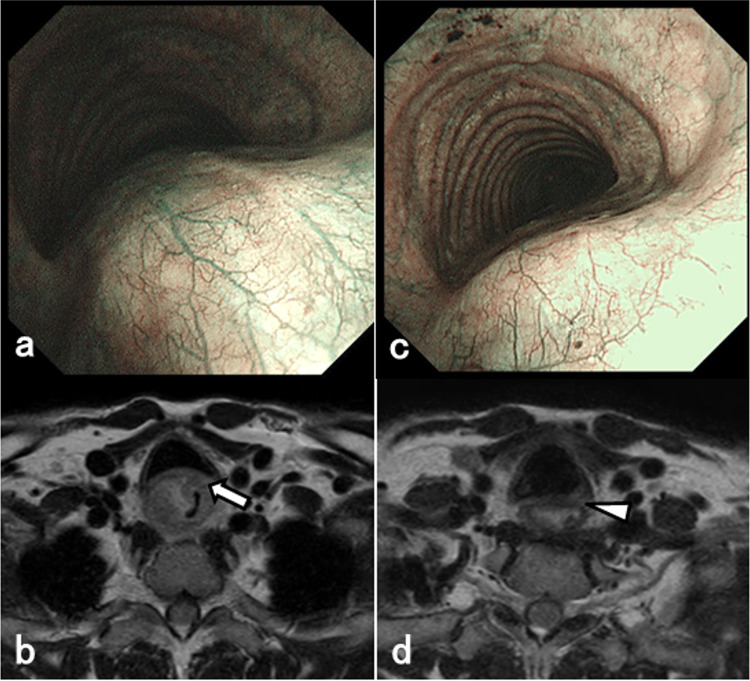
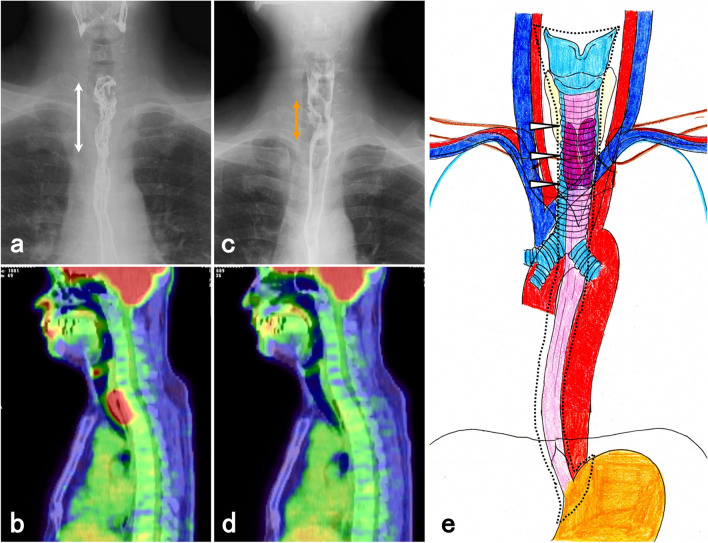
Cervical esophageal adenocarcinoma has a low incidence rate and its treatment involves various strategies. We report a patient with locally advanced cervical to upper esophageal adenocarcinoma who was able to undergo induction chemotherapy and radical surgery. A 55-year-old man was diagnosed with a poorly differentiated adenocarcinoma between the cervical and upper thoracic esophagus. The primary lesion had infiltrated into the tracheal membrane and had metastasized into the cervical lymph nodes. The initial diagnosis was T4bN1M1 stage IVB. The lower edge of the tumor was close to the tracheal bifurcation, making it difficult to create a longitudinal tracheal foramen during surgery. Therefore, when biweekly-DCF therapy was performed as induction chemotherapy, the tumor shrank sufficiently and its infiltration into the tracheal membrane decreased subsequently. We performed total laryngopharyngoesophagectomy with three-field lymph node dissection and reconstruction using free jejunal grafts and subtotal stomach via a posterior mediastinum route and a permanent tracheal foramen as a radical surgery. The pathological diagnosis was T2/MP, N1, and the effect of chemotherapy was grade 2. Cervical esophageal adenocarcinoma was rare, but technically reliable and safe oncologic surgery was possible after induction chemotherapy.

Keywords: Cervical esophageal adenocarcinoma; Induction chemotherapy; Total laryngopharyngoesophagectomy.


Conflict of interest statement

Conflict of interestDr. Yoshida reports receipt of grants, personal fees and non-financial support from EA Pharma Co., Ltd., Sanofi, Yakult Honsha Co., Ltd., Chugai Pharmaceutical Co., Ltd., Taiho Pharmaceutical Co., Ltd., Takeda Pharmaceutical Co., Ltd., Eli Lilly Japan K.K., Daiichi Sankyo Co., Ltd., Ono Pharmaceutical Co., Ltd., Merck Serono Co., Ltd., and Novartis Pharma K.K.; and grants from Kyowa Hakko Kirin Co., Ltd. outside of the submitted work. Other authors have no conflict of interest to disclose.

Figures

Fig. 1
Fig. 1
EGD and Contrast-enhanced CT findings. a EGD showed circumferential type 3 tumor at cervical to upper thoracic esophagus. b Enhanced CT showed thickened wall of the cervical esophagus (arrow). c Tumor decreased in size after induction chemotherapy. d Thickened wall improved after chemotherapy (arrowhead)
Fig. 2
Fig. 2
Bronchoscopy and MRI findings. a Bronchoscopy showed tracheal membrane raising by the tumor. b MRI showed tracheal membrane infiltration of the tumor (arrow). c Trachea membrane raising caused by the tumor improved. d The tumor shrank and its infiltration into the trachea membrane reduced (arrowhead)
Fig. 3
Fig. 3
UGI and PET-CT findings. a UGI showing the tumor at the cervical and upper thoracic esophagus, measuring 60 mm in diameter (arrow). b PET-CT showing high FDG accumulation of tumor at cervical and upper thoracic esophagus. c The tumor shrank from the upper thoracic esophagus toward the cervical esophagus after induction chemotherapy (orange arrow). d FDG accumulation in the tumor disappeared after chemotherapy. e Preoperative schema: the tumor (arrowhead) and the extent of resection (dot-line)
Fig. 4
Fig. 4
Operation findings. a Total laryngopharyngoesophagectomy with three-field lymph node dissection and reconstruction using free jejunal grafts and subtotal stomach via a posterior mediastinum route, a permanent tracheal foramen (arrowhead) and jejunostomy were performed. b The bilateral bronchial arteries were preserved to maintain blood flow to the oral side of the trachea. The right bronchial artery diverged from the right subclavian artery (arrow). The tumor was detached from the tracheal membrane (orange arrowhead). c The trachea was cut between the 5-6th tracheal cartilage, and a permanent tracheal foramen was constructed at the upper sternal margin level (dot-line). d A free jejunum was placed between the mesopharynx and the subtotal stomach (yellow arrow)
Fig. 5
Fig. 5
Resected specimen and histopathological findings. a The resected specimen showed a 2 cm scar-like tumor on the cervical esophagus (arrow). b Tumor was not stained with lugol (yellow arrow). c Histopathological findings were of poorly and moderately differentiated adenocarcinoma. d The background mucosa adjacent to the adenocarcinoma cells showed ectopic gastric mucosa (EGM)

References

    1. Yamaji T, Tsugane S. Epidemiology of esophageal squamous cell carcinoma. In: Ando N, editor. Esophageal squamous cell carcinoma. Singapore: Springer; 2020. pp. 1–13.
    1. Siewert JR, Ott K. Are squamous and adenocarcinomas of the esophagus the same disease? Semin Radiat Oncol. 2007;17:38–44. doi: 10.1016/j.semradonc.2006.09.007. - DOI - PubMed
    1. Ilson DH. Adenocarcinoma of the esophagus: controversies and consensus. Chin Clin Oncol. 2017;6:52. doi: 10.21037/cco.2017.09.02. - DOI - PubMed
    1. Rubenstein JH, Shaheen NJ. Epidemiology, diagnosis, and management of esophageal adenocarcinoma. Gastroenterology. 2015;149:302–317.e1. doi: 10.1053/j.gastro.2015.04.053. - DOI - PMC - PubMed
    1. von Rahden BH, Stein HJ, Becker K, Siewert RJ. Esophageal adenocarcinomas in heterotopic gastric mucosa: review and report of a case with complete response to neoadjuvant radiochemotherapy. Dig Surg. 2005;22:107–112. doi: 10.1159/000085448. - DOI - PubMed

Publication types

LinkOut - more resources