. 2021 Sep 10:11:739874.
doi: 10.3389/fcimb.2021.739874. eCollection 2021.

S100B Inhibition Attenuates Intestinal Damage and Diarrhea Severity During Clostridioides difficile Infection by Modulating Inflammatory Response

Affiliations

S100B Inhibition Attenuates Intestinal Damage and Diarrhea Severity During Clostridioides difficile Infection by Modulating Inflammatory Response

Deiziane V S Costa et al. Front Cell Infect Microbiol. .

Abstract

The involvement of the enteric nervous system, which is a source of S100B, in Clostridioides difficile (C. difficile) infection (CDI) is poorly understood although intestinal motility dysfunctions are known to occur following infection. Here, we investigated the role of S100B in CDI and examined the S100B signaling pathways activated in C. difficile toxin A (TcdA)- and B (TcdB)-induced enteric glial cell (EGC) inflammatory response. The expression of S100B was measured in colon tissues and fecal samples of patients with and without CDI, as well as in colon tissues from C. difficile-infected mice. To investigate the role of S100B signaling in IL-6 expression induced by TcdA and TcdB, rat EGCs were used. Increased S100B was found in colonic biopsies from patients with CDI and colon tissues from C. difficile-infected mice. Patients with CDI-promoted diarrhea exhibited higher levels of fecal S100B compared to non-CDI cases. Inhibition of S100B by pentamidine reduced the synthesis of IL-1β, IL-18, IL-6, GMCSF, TNF-α, IL-17, IL-23, and IL-2 and downregulated a variety of NFκB-related genes, increased the transcription (SOCS2 and Bcl-2) of protective mediators, reduced neutrophil recruitment, and ameliorated intestinal damage and diarrhea severity in mice. In EGCs, TcdA and TcdB upregulated S100B-mediated IL-6 expression via activation of RAGE/PI3K/NFκB. Thus, CDI appears to upregulate colonic S100B signaling in EGCs, which in turn augment inflammatory response. Inhibition of S100B activity attenuates the intestinal injury and diarrhea caused by C. difficile toxins. Our findings provide new insight into the role of S100B in CDI pathogenesis and opens novel avenues for therapeutic interventions.

Keywords: Clostridioides difficile; S100B; diarrhea; enteric glia; inflammation.


Conflict of interest statement

The authors declare that the research was conducted in the absence of any commercial or financial relationships that could be construed as a potential conflict of interest.

Figures

Figure 1
Figure 1
C. difficile infection increases S100B in fecal samples and in colon from humans and mice. (A) Representative immunohistochemical images of S100Bexpression in human colonic biopsies with active C. difficile infection (CDI) and healthy subjects (Control). Increased S100B expression (arrowhead or rectangle) was found in colonic mucosal (center panel, red arrowhead), submucosal (center panel, black arrowhead), and myenteric plexus (right panel, black rectangle). Scale bars, 200 (left panels), 100 (center panels), and 50 (right panels) μm. Left panels are showing colon tissues from control and CDI patients in a low magnification. (B) Quantification of percentage (mean ± s.e.m.) of S100B-immunopositive area in colons from human control and C. difficile-infected subjects in 15–20 microscope fields per sample (n = 4 subjects per group). Unpaired two-tailed Student t test. (C) S100B levels in fecal samples of patients with diarrhea caused by CDI (n = 53) and non-CDI (n = 27) evaluated by ELISA. Data are median ± s.d. Two-tailed non-parametric Mann–Whitney U-test. (D) Schematic diagram of CDI experimental model in mice. (E) Representative Western blot (WB) bands of S100B and α-tubulin in colonic tissues from mice infected with C. difficile (CDI group) and non-infected (control group) at days 1 and 3 postinfection (p.i.). (F) WB analysis of S100B (mean ± s.e.m.) in colonic tissues from CDI and control group (n = 3 mice per group). α-Tubulin was used to normalize the levels of S100B. Unpaired two-tailed Student’s t test. (G) Representative immunohistochemical images of S100B immunostaining in colonic tissues of mice with CDI and control (non-infected mice). (H) Quantification of percentage (mean ± s.e.m.) of S100B-immunopositive area in colon from mice with CDI and control (non-infected mice) (n = 5 mice per group). Unpaired two-tailed Student’s t test.
Figure 2
Figure 2
S100B inhibition decreases diarrhea severity, intestinal damage, and neutrophil recruitment during CDI in mice. Mice were infected with 105 vegetative C. difficile (VPI10463 strain) and treated with a S100B inhibitor, pentamidine (40 mg/kg, i.p., once daily for 3 days, CDI+pentamidine group) or non-treated (CDI group). (A) Quantification of C. difficile shedding (mean ± s.e.m.) in stools by amplifying the tcdB gene by qPCR. Unpaired two-tailed Student’s t test. (B) Diarrhea score (median ± s.d) of CDI and CDI+pentamidine mice. Two-tailed non-parametric Mann–Whitney U-test. (C) Representative H&E stains of cecal and colonic tissues collected from CDI and CDI+pentamidine mice at day 3 postinfection (p.i.). CDI promotes damage of colonic and cecal epithelium (black arrow), edema (green arrow), and inflammatory cell infiltration (red arrow) in mice infected by C. difficile (CDI group). Scale bars, 100 µm. (D) Histopathologic score (median, 0-no damage, and 9-intense damage) based on epithelial damage, submucosal edema, and infiltration of inflammatory cells. Two-tailed non-parametric Mann–Whitney U-test. (E) MPO levels (mean ± s.e.m.) in cecum content, cecum, and colon samples from CDI and CDI+pentamidine mice at day 3 p.i. measured by ELISA. Unpaired two-tailed Student’s t test.
Figure 3
Figure 3
S100B modulates the release of pro-inflammatory mediators and tissue repair cytokines during CDI in mice. Levels of (A) IL-1β, (B) IL-18, (C) IL-6, (D) GMCSF, (E) TNF-α, (F) IL-17, (G) IL-23, (H) IL-2, (I) IL-33, and (J) IL-22 in colonic tissues from uninfected (control), uninfected receiving pentamidine (40 mg/kg, pentamidine), non-pretreated C. difficile-infected (CDI), and pentamidine-pretreated C. difficile-infected (CDI+pentamidine) mice at day 3 p.i. were measured by ELISA. Data are mean ± s.e.m. **ANOVA followed by Turkey test was used. *Unpaired two-tailed Student’s t test.
Figure 4
Figure 4
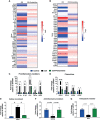
S100B inhibition upregulates anti-inflammatory (SOCS2) and antiapoptotic mediators (Bcl2) and downregulates inflammatory mediators during CDI. (A, B) Heat map of the cDNA microarray analysis of colonic tissues from non-pretreated C. difficile-infected (CDI) and pentamidine-pretreated C. difficile-infected (CDI+pentamidine) mice at day 3 p.i. Expression of the genes is normalized to median of the control, log 2 scale. TaqMan qPCR analysis of (C) proinflammatory (D) chemokine, (E) cellular recruitment (SELP), (F) anti-inflammatory SOCS2, and (G) antiapoptotic Bcl2 mediators. Data are mean ± s.e.m. (C–E) #p < 0.05, *p < 0.01, **p < 0.001. ANOVA followed by Turkey test was used.
Figure 5
Figure 5
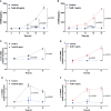
TcdA and TcdB increase S100B-dependent upregulation of IL-6 expression in enteroglial cells (EGC/PK060399). (A, B) Levels of S100B (mean ± s.e.m) released by ELISA, (C, D) S100B, and (E, F) IL-6 gene expression (mean ± s.e.m) by qPCR in enteroglial cells (EGC/PK060399) challenged with (A, C, E) TcdA and (B, D, F) TcdB (n = 4) with TcdA and TcdB (n = 4). (A–F) Cells receiving only supplemented DMEM were applied as a control. (A–F) ANOVA followed by Sidak’s multiple-comparison test was used.
Figure 6
Figure 6
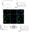
Blockade of the S100B receptor and PI3K decreases C. difficile toxin-induced IL-6 upregulation in EGCs. (A) RAGE gene expression (mean ± s.e.m) by qPCR in enteroglial cells (EGC/PK060399) challenged with TcdA and TcdB (n = 4). (B) Analysis of IL-6 gene expression (mean ± s.e.m) by qPCR in enteroglial cells (EGC/PK060399) challenged with TcdA and TcdB for 18 h in the presence or absence of 30 μM FPSZM1 (a RAGE antagonist) which was added 1 h prior to C. difficile toxin challenge. (C) Representative photomicrographs of NFκBp65 (green) immunostaining and DAPI (blue) nuclear staining in enteroglial cells (EGC/PK060399) exposed to TcdA and TcdB after 18 h of incubation. (D) Percentages of cells (mean ± s.e.m) with positive nuclear NFκBp65 staining under different experimental conditions at 18 h of incubation with TcdA and TcdB. (E) Western blot (WB) bands of NFκB p65 and PCNA in nuclear extract fraction of enteroglial cells (EGC/PK060399) exposed to TcdA and TcdB at 18 h of incubation. (F) Analysis of IL-6 gene expression (mean ± s.e.m) by qPCR in enteroglial cells (EGC/PK060399) challenged with TcdA and TcdB for 18 h in the presence or absence of 10 μM LY294002 (a PI3K inhibitor) which was added 1 h prior to C. difficile toxin challenge. (A–F) Cells receiving only supplemented DMEM was applied as a control. (B, F) Experiments were performed with the same negative (Control) and positive controls (TcdA and TcdB). **p < 0.0001. ANOVA followed by (B, D, F) Turkey test was used. *p < 0.01.
Figure 7
Figure 7
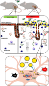
Schematic diagram of the hypothetical role of S100B during C. difficile infection. C. difficile releases TcdA and TcdB which in turn promote epithelial damage and release of S100B by colonic EGCs. S100B stimulates the synthesis and secretion of inflammatory mediators (IL-1β, TNF-α, IL-18, IL-6, GMCSF, IL-17, IL-23, IL-2) promoting recruitment of immune cells, such as neutrophils, macrophages, and T cells, resulting in amplification of the C. difficile toxin-induced colonic damage. TcdA and TcdB induce IL-6 expression via S100B/RAGE/PI3K/NFκB. S100B also impairs epithelial integrity during CDI by decreasing IL-22 production, thereby hampering repair of the epithelium. Inhibition of S100B by pentamidine (PEN) blocks these events mediated by S100B during CDI.

References

    1. Abhyankar M. M., Ma J. Z., Scully K. W., Nafziger A. J., Frisbee A. L., Saleh M. M., et al. . (2020). Immune Profiling To Predict Outcome of Clostridioides Difficile Infection. mBio 11, 1–10. doi: 10.1128/mBio.00905-20 - DOI - PMC - PubMed
    1. Bauman B. D., Meng J., Zhang L., Louiselle A., Zheng E., Banerjee S., et al. . (2017). Enteric Glial-Mediated Enhancement of Intestinal Barrier Integrity Is Compromised by Morphine. J. Surg. Res. 219, 214–221. doi: 10.1016/j.jss.2017.05.099 - DOI - PMC - PubMed
    1. Bianchi R., Kastrisianaki E., Giambanco I., Donato R. (2011). S100B Protein Stimulates Microglia Migration via RAGE-Dependent Up-Regulation of Chemokine Expression and Release. J. Biol. Chem. 286, 7214–7226. doi: 10.1074/jbc.M110.169342 - DOI - PMC - PubMed
    1. Biondo C., Mancuso G., Midiri A., Signorino G., Domina M., Lanza Cariccio V., et al. . (2014). The Interleukin-1beta/CXCL1/2/neutrophil Axis Mediates Host Protection Against Group B Streptococcal Infection. Infect. Immun. 82, 4508–4517. doi: 10.1128/IAI.02104-14 - DOI - PMC - PubMed
    1. Body-Malapel M., Djouina M., Waxin C., Langlois A., Gower-Rousseau C., Zerbib P., et al. . (2019). The RAGE Signaling Pathway Is Involved in Intestinal Inflammation and Represents a Promising Therapeutic Target for Inflammatory Bowel Diseases. Mucosal Immunol. 12, 468–478. doi: 10.1038/s41385-018-0119-z - DOI - PubMed

Publication types

LinkOut - more resources