. 2021 Sep 9:9:715717.
doi: 10.3389/fbioe.2021.715717. eCollection 2021.

Reconstruction of the Swine Pulmonary Artery Using a Graft Engineered With Syngeneic Cardiac Pericytes

Affiliations

Reconstruction of the Swine Pulmonary Artery Using a Graft Engineered With Syngeneic Cardiac Pericytes

Valeria Vincenza Alvino et al. Front Bioeng Biotechnol. .

Abstract

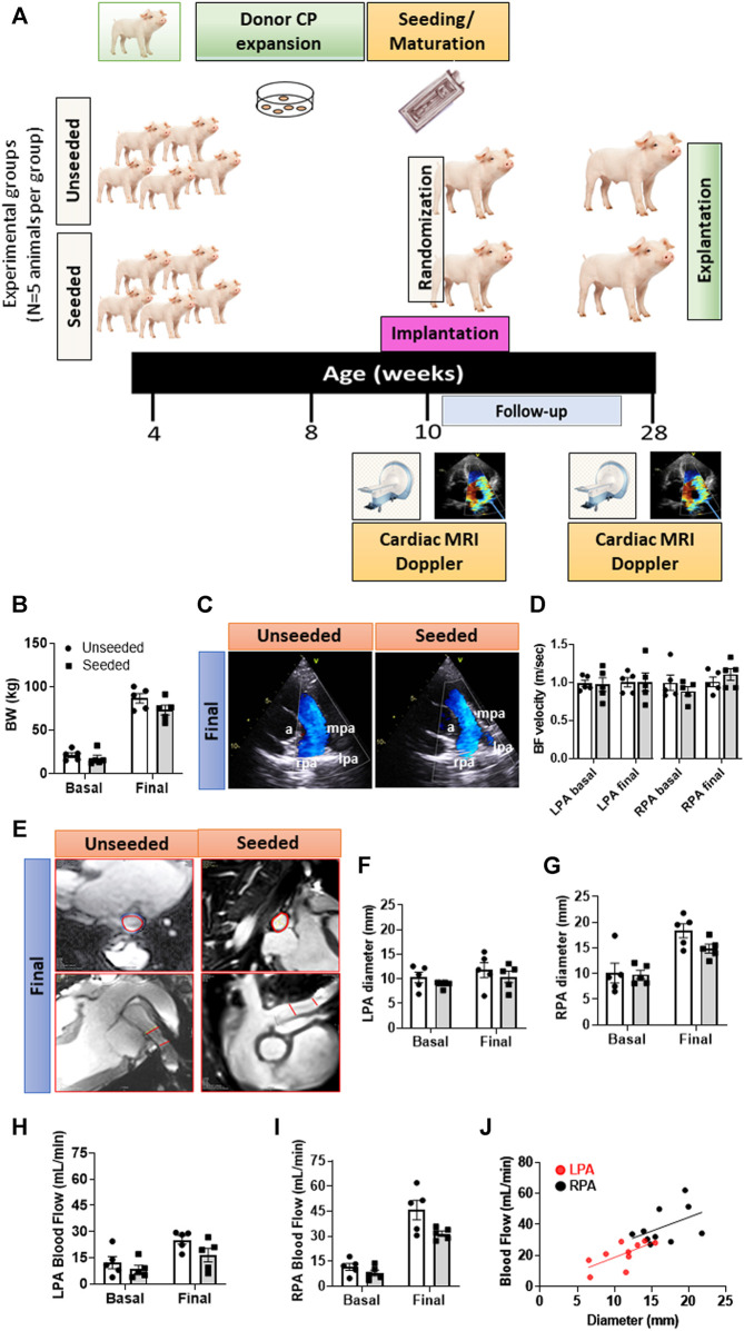
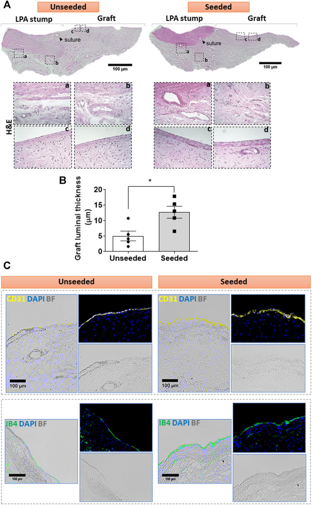
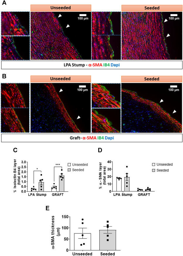
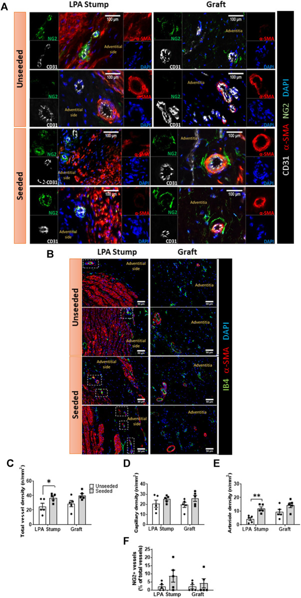
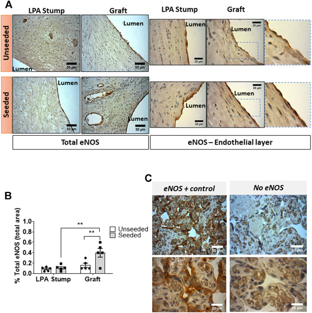
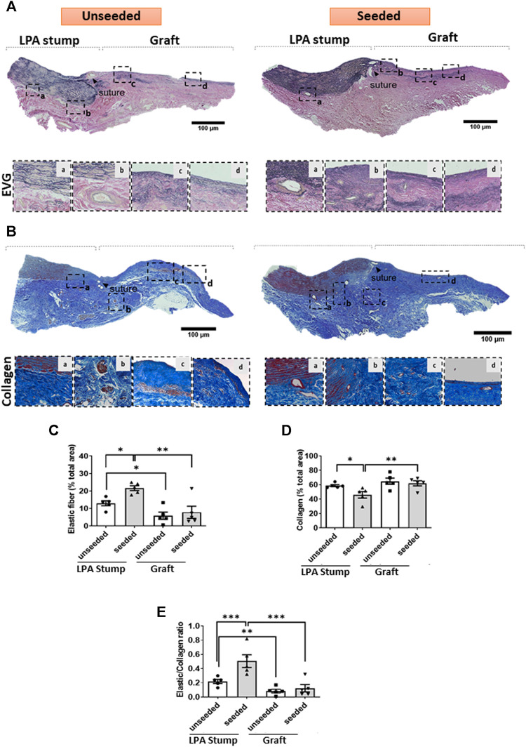
The neonatal heart represents an attractive source of regenerative cells. Here, we report the results of a randomized, controlled, investigator-blinded preclinical study, which assessed the safety and effectiveness of a matrix graft cellularized with cardiac pericytes (CPs) in a piglet model of pulmonary artery (PA) reconstruction. Within each of five trios formed by 4-week-old female littermate piglets, one element (the donor) was sacrificed to provide a source of CPs, while the other two elements (the graft recipients) were allowed to reach the age of 10 weeks. During this time interval, culture-expanded donor CPs were seeded onto swine small intestinal submucosa (SIS) grafts, which were then shaped into conduits and conditioned in a flow bioreactor. Control unseeded SIS conduits were subjected to the same procedure. Then, recipient piglets were randomized to surgical reconstruction of the left PA (LPA) with unseeded or CP-seeded SIS conduits. Doppler echocardiography and cardiac magnetic resonance imaging (CMRI) were performed at baseline and 4-months post-implantation. Vascular explants were examined using histology and immunohistochemistry. All animals completed the scheduled follow-up. No group difference was observed in baseline imaging data. The final Doppler assessment showed that the LPA's blood flow velocity was similar in the treatment groups. CMRI revealed a mismatch in the average growth of the grafted LPA and contralateral branch in both treatment groups. Histology of explanted arteries demonstrated that the CP-seeded grafts had a thicker luminal cell layer, more intraparietal arterioles, and a higher expression of endothelial nitric oxide synthase (eNOS) compared with unseeded grafts. Moreover, the LPA stump adjacent to the seeded graft contained more elastin and less collagen than the unseeded control. Syngeneic CP engineering did not accomplish the primary goal of supporting the graft's growth but was able to improve secondary outcomes, such as the luminal cellularization and intraparietal vascularization of the graft, and elastic remodeling of the recipient artery. The beneficial properties of neonatal CPs may be considered in future bioengineering applications aiming to reproduce the cellular composition of native arteries.

Keywords: congenital heart disease; grafts; pericytes; pulmonary artery; tissue engineering.


Conflict of interest statement

The authors declare that the research was conducted in the absence of any commercial or financial relationships that could be construed as a potential conflict of interest.

Figures

FIGURE 1
FIGURE 1
General outcome and imaging endpoints of the randomized, controlled preclinical study. (A) Schematic of the study. (B) Body weight of animals randomized to the two groups. (C) Representative images of Doppler echocardiography. Vascular structures are highlighted in the images: a = aorta, lpa = left pulmonary artery, mpa = main pulmonary artery, rpa = right pulmonary artery. (D) Column graph and individual Doppler data in the two groups at baseline and 4-moths follow-up. (E) Representative images of the cardiac MRI showing transversal and longitudinal sections of the site of grant implantation delimited by the red lines. (F–I) Column graph and individual MRI data (diameter and blood flow) in the two groups at baseline and 4-months follow-up regarding the LPA (F,H) and RPA (G,I). (J) Correlation between diameter and blood flow regarding LPA and RPA. Abbreviations: BF = blood flow, BW = body weight. N = 5 biological replicates per experimental group. Data are expressed as mean ± SEM.
FIGURE 2
FIGURE 2
Cellular composition of the graft: (A) H&E staining of unseeded and seeded grafts. Tiled images and inserts show the general architecture and details of explanted grafts, including the distribution of cells (cell nuclei are dark blue and eosinophilic compounds in the cytoplasm are pink) and the ECM underneath the luminal cell layer, containing fibroblasts and microvessels. (B) Bar graph showing the thickness of the luminal layer. N = 5 biological replicates per experimental group. Data are represented as mean ± SEM. *p < 0.05. (C) Immunofluorescent and bright-field images display the presence of CD31 (yellow) and IB4 (green) markers in the luminal endothelium of the grafts. Representative images are acquired using 2.5x, 10x, and 20x objectives.
FIGURE 3
FIGURE 3
Multicolor fluorescent microscopy shows the luminal layer of the graft is composed of endothelial and smooth muscle cells. (A,B) Images showing the layer of luminal endothelial cells (green) sitting on several sheets of α-SMA + VSMCs (red) at the level of the LPA stump (A) and the grafts (B). Bar graphs showing the quantification of IB4 (C) and α-SMA staining measured as percentage of positive expression in the internal layer of total section area (D). α-SMA was also calculated as thickness layer expressed in μm (E). The images were visualized using a fluorescent microscope with 10x objectives and bigger inserts were added for better visualization of the details. N = 5 biological replicates per experimental group. Data are expressed as mean ± SEM. *p < 0.05, ***p < 0.0005.
FIGURE 4
FIGURE 4
Microvascular composition of the implanted artery. (A,B) Immunofluorescent microscopy image of the vascularization at the level of the LPA stump and grafts. In panel A, endothelial cells were stained with CD31 (white), vascular smooth muscle cells with a-SMA (red), pericytes with NG2 (green), and nuclei with DAPI (blue). In panel B, endothelial cells stained with IB4 (green), vascular smooth muscle cells with α-SMA (red), and nuclei with DAPI (blue). The location of the neo-adventitia is indicated in each image. Vascular smooth muscle cells are visualized in the tunica media of the LPA stump, the graft structure, and within intraparietal arterioles. (C–F) Bar graphs showing the vascular density and pericyte coverage in the two groups at the level of the LPA stump and grafts (C) Total vascular density, (D) Capillary density, (E) arteriole density, (F) NG2+ percentage. N = 5 biological replicates per experimental group. Data are expressed as mean ± SEM. *p < 0.05, **p < 0.01.
FIGURE 5
FIGURE 5
Expression of eNOS expression in the implanted artery. (A) Light microscopy images displaying the expression of eNOS in the LPA stump and graft in the form of brown precipitates. An intense staining, continuous staining was detected in endothelial cells of the seeded grafts (both at the luminal side and in intraparietal microvessels) compared with unseeded grafts. Moreover, weak positive staining could be appreciated in muscular cells underneath the luminal endothelial cells of the seeded grafts. (B) Bar graph showing the quantification of eNOS staining measured as the percentage of total section area. Images were quantified using a light microscope with 40x and 100x objectives. N = 5 biological replicates per experimental group. Data are expressed as mean ± SEM. **p < 0.01 (C) Cross-sections of human placental villi (used as a positive control) show the strong expression of eNOS in the syncytiotrophoblasts.
FIGURE 6
FIGURE 6
ECM remodeling of the implanted artery. (A,B) Tiled images and inserts at higher magnification display the composition of elastic fibers of the LPA and graft, as assessed using EVG (A) and Azan Mallory (B) stainings. (C–E) Bar graphs showing the results of the quantification of elastin and collagen and their ratio. Images were visualized using a light microscope with 2.5x, 10x and 20x objectives. N = 5 biological replicates per experimental group. per experimental group. Data are expressed as mean ± SEM. *p < 0.05, **p < 0.01, and ***p < 0.001.

References

    1. Akentjew T. L., Terraza C., Suazo C., Maksimcuka J., Wilkens C. A., Vargas F., et al. (2019). Rapid Fabrication of Reinforced and Cell-Laden Vascular Grafts Structurally Inspired by Human Coronary Arteries. Nat. Commun. 10, 3098. 10.1038/s41467-019-11090-3 - DOI - PMC - PubMed
    1. Albertario A., Swim M. M., Ahmed E. M., Iacobazzi D., Yeong M., Madeddu P., et al. (2019). Successful Reconstruction of the Right Ventricular Outflow Tract by Implantation of Thymus Stem Cell Engineered Graft in Growing Swine. JACC: Basic Transl. Sci. 4, 364–384. 10.1016/j.jacbts.2019.02.001 - DOI - PMC - PubMed
    1. Alvino V. V., Kilcooley M., Thomas A. C., Carrabba M., Fagnano M., Cathery W., et al. (2020). In Vitro and In Vivo Preclinical Testing of Pericyte-Engineered Grafts for the Correction of Congenital Heart Defects. J. Am. Heart Assoc. 9, e014214. 10.1161/JAHA.119.014214 - DOI - PMC - PubMed
    1. Avolio E., Caputo M., Madeddu P. (2015). Stem Cell Therapy and Tissue Engineering for Correction of Congenital Heart Disease. Front. Cel Dev. Biol. 3, 39. 10.3389/fcell.2015.00039 - DOI - PMC - PubMed
    1. Avolio E., Rodriguez-Arabaolaza I., Spencer H. L., Riu F., Mangialardi G., Slater S. C., et al. (2015). Expansion and Characterization of Neonatal Cardiac Pericytes Provides a Novel Cellular Option for Tissue Engineering in Congenital Heart Disease. J. Am. Heart Assoc. 4, e002043. 10.1161/JAHA.115.002043 - DOI - PMC - PubMed