. 2021 Sep 9:9:729444.
doi: 10.3389/fcell.2021.729444. eCollection 2021.

WW, PH and C-Terminal Domains Cooperate to Direct the Subcellular Localizations of PLEKHA5, PLEKHA6 and PLEKHA7

Affiliations

WW, PH and C-Terminal Domains Cooperate to Direct the Subcellular Localizations of PLEKHA5, PLEKHA6 and PLEKHA7

Sophie Sluysmans et al. Front Cell Dev Biol. .

Abstract

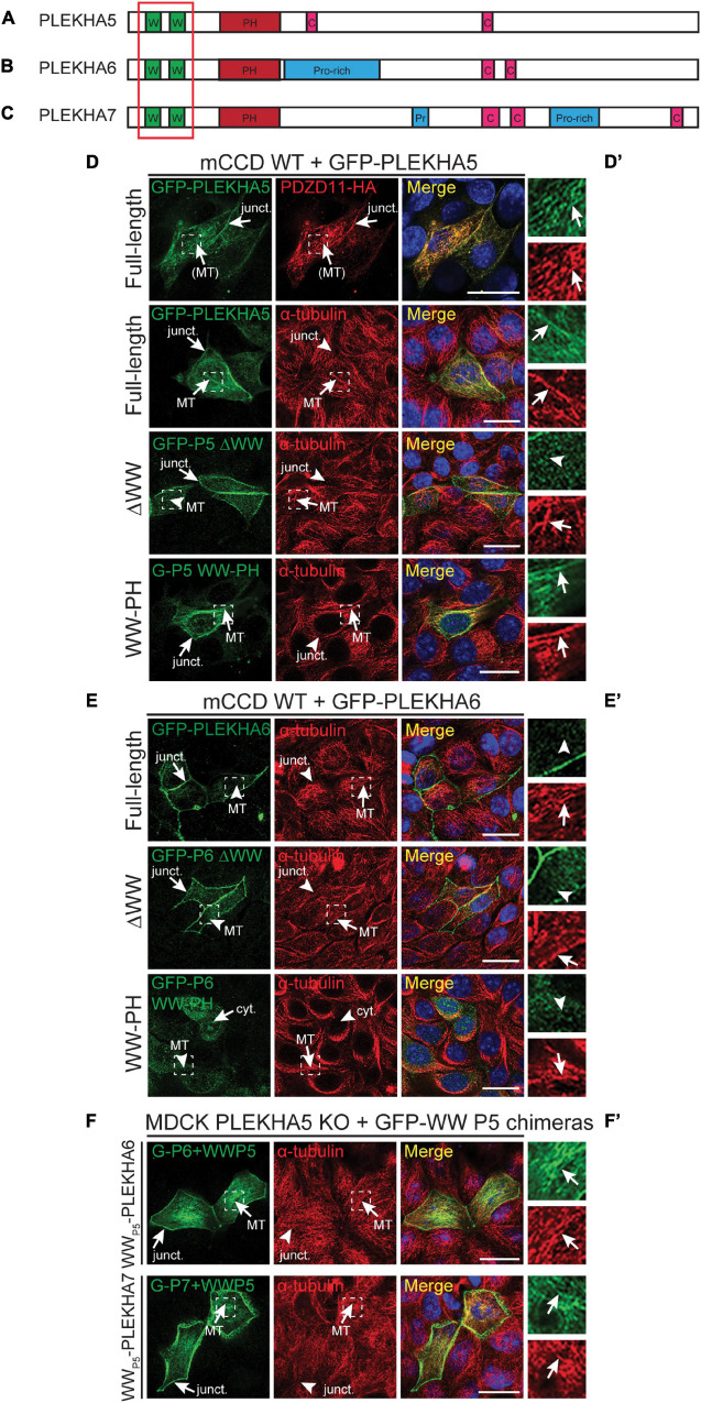
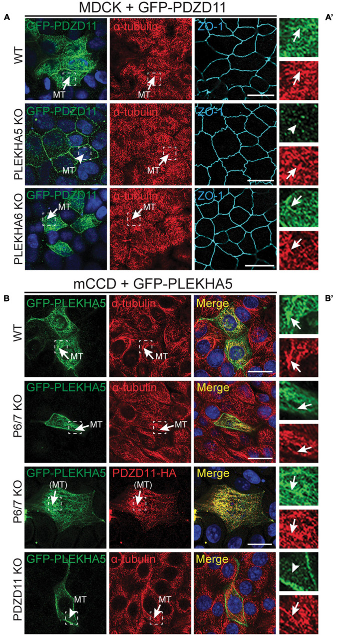
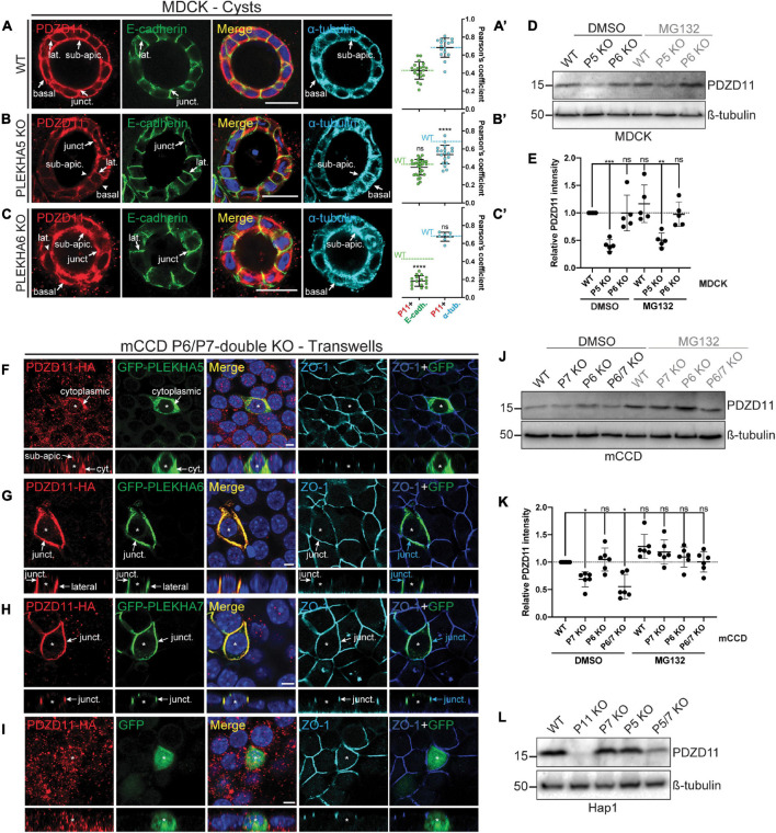
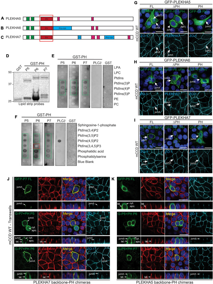
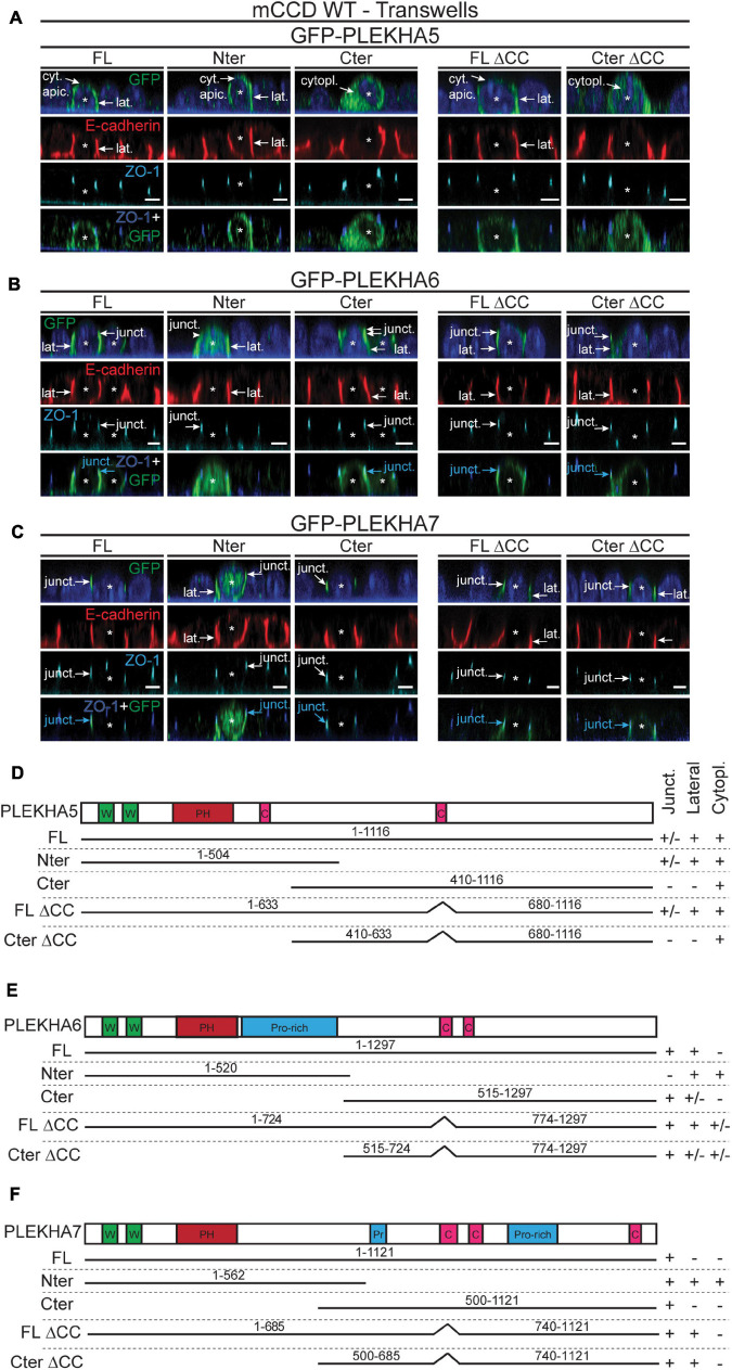
PLEKHA5, PLEKHA6, and PLEKHA7 (WW-PLEKHAs) are members of the PLEKHA family of proteins that interact with PDZD11 through their tandem WW domains. WW-PLEKHAs contribute to the trafficking and retention of transmembrane proteins, including nectins, Tspan33, and the copper pump ATP7A, at cell-cell junctions and lateral membranes. However, the structural basis for the distinct subcellular localizations of PLEKHA5, PLEKHA6, and PLEKHA7 is not clear. Here we expressed mutant and chimeric proteins of WW-PLEKHAs in cultured cells to clarify the role of their structural domains in their localization. We found that the WW-mediated interaction between PLEKHA5 and PDZD11 is required for their respective association with cytoplasmic microtubules. The PH domain of PLEKHA5 is required for its localization along the lateral plasma membrane and promotes the lateral localization of PLEKHA7 in a chimeric molecule. Although the PH domain of PLEKHA7 is not required for its localization at the adherens junctions (AJ), it promotes a AJ localization of chimeric proteins. The C-terminal region of PLEKHA6 and PLEKHA7 and the coiled-coil region of PLEKHA7 promote their localization at AJ of epithelial cells. These observations indicate that the localizations of WW-PLEKHAs at specific subcellular sites, where they recruit PDZD11, are the result of multiple cooperative protein-lipid and protein-protein interactions and provide a rational basis for the identification of additional proteins involved in trafficking and sorting of WW-PLEKHAs.

Keywords: PDZD11; PLEKHA5; PLEKHA6; PLEKHA7; WW domain; pleckstrin homology domain-containing family A.


Conflict of interest statement

The authors declare that the research was conducted in the absence of any commercial or financial relationships that could be construed as a potential conflict of interest.

Figures

FIGURE 1
FIGURE 1
The tandem WW domains of PLEKHA5 stabilize its localization along microtubules. (A–C) Scheme of the structural organization of PLEKHA5 (A), PLEKHA6 (B), and PLEKHA7 (C) showing the domains: WW (Trp-Trp) (W) in green, PH (pleckstrin homology) in red, Proline-rich (Pro-rich or Pr) in blue, coiled-coil (C) in pink. Red box indicates the structural domains analyzed. (D,E) IF analysis of the localization of GFP-tagged PLEKHA5 (D) and PLEKHA6 (E) constructs, either full-length, or a deletion lacking the WW domains (ΔWW), or the isolated N-terminal WW-PH region (WW-PH) after transfection in WT mCCD cells. (F) IF analysis of the localization of GFP-tagged chimeras of either PLEKHA6 or PLEKHA7, containing the WW domains of PLEKHA5 (P5), expressed in PLEKHA5 KO MDCK cells. α-tubulin is used as a marker for microtubules. In panels (D–F), squares correspond to enlarged areas in panels (D′–F′). Arrows show labeling, arrowheads indicate low/undetectable labeling. Junctional (junct.), fibrillar microtubules-like [(MT)], microtubules (MT) and cytoplasmic (cyt.) localizations are indicated. Scale bar = 20 μm.
FIGURE 2
FIGURE 2
The interaction between the tandem WW domains of PLEKHA5 and PDZD11 is necessary and sufficient for their localization along cytoplasmic microtubules. (A) IF analysis of the localization of GFP-tagged PDZD11 in MDCK cells, either WT, or PLEKHA5 KO, or PLEKHA6 KO. α-tubulin is used as a marker for microtubules and ZO-1 as a marker for cell-cell junctions. (B) IF analysis of the localization of GFP-tagged PLEKHA5 full-length in mCCD WT, PLEKHA6-PLEKHA7 double KO (P6/7 KO) and PDZD11 KO. α-tubulin is used as a marker for microtubules. Squares correspond to enlarged areas of green and red channels in panels (A′,B′). Microtubules (MT) and microtubules-like fibrils [(MT)] are indicated, with arrows showing labeling and arrowheads low/undetectable labeling. Scale bar = 20 μm.
FIGURE 3
FIGURE 3
PLEKHA5, PLEKHA6, and PLEKHA7 define and stabilize cellular pools of PDZD11 with distinct localizations. (A–C) IF analysis of endogenous PDZD11 localization in MDCK WT (A), PLEKHA5 KO (B), and PLEKHA6 KO (C) cells grown in Matrigel as 3D cysts. α-tubulin and E-cadherin show microtubular and lateral/junctional staining, respectively. Basal, junctional (junct.), lateral (lat.) and sub-apical (sub-apic.) localizations are indicated, with arrows showing labeling and arrowheads low/undetectable labeling. In (A′–C′),colocalization between PDZD11 (P11) and E-cadherin (E-cadh., green) or α-tubulin (α-tub., cyan) is quantified by Pearson’s correlation coefficients. Dashed lines indicate the mean of the WT cells for P11-E-cadh. (green) or P11-α-tub. (cyan). Dots show replicates corresponding to individual cysts (n: E-cadh.: WT: 20, PLEKHA5 KO: 29, PLEKHA6 KO: 16; α-tub.: WT: 16, PLEKHA5 KO: 20, PLEKHA6 KO: 7), and bars represent mean ± SD. Ordinary one-way ANOVA with post hoc Dunnett’s test to compare to WT (ns, not significant, ****p < 0.0001). (D,E,J–L) IB analysis (D,J,L) and quantification (E,K) of PDZD11 protein expression in WT, PLEKHA5 (P5) KO and PLEKHA6 (P6) KO MDCK cells (D,E), in WT, PLEKHA7 (P7) KO, P6 KO, and P6/P7 double KO mCCD cells (J,K), and in WT, PDZD11 (P11) KO, P7 KO, P5 KO, and P5/7 double KO Hap1 cells (L), upon treatment with the proteasome inhibitor MG132 (DMSO as control; ß-tubulin serves as loading normalization). In panels (E,K) dots show replicates (ß-tubulin-normalized PDZD11 signals relative to DMSO-treated WT cells) and bars represent mean ± SD. Repeated measures (RM) one-way ANOVA with post hoc Dunnett’s test (ns, not significant; *p < 0.05, **p < 0.01, and ***p < 0.001). (F–I) IF analysis of the localization of exogenous HA-tagged PDZD11 (PDZD11-HA) upon co-transfection either with GFP-PLEKHA5 (F), or GFP-PLEKHA6 (G), or GFP-PLEKHA7 (H), or GFP alone (I) in PLEKHA6-PLEKHA7 double KO mCCD cells grown on transwells. Bottom panels show XZ sections taken at the horizontal middle of the XY plane. Cytoplasmic (cyt.), junctional (junct.), lateral and sub-apical (sub-apic.) localizations are indicated. Asterisks show transfected cells. ZO-1 is used as a junctional marker, and merge panels between ZO-1 (in blue) and GFP (in green) show junctional localization (pointed by cyan arrows). Scale bars = 20 μm (A–C) or 5 μm (F–I).
FIGURE 4
FIGURE 4
Phospholipid binding and cellular localizations of the PH domains of WW-PLEKHAs. (A–C) Scheme of the structural organization of PLEKHA5 (A), PLEKHA6 (B), and PLEKHA7 (C) showing the domains: WW (Trp-Trp) (W) in green, PH (pleckstrin homology) in red, Proline-rich (Pro-rich or Pr) in blue, coiled-coil (C) in pink. Red box indicates the structural domain analyzed. (D–F) In vitro interaction of PH domains of WW-PLEKHAs with phospholipids. Coomassie staining (D) of purified GST fusions of PH domains of PLEKHA5 (P5), PLEKHA6 (P6) and PLEKHA7 (P7) (framed bands), and (E,F) IB analysis of lipid-protein overlay assay (GST-PH domain of phospholipase C-∂1 (PLC∂) as positive control, GST alone as negative control). Green frames highlight binding, red frames indicate low/undetectable interaction. LPA, lysophosphatidic acid; LPC, lysophosphocholine; PtdIns, phosphatidylinositol; P, phosphate; P2, biphosphate; P3, triphosphate; PE, phosphatidylethanolamine; PC, phosphatidylcholine. (G–I) IF analysis of the localization of exogenous GFP-tagged constructs of PLEKHA5 (G), PLEKHA6 (H), and PLEKHA7 (I) in WT mCCD cells. Either full-length (FL), or FL with a deletion of the PH domain (ΔPH), or PH domain alone (PH) were used. ZO-1 and PLEKHA7 are used as junctional markers. Junctional (junct.) and fibrillar microtubules-like [(MT)] localizations are indicated. Arrows show labeling, arrowheads indicate low/undetectable labeling. (J,K) IF analysis of the localization of either GFP-tagged PLEKHA7 (P7) (J) or PLEKHA5 (P5) (K) constructs in WT mCCD cells. Either full-length (FL) proteins, or chimeras where the PH domain was replaced [either from P5 or P6 in PLEKHA7 (J), or from P6 or P7 for PLEKHA5 (K)] are shown. mCCD cells were polarized on transwells and XZ section were taken at the horizontal middle of the XY plane (square panels). Cytoplasmic sub-apical (cyt. apic.), junctional (junct.), and lateral (lat.) localizations are indicated. E-cadherin and ZO-1 are used as lateral and junctional markers, respectively. Scale bar = 20 μm.
FIGURE 5
FIGURE 5
The N-terminal half of PLEKHA5 and the C-terminal halves of PLEKHA6 and PLEKHA7, respectively, are the major determinants of their distinct subcellular localizations. (A–C) IF analysis of the localization of GFP-tagged exogenous PLEKHA5 (A), PLEKHA6 (B), and PLEKHA7 (C) after transfection in WT mCCD cells grown on transwells. Either full-length (FL), N-terminal fragment (Nter), C-terminal fragment (Cter), or mutants of FL and Cter fragments lacking the coiled-coil region (ΔCC) were used. Asterisks show transfected cells. Cytoplasmic sub-apical (cyt. apic.), cytoplasmic (cytopl.), junctional (junct.) and lateral (lat.) localizations are indicated, with arrows showing labeling and arrowheads low/undetectable labeling. E-cadherin and ZO-1 are used as lateral and junctional markers, respectively, and merge panels between ZO-1 (in blue) and GFP (in green) show junctional localization (pointed by cyan arrows). Scale bar = 5 μm. (D–F) Schemes of the domain organization of PLEKHA5 (D), PLEKHA6 (E), and PLEKHA7 (F) and details (amino acid numbers are indicated) of the constructs used for transfection (N-terminal GFP is not shown for clarity), and of their localization (Junct. = junctional, Lateral, Cytopl. = cytoplasmic). W, WW domain (Trp-Trp); PH, Pleckstin Homology domain; C, coiled-coil region; Pr/Pro-rich, proline-rich region.

References

    1. Barbitoff Y. A., Serebryakova E. A., Nasykhova Y. A., Predeus A. V., Polev D. E., Shuvalova A. R., et al. (2018). Identification of novel candidate markers of type 2 diabetes and obesity in russia by exome sequencing with a limited sample size. Genes (Basel) 9:415. 10.3390/genes9080415 - DOI - PMC - PubMed
    1. Cox L. L., Cox T. C., Moreno Uribe L. M., Zhu Y., Richter C. T., Nidey N., et al. (2018). Mutations in the epithelial cadherin-p120-catenin complex cause mendelian non-syndromic cleft lip with or without cleft palate. Am. J. Hum. Genet. 102 1143–1157. - PMC - PubMed
    1. Dowler S., Currie R. A., Campbell D. G., Deak M., Kular G., Downes C. P., et al. (2000). Identification of pleckstrin-homology-domain-containing proteins with novel phosphoinositide-binding specificities. Biochem. J. 351 19–31. 10.1042/0264-6021:3510019 - DOI - PMC - PubMed
    1. Fromer M., Pocklington A. J., Kavanagh D. H., Williams H. J., Dwyer S., Gormley P., et al. (2014). De novo mutations in schizophrenia implicate synaptic networks. Nature 506 179–184. - PMC - PubMed
    1. Godi A., Di Campli A., Konstantakopoulos A., Di Tullio G., Alessi D. R., Kular G. S., et al. (2004). FAPPs control Golgi-to-cell-surface membrane traffic by binding to ARF and PtdIns(4)P. Nat. Cell. Biol. 6 393–404. - PubMed

LinkOut - more resources