. 2021 Sep 7;6(37):24166-24175.
doi: 10.1021/acsomega.1c03729. eCollection 2021 Sep 21.

pH-Dependent Conformations of an Antimicrobial Spider Venom Peptide, Cupiennin 1a, from Unbiased HREMD Simulations

Affiliations

pH-Dependent Conformations of an Antimicrobial Spider Venom Peptide, Cupiennin 1a, from Unbiased HREMD Simulations

Jokent T Gaza et al. ACS Omega. .

Abstract

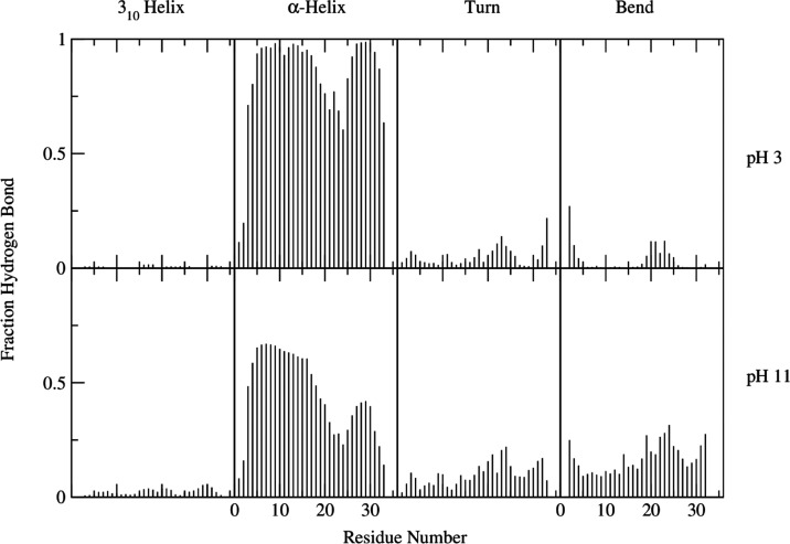
Cupiennin 1a is an antimicrobial peptide found in the venom of the spider Cupiennius salei. A highly cationic peptide, its cell lysis activity has been found to vary between neutral and charged membranes. In this study, Hamiltonian replica-exchange molecular dynamics (HREMD) was used to determine the conformational ensemble of the peptide in both charged (pH 3) and neutral (pH 11) states. The obtained free energy landscapes demonstrated the conformational diversity of the neutral peptide. At high pH, the peptide was found to adopt helix-hinge-helix and disordered structures. At pH 3, the peptide is structured with a high propensity toward α-helices. The presence of these α-helices seems to assist the peptide in recognizing membrane surfaces. These results highlight the importance of the charged residues in the stabilization of the peptide structure and the subsequent effects of pH on the peptide's conformational diversity and membrane activity. These findings may provide insights into the antimicrobial activity of Cupiennin 1a and other amphipathic linear peptides toward different cell membranes.


Conflict of interest statement

The authors declare no competing financial interest.

Figures

Figure 1
Figure 1
Helix–hinge–helix conformation of Cupiennin 1a obtained from the Hamiltonian replica-exchange molecular dynamics (HREMD) simulations. The eight lysine residues are represented by the purple stick model. Nonlysine hydrogen atoms were omitted for clarity purposes. The structure was visualized using UCSF Chimera software. The initial structure for the HREMD simulations was obtained from the protein data bank (ID 2K38). Sequence: GFGAL FKFLA KKVAK TVAKQ AAKQG AKYVV NKQME.
Figure 2
Figure 2
Free energy landscape of the two protein systems ((a) pH 3; (b) pH 11) as sampled by the HREMD simulation. The contours represent the estimated energy for each possible SASA (solvent-accessible surface area) and Rg (radius of gyration) pair. The difference in landscape sizes demonstrates the flexibility of the peptide at pH 11.
Figure 3
Figure 3
End-to-end distance values reported as probability distributions. The distances were calculated as the degree of separation between the α-carbons of the terminal residues 1 and 35. The calculation was done for all sampled conformations. The dashed brown lines correspond to distance values of 10, 33, and 48 Å. For each line, corresponding representative structures for both pH values were embedded. The dashed black boxes are for the pH 3 system, while the dashed red boxes are for pH 11.
Figure 4
Figure 4
Secondary structures of each residue as obtained using the DSSP method. Calculations were done for all sampled conformations.
Figure 5
Figure 5
Network of conformations in the (a) pH 3 and (b) pH 11 systems and the corresponding representative structures of each major node. Node 183 was included to represent the disordered structure of the neutral peptide. The structure for each node of the conformational cluster transition network (CCTN) was determined using cpptraj and visualized through Python’s graph-tool.
Figure 6
Figure 6
Coulombic surfaces of (a) full helical and (b) helix–hinge–helix structures. The color of the surfaces indicates the magnitude of the electrostatic potentials—red is negative (set at -10 kcal/mol), white is neutral, and blue is positive (10 kcal/mol). Chloride ions are represented by green circles. The interaction between the charged lysine residues and the negatively charged ions seems to stabilize the helices in both conformations. The structures were visualized using UCSF Chimera.
Figure 7
Figure 7
(a) Initial structure of the protein-membrane system for the full helical peptide at pH 3. Ions and water molecules were removed for clarity purposes. (b) Minimum distance values between lysine residues and membrane. The distance values were calculated between the nearest lysine residue and the membrane atom. (c) Last conformation from the MD simulation. Hydrogen bonds (dashed red line) were seen between the phosphate heads of the lipids and lysine residues 7 and 32.

References

    1. Ferreira F. R. B.; da Silva P. M.; Soares T.; Machado L. G.; de Araújo L. C. C.; da Silva T. G.; de Mello G. S. V.; da Rocha Pitta M. G.; de Melo Rego M. J. B.; Pontual E. V.; Zingali R. B.; Napoleão T. H.; Paiva P. M. G. Evaluation of antimicrobial, cytotoxic, and hemolytic activities from venom of the spider Lasiodora sp. Toxicon 2016, 122, 119–126. 10.1016/j.toxicon.2016.09.019. - DOI - PubMed
    1. Mourão C. B. F.; Heghinian M. D.; Barbosa E. A.; Marí F.; Bloch C.; Restano-Cassulini R.; Possani L. D.; Schwartz E. F. Characterization of a Novel Peptide Toxin from Acanthoscurria paulensis Spider Venom: A Distinct Cysteine Assignment to the HWTX-II Family. Biochemistry 2013, 52, 2440–2452. 10.1021/bi4000035. - DOI - PubMed
    1. Sanggaard K. W.; Bechsgaard J. S.; Fang X.; Duan J.; Dyrlund T. F.; Gupta V.; Jiang X.; Cheng L.; Fan D.; Feng Y.; et al. Spider genomes provide insight into composition and evolution of venom and silk. Nat. Commun. 2014, 5, 376510.1038/ncomms4765. - DOI - PMC - PubMed
    1. Undheim E.; Sunagar K.; Herzig V.; Kely L.; Low D.; Jackson T.; Jones A.; Kurniawan N.; King G.; Ali S.; Antunes A.; Ruder T.; Fry B. A Proteomics and Transcriptomics Investigation of the Venom from the Barychelid Spider Trittame loki (Brush-Foot Trapdoor). Toxins 2013, 5, 2488–2503. 10.3390/toxins5122488. - DOI - PMC - PubMed
    1. Vassilevski A. A.; Kozlov S. A.; Grishin E. V. Molecular diversity of spider venom. Biochemistry 2009, 74, 1505–1534. 10.1134/S0006297909130069. - DOI - PubMed