. 2021 Sep 13;6(37):24218-24232.
doi: 10.1021/acsomega.1c03816. eCollection 2021 Sep 21.

Preparation of pH-Responsive Vesicular Deferasirox: Evidence from In Silico, In Vitro, and In Vivo Evaluations

Affiliations

Preparation of pH-Responsive Vesicular Deferasirox: Evidence from In Silico, In Vitro, and In Vivo Evaluations

Mahmood Barani et al. ACS Omega. .

Abstract

pH-sensitive nanocarriers can effectively deliver anticancer drugs to tumors and reduce the adverse effects of conventional chemotherapy. In this light, we prepared a novel pH-responsive deferasirox (DFX)-loaded vesicle and comprehensively performed in silico, in vitro, and in vivo studies to examine the properties of the newly synthesized formulation. Physiochemical assessment of the developed formulations showed that they have an average size (107 ± 2 nm), negative zeta potential (-29.1 ± 1.5 mV), high encapsulation efficiency (84.2 ± 2.6%), and a pH-responsive release. Using the molecular dynamics simulation, the structural and dynamic properties of ergosterol-containing niosomes (ST60/Ergo) in the presence of DFX molecules were analyzed and showed a good interaction between DFX and vesicle components. Cytotoxic assessment showed that niosomal DFX exhibited a greater cytotoxic effect than free DFX in both human cancer cells (MCF-breast cancer and Hela cervical cancer) and induced evident morphological features of apoptotic cell death. No marked difference between the ability of free and niosomal DFX was found in activating caspase-3 in Hela cells. Eight weeks of intraperitoneal administrations of free DFX at three doses caused a significant increase in serum biochemical parameters and liver lipid peroxidation. Treatment with 5 mg/kg dose of niosomal DFX caused a significant increase in serum creatinine (P < 0.05); however, other parameters remained unchanged. On the other hand, administration of niosomal DFX at the highest dose (10 mg/kg) significantly increased serum creatinine (P < 0.05), BUN, and serum liver enzymes compared to the control rats (P < 0.001). Based on the results, the application of pH-responsive DFX-loaded niosomes, as a novel drug delivery platform, may yield promising results in cancer treatment.


Conflict of interest statement

The authors declare no competing financial interest.

Figures

Figure 1
Figure 1
Mass density distributions for the headgroup (blue), ester group (black), acyl group (green), and water (red) of (a) Span 60 and (b) Tween 60 as a function of z-direction normalized to the bilayer plane.
Figure 2
Figure 2
Molecular structure of DFX at the edge of lipid bilayer as facial (right) and lateral representation (left).
Figure 3
Figure 3
Optical microscopy imaging of pH-responsive DFX-niosome before sonication and filtration procedure.
Figure 4
Figure 4
(A) Physical appearance, (B) transmission electron microscopy (TEM) image, (C) size distribution, and (D) zeta potential of pH-responsive DFX-loaded niosomes.
Figure 5
Figure 5
In vitro release curve of pH-responsive DFX-niosomes at pH 5.4 and 7.4 in phosphate buffer at 37 °C (mean ± SD, n = 3).
Figure 6
Figure 6
Cytotoxic effects of unloaded niosomes and free and niosomal DFX on malignant (MCF7 and Hela) and normal (HUVEC) human cells evaluated via the MTT assay. (**P < 0.05 compared with untreated cells).
Figure 7
Figure 7
Morphological examination of MCF7 cancer cells treated with different concentrations of free and niosomal DFX for 48 h.
Figure 8
Figure 8
Morphological examination of Hela cancer cells treated with different concentrations of free and niosomal DFX for 48 h.
Figure 9
Figure 9
Effects of free and niosomal DFX on caspase-3 activity in Hela cells. (**P < 0.05 compared with untreated cells).
Figure 10
Figure 10
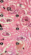
Kidney histopathology of control and experimental groups. (A) Normal kidney histopathology of control rats; (B) normal features in the kidneys of rats treated with 2.5 mg/kg of niosomal DFX; (C) normal histology of kidneys of rats treated with 5 mg/kg of niosomal DFX; (D) some degree of renal damages including tubular swelling in rats treated with 2.5 mg/kg of niosomal DFX (arrow); (E) abnormal features in the kidneys of rats subjected to free DFX 2.5 mg/kg; (F) photomicrograph of a rat treated with 5 mg/kg of free DFX, some degree of renal damages including hemorrhage (arrow), tubular swelling, and glomerular atrophy. (G) Histopathological changes such as hemorrhage in rats treated with 10 mg/kg of niosomal DFX including hemorrhage (arrow).
Figure 11
Figure 11
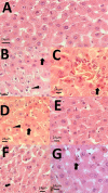
Liver histopathology of control and experimental groups. Hematoxylin–eosin (H&E) staining. (A) Normal liver micrograph of control rats; (B) necrosis (arrow) and fatty change (arrow head) in the liver of rats treated with DFX 2.5 mg/kg; (C) normal histology of liver of rats treated with free DFX 5 mg/kg; (D) some degree of liver damages including cytoplasmic swelling in rats treated with free DFX 10 mg/kg (arrowhead); (E) liver histopathology of liver of rats subjected to free DFX 0.25 niosome mg/kg showing normal appearance; and (F) photomicrograph of a rat treated with niosomal DFX 5 mg/kg signs of mild liver damage including cytoplasmic vacuolation. (G) Some degree of histopathological changes including necrosis (black arrow) in rats treated with DFX niosome 10 mg/kg.
Scheme 1
Scheme 1. Schematic Representation for pH-Responsive DFX-Niosome and Its In Silico, In Vitro, and In Vivo Evaluations
Figure 12
Figure 12
(a) Molecular structure of deferasirox, Span 60, Tween 60, and ergosterol with the numbering of atoms based on the amber nomenclature. (b) Starting structure of niosome bilayer containing DFX drugs at the top. Span 60, Tween 60, and ergosterol lipids are colored as tan, cyan, and green, respectively. The DFX molecules are represented as the space-filling model.

References

    1. Rahdar A.; Hajinezhad M. R.; Sargazi S.; Barani M.; Bilal M.; Kyzas G. Z. Deferasirox-loaded pluronic nanomicelles: Synthesis, characterization, in vitro and in vivo studies. J. Mol. Liq. 2021, 323, 114605.10.1016/j.molliq.2020.114605. - DOI
    1. Taghavi F.; Saljooghi A. S.; Gholizadeh M.; Ramezani M. Deferasirox-coated iron oxide nanoparticles as a potential cytotoxic agent. MedChemComm 2016, 7, 2290–2298. 10.1039/c6md00293e. - DOI
    1. Taghavi F.; Gholizadeh M.; Saljooghi A. S. Deferasirox loaded on fumed silica nanoparticles used in cancer treatment. New J. Chem. 2016, 40, 2696–2703. 10.1039/c5nj02790j. - DOI
    1. Moukalled N. M.; Bou-Fakhredin R.; Taher A. T. Deferasirox: over a decade of experience in thalassemia. J. Hematol. 2018, 10, e201806610.4084/MJHID.2018.066. - DOI - PMC - PubMed
    1. Bedford M. R.; Ford S. J.; Horniblow R. D.; Iqbal T. H.; Tselepis C. Iron chelation in the treatment of cancer: a new role for deferasirox?. J. Clin. Pharmacol. 2013, 53, 885–891. 10.1002/jcph.113. - DOI - PubMed