. 2021 Sep 20;4(9):7234-7242.
doi: 10.1021/acsabm.1c00766. Epub 2021 Aug 31.

Tissue Engineering the Pinna: Comparison and Characterization of Human Decellularized Auricular Biological Scaffolds

Affiliations

Tissue Engineering the Pinna: Comparison and Characterization of Human Decellularized Auricular Biological Scaffolds

Zaid Al-Qurayshi et al. ACS Appl Bio Mater. .

Abstract

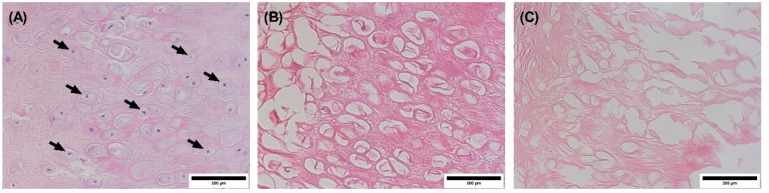
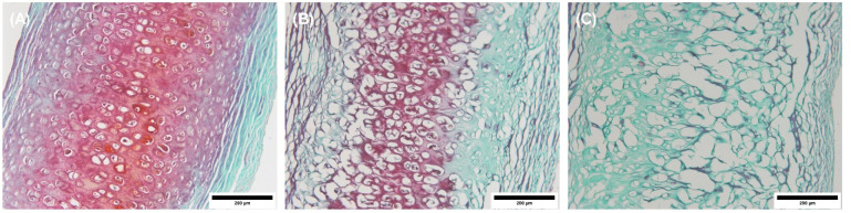
Decellularization is one of the promising techniques in tissue engineering used to create a biological scaffold for subsequent repopulation with the patient's own cells. This study aims to compare two different decellularization protocols to optimize the process of auricle decellularization by assessing and characterizing the decellularization effects on human auricular cartilage. Herein, 12 pairs (8 females, 4 males) of freshly frozen adult human cadaveric auricles were de-epithelialized and defatted leaving only the cartilaginous framework. An auricle from each pair was randomly assigned to either protocol A (latrunculin B-based decellularization) or protocol B (trypsin-based decellularization). Gross examination of the generated scaffolds demonstrated preservation of the auricles' contours and a change in color from pinkish-white to yellowish-white. Hematoxylin and eosin staining demonstrated empty cartilaginous lacunae in both study groups, which confirms the depletion of cells. However, there was greater preservation of the extracellular matrix in auricles decellularized with protocol A as compared to protocol B. Comparing protocol A to protocol B, Masson's trichrome and Safranin-O stains also demonstrated noticeable preservation of collagen and proteoglycans, respectively. Additionally, scanning electron micrographs demonstrated preservation of the cartilaginous microtopography in both study groups. Biomechanical testing demonstrated a substantial decrease in Young's modulus after decellularization using protocol B (1.3 MPa), albeit not significant (P-value > 0.05) when compared to Young's modulus prior to decellularization (2.6 MPa) or after decellularization with protocol A (2.7 MPa). A DNA quantification assay demonstrated a significant drop (P-value < 0.05) in the DNA content after decellularization with protocol A (111.0 ng/mg) and protocol B (127.6 ng/mg) in comparison to before decellularization (865.3 ng/mg). Overall, this study demonstrated effective decellularization of human auricular cartilage, and it is concluded that protocol A provided greater preservation of the extracellular matrix and biomechanical characteristics. These findings warrant proceeding with the assessment of inflammation and cell migration in a decellularized scaffold using an animal model.


Conflict of interest statement

The authors declare no competing financial interest.

Figures

Figure 1
Figure 1
Schematic representation of the external ear anatomy showing the structural differences between the normal healthy auricle and the deformed auricle (microtia).
Figure 2
Figure 2
Gross structural characteristics of auricles before and after decellularization. Images showing one of the auricles (A) before (native) and (B) after decellularization using protocol A; and one of the auricles (C) before and (D) after decellularization using protocol B.
Figure 3
Figure 3
Images of H&E stained sections visualized by a light microscope. (A) Auricle before decellularization (native auricle, arrows point to nuclei); (B) and (C) represent the decellularized auricles using protocol A and protocol B, respectively. The nuclei are stained blue (hematoxylin), while the extracellular matrix and cytoplasm are stained with varying degrees of pink (eosin). Images were captured at 40× objective magnification. Scale bar = 200 μm.
Figure 4
Figure 4
Images of Masson’s trichrome stained sections of punch biopsies from auricles. (A) Auricle before decellularization (native auricle); (B) and (C) represent the decellularized auricles using protocol A and protocol B, respectively. The blue-stained areas indicate the presence of collagen. Images were captured at 20× objective magnification. Scale bar = 200 μm.
Figure 5
Figure 5
Images of Safranin-O stained sections of punch biopsies from auricles. (A) The auricle before decellularization (native auricle); (B) and (C) represent the decellularized auricles using protocol A and protocol B, respectively. The red-stained areas indicate the presence of proteoglycan. Images were captured at 20× objective magnification. Scale bar = 200 μm.
Figure 6
Figure 6
Scanning electron micrographs illustrating histological features of the human auricle. (A) The auricle before decellularization (native auricle); (B) and (C) represent the decellularized auricles using protocol A and protocol B, respectively. Images were captured at 300× magnification. Scale bar = 100 μm.
Figure 7
Figure 7
Characteristics of the human auricles before and after two different decellularization methodologies. (A) Indentation test to evaluate the stress. (B) DNA content. (C) sGAG content. Data were plotted as mean ± standard deviation. ns, not statistically significant (i.e., p > 0.05), *p < 0.05. Baseline (n = 6) represents native auricles; protocol A (n = 3); protocol B (n = 3).

References

    1. Bly R. A.; Bhrany A. D.; Murakami C. S.; Sie K. C. Microtia Reconstruction. Facial Plast Surg Clin North Am. 2016, 24 (4), 577–591. 10.1016/j.fsc.2016.06.011. - DOI - PMC - PubMed
    1. Luquetti D. V.; Heike C. L.; Hing A. V.; Cunningham M. L.; Cox T. C. Microtia: epidemiology and genetics. Am. J. Med. Genet., Part A 2012, 158A (1), 124–39. 10.1002/ajmg.a.34352. - DOI - PMC - PubMed
    1. Harris J.; Källén B.; Robert E. The epidemiology of anotia and microtia. Journal of Medical Genetics 1996, 33 (10), 809–813. 10.1136/jmg.33.10.809. - DOI - PMC - PubMed
    1. Mastroiacovo P.; Corchia C.; Botto L. D.; Lanni R.; Zampino G.; Fusco D. Epidemiology and genetics of microtia-anotia: a registry based study on over one million births. J. Med. Genet 1995, 32 (6), 453–7. 10.1136/jmg.32.6.453. - DOI - PMC - PubMed
    1. Forrester M. B.; Merz R. D. Descriptive epidemiology of anotia and microtia, Hawaii, 1986–2002. Congenital Anomalies 2005, 45 (4), 119–24. 10.1111/j.1741-4520.2005.00080.x. - DOI - PubMed

Publication types