. 2021 Apr;2(4):429-443.
doi: 10.1038/s43018-021-00174-z. Epub 2021 Mar 1.

Distinct CDK6 complexes determine tumor cell response to CDK4/6 inhibitors and degraders

Affiliations

Distinct CDK6 complexes determine tumor cell response to CDK4/6 inhibitors and degraders

Xuewei Wu et al. Nat Cancer. 2021 Apr.

Abstract

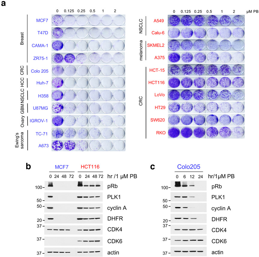
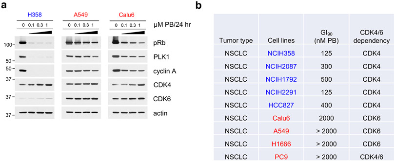
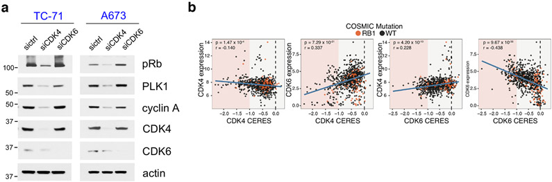
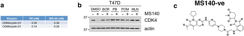
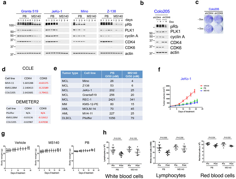
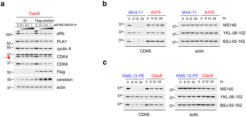
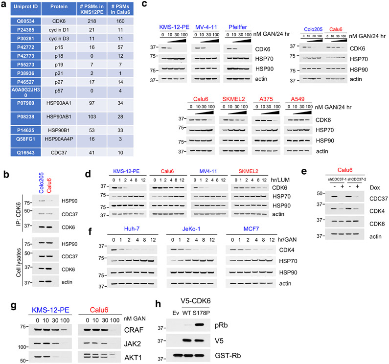
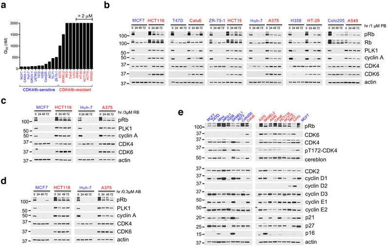
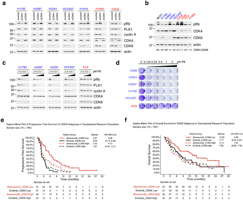
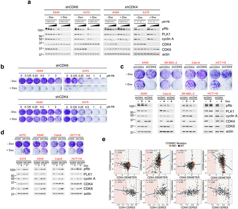
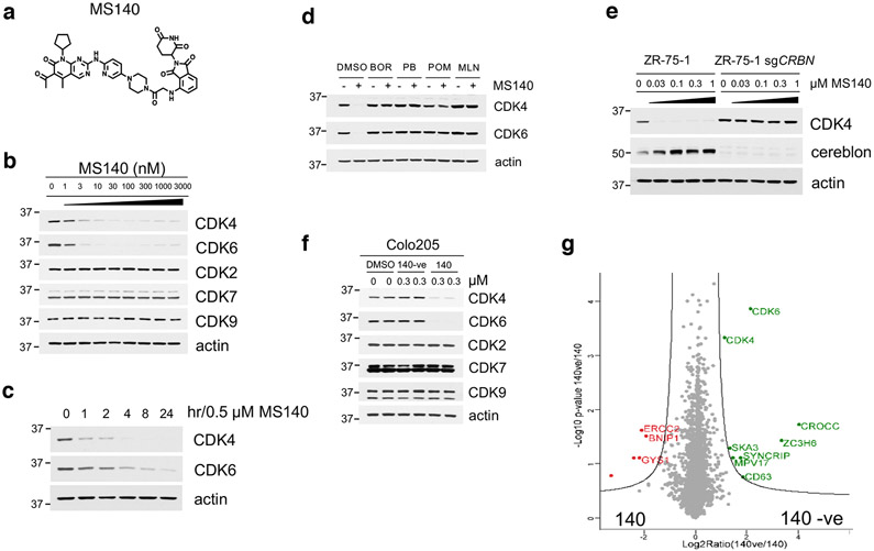
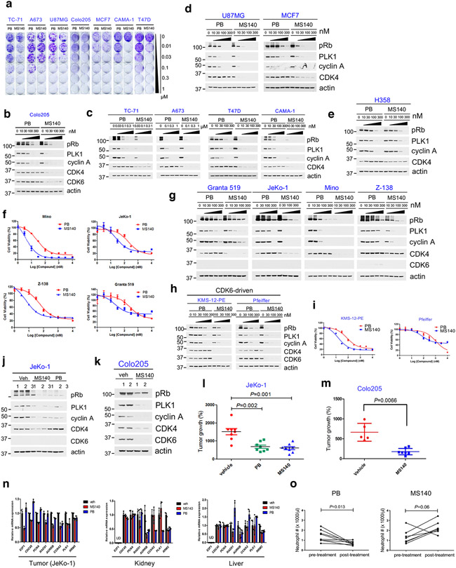
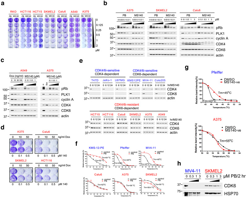
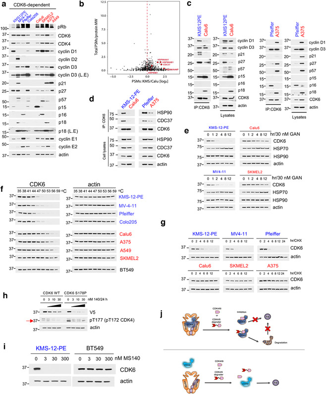
CDK4/6 inhibitors (CDK4/6i) are effective in metastatic breast cancer, but they have been only modestly effective in most other tumor types. Here we show that tumors expressing low CDK6 rely on CDK4 function, and are exquisitely sensitive to CDK4/6i. In contrast, tumor cells expressing both CDK4 and CDK6 have increased reliance on CDK6 to ensure cell cycle progression. We discovered that CDK4/6i and CDK4/6 degraders potently bind and inhibit CDK6 selectively in tumors in which CDK6 is highly thermo-unstable and strongly associated with the HSP90/CDC37 complex. In contrast, CDK4/6i and CDK4/6 degraders are ineffective in antagonizing tumor cells expressing thermostable CDK6, due to their weaker binding to CDK6 in these cells. Thus, we uncover a general mechanism of intrinsic resistance to CDK4/6i and CDK4/6i-derived degraders and the need for novel inhibitors targeting the CDK4/6i-resistant, thermostable form of CDK6 for application as cancer therapeutics.


Figures

Extended Data Fig. 1
Extended Data Fig. 1. Intrinsic resistance to CDK4/6i is associated with incomplete inhibition of Rb/E2F and expression of CDK6
a, Cell growth crystal violet assay for the indicated cell lines treated with increasing concentrations of PB for 10-16 days and stained with crystal violet. CDK4/6i-sensitive cell lines were highlighted in blue, CDK4/6i-resistant cell lines were in red. b, MCF7 and HCT116 were treated with 1 μM PB for 24, 48 and 72 hr and lysates were immunoblotted with the indicated antibodies. c, Colo205 cells were treated with 1 μM PB at the indicated time points. Cell lysates were immunoblotted with the indicated antibodies.
Extended Data Fig. 2
Extended Data Fig. 2. Low expression of CDK6 predicts for sensitivity to CDK4/6i in NSCLC
a, The indicated cell lines were treated with increasing concentrations of PB for 24 hr. Lysates were immunoblotted with the indicated antibodies. b, GI90 values of PB and CDK4/6 dependency in NSCLC cell lines.
Extended Data Fig. 3
Extended Data Fig. 3. Tumors expressing both CDK4 and CDK6 depend selectively on CDK6
a, A673 and TC-71 cells were transfected with non-targeting control or siCDK4 or siCDK6 for 72 hr. Lysates were immunoblotted with the indicated antibodies. b, Relationship between CDK4 and CDK6 expression (CCLE RNA-seq) and DepMap CRISPR–Cas9 single-gene knockout scores (CERES; 20Q1 public dataset). All expression values are in log2(TPM +1). Cell lines harboring COSMIC hotspot mutations to RB1 are annotated in orange. P-values were calculated based on linear regression analysis.
Extended Data Fig. 4
Extended Data Fig. 4. Development of MS140, a potent and selective CDK4/6-degrader (PROTAC)
a. IC50 of in vitro kinase activity assays for PB and MS140 against CDK4/cyclin D1 and CDK6/cyclin D1. b, T47D cells were pretreated with either the proteasome inhibitor 100 nM bortezomib (BOR), 10 μM PB, 10 μM pomalidomide (POM) or 1 μM MLN4924 (MLN) for 4 hr, followed by treatment with MS140 (100 nM/3 hr). Lysates were subjected to immunoblotting with the indicated antibodies. c, Chemical structure of the MS140 negative control (MS140-ve) that does not bind CRBN.
Extended Data Fig. 5
Extended Data Fig. 5. CDK4/6-directed degradation is more effective than CDK4/6i in CDK4/6i-S tumor cells
a, MCL cell lines were treated with 0.1 μM PB or MS140 at different time points. Lysates were immunoblotted with the indicated antibodies. b, Colo205 cells expressing Dox-inducible shCDK4 or shCDK6 were treated with or without 0.1 μg/ml doxycycline for 72 hr and cell lysates were subjected to immunoblotting with the indicated antibodies. c, Colo205 cells expressing Dox-inducible shCDK4 or shCDK6 were treated with or without 0.1 μg/ml doxycycline for 10 days followed by crystal violet staining. d, Dependency score of CDK4 and CDK6 from cancer cell line encyclopedia (CCLE) and Depmap portal database. e, GI50 values of PB and MS140 in hematologic cancer cell lines. f, Growth curve for an efficacy assay in JeKo-1 tumor xenografts in nude mice treated with vehicle or MS140 (25 mg/kg, b.i.d) or PB (50 mg/kg, q.d.) for 21 days. Each treatment contained 8 animals (n=8). Data represent mean ± SEM. g, Body weight in mice bearing JeKo-1 tumors treated with vehicle (n=8) or PB (50 mg/kg, q.d., n=8) or MS140 (25 mg/kg, b.i.d., n=8) in the course of the experiment (21 days). Data are presented as mean ± S.D. h, White blood cell, lymphocytes and red blood cell counts in C57BL/6 mice before treatment and post treatment with PB (50 mg/kg, q.d., n=8) or MS140 (25 mg/kg, b.i.d., n=7) for 21 days. Data are presented as mean ± S.D. Statistical significance was determined by paired two-tailed Student’s t-test.
Extended Data Fig. 6
Extended Data Fig. 6. In CDK4/6-R cells, CDK4/6 degraders fail to degrade CDK6 due to weak binding of compound
a, Calu6 cells transiently expressing pcDNA3 (Ev) or pcDNA3-Flag-CRBN were treated with increasing concentrations of MS140 for 24 hr. Lysates were subjected to immunoblotting with the indicated antibodies. b, MV4-11 and A375 were treated MS140 (3 nM) or YKL-06-102 (3 nM) or BSJ-02-162 (3 nM) at different time points. Lysates were subjected to immunoblotting with the indicated antibodies. c, KMS-12-PE and Calu6 were treated MS140 (3 nM) or YKL-06-102 (3 nM) or BSJ-02-162 (3 nM) at different time points. Lysates were subjected to immunoblotting with the indicated antibodies.
Extended Data Fig. 7
Extended Data Fig. 7. CDK4/6i-resistant cells express CDK6 as a thermostable, weak HSP90 client protein
a, Comparison of total peptide-spectrum match (PSM) for CDK6-interacting proteins by mass spectrometry in KMS-12-PE and Calu6. b, Cell lysates from Colo205 and Calu6 were either subjected to Co-IP with a CDK6 antibody followed by immunoblotting with HSP90, CDC37 and CDK6, or immunoblotted with the indicated antibodies. c, The indicated cell lines were treated with increasing concentrations of Ganetespib (GAN) for 24 hr. Lysates were subjected to immunoblotting with the indicated antibodies. d, The indicated cell lines were treated with 40 nM Luminespib (LUM) at the indicated time points. Lysates were subjected to immunoblotting with the indicated antibodies. e. Calu6 cell line expressing Dox-inducible shCDC37 were treated with or without 0.1 μg/ml doxycycline for 72 hr and cell lysates were subjected to immunoblotting with the indicated antibodies. f, CDK4-dependent cell lines were treated with 30 nM GAN for the indicated time points. Lysates were subjected to immunoblotting with the indicated antibodies. g, KMS-12-PE and Calu6 were treated with increasing concentrations of GAN for 24 hr. Lysates were subjected to immunoblotting with the indicated antibodies. h, Lysates from A375 cells ectopically expressing V5-CDK6 or V5-CDK6 S178p were immunoprecipited with a V5 antibody. The immunoprecipitates were subjected to kinase assay with recombinant Rb protein as substrate.
Fig. 1.
Fig. 1.. Intrinsic resistance to CDK4/6i is associated with incomplete inhibition of Rb/E2F and expression of CDK6
a, Bar graph of GI90 values based on cell growth crystal violet assays for the indicated cell lines treated with increasing concentrations of PB for 10-15 days. CDK4/6i-sensitive cell lines were highlighted in blue, CDK4/6i-resistant cell lines were in red. b, The indicated cell lines were treated with 1 μM PB for 24, 48 and 72 hr and lysates were immunoblotted with the indicated antibodies. c, The indicated cell lines were treated with 3 μM Ribociclib (RB) for 24, 48 and 72 hr and lysates were immunoblotted with the indicated antibodies. d, The indicated cell lines were treated with 0.3 μM Abemaciclib (AB) for 24, 48 and 72 hr and lysates were immunoblotted with the indicated antibodies. e, Expression of known cell cycle regulators were detected by immunoblotting with the indicated antibodies. All data represent single experiments.
Fig. 2.
Fig. 2.. Low expression of CDK6 predicts for sensitivity to CDK4/6i in NSCLC
a, The indicated NSCLC cell lines were transfected with control siRNA, siCDK4 and siCDK6 for 72 hr and lysates were immunoblotted with the indicated antibodies. b, Total cell lysates of the indicated NSCLC cell lines were subjected to immunoblotting with the indicated antibodies. The ratio of CDK4/CDK6 was calculated by band intensity using Image J. The samples are derived from the same experiment and the images were processed in parallel. c, The indicated NSCLC cell lines were treated with increasing concentrations of PB for 24 hr and lysates were immunoblotted with the indicated antibodies. d, Cell growth crystal violet assay for the indicated NSCLC cell lines treated with increasing concentrations of PB for 10-15 days and stained with crystal violet. CDK4/6i-sensitive cell lines were in blue, CDK4/6i-resistant cell lines were in red. e, Progression-free survival analysis of NSCLC/RAS-mutant patients that received abemaciclib in the JUNIPER trial based on CDK6-low versus CDK6-high tumors. (Abemaciclib: n=79 patients. n=43 patients in CDK6-low arm, n=36 patients in CDK6-high arm; Erlotinib: n=69 patients. n=37 patients in CDK6-low arm, n=32 patients in CDK6-high arm). f, Overall survival analysis of NSCLC/RAS-mutant patients that received abemaciclib in the JUNIPER trial based on CDK6-low versus CDK6-high tumors. (Abemaciclib: n=79 patients. n=43 patients in CDK6-low arm, n=36 patients in CDK6-high arm; Erlotinib: n=69 patients. n=37 patients in CDK6-low arm, n=32 patients in CDK6-high arm). All data represent single experiments.
Fig. 3.
Fig. 3.. Tumors expressing both CDK4 and CDK6 depend selectively on CDK6
a, A549 and A375 expressing Dox-inducible shCDK4 or shCDK6 were treated with 0.1 μg/ml doxycycline for 36 hr, followed by PB for 24 hr. Cell lysates were immunoblotted with the indicated antibodies. b, Cell growth crystal violet assay for A549 and A375 expressing Dox-inducible shCDK4 or shCDK6 in the presence or absence of 0.1 μg/ml doxycycline and increasing concentrations of PB for 10 days. c, Top panel: The indicated cell lines expressing Dox-inducible shCDK4 or shCDK6 were treated with or without 0.1 μg/ml doxycycline for 10 days followed by crystal violet staining. Bottom panel: cell lines were treated with or without 0.1 μg/ml doxycycline for 72 hr and cell lysates were subjected to immunoblotting with the indicated antibodies. d, Top panel: The indicated cell lines co-expressing Dox-inducible shCDK6 and shRNA-resistant form of V5-CDK6 were treated with or without 0.05 μg/ml doxycycline for 10 days followed by crystal violet staining. Bottom panel: cell lines were treated with or without 0.05 μg/ml doxycycline for 72 hr and cell lysates were subjected to immunoblotting with the indicated antibodies. e, Top panel: Scatterplots of DEMETER score (DepMap RNAi; DEMETER2 Data v5) and expression for CDK4 and CDK6. Bottom panel: Scatterplots of CERES (DepMap CRISPR; Public 20Q1) score and the mass spectrometry-based proteomics levels of CDK4 and CDK6 (PMID:31978347). All expression values are in log2(TPM +1). Proteomic levels are shown as normalized log2-transformed ratios to the bridge sample in each Tandem Mass Tags (TMT) 10-plex as previously described (BIORXIV: doi:10.1101/2020.02.03.932384). Cell lines harboring COSMIC hotspot mutations to RB1 are annotated in orange. P-values were calculated based on linear regression analysis. All data represent single experiments.
Fig. 4.
Fig. 4.. Development of MS140, a potent and selective CDK4/6-degrader (PROTAC)
a, Chemical structure of the bifunctional CDK4/6 inhibitor-degrader MS140. b, Colo205 cells were treated with increasing concentrations of MS140 for 5 hr. Lysates were immunoblotted with the indicated antibodies. c, Colo205 cells were treated with MS140 (0.5 μM) for the indicated time points. Lysates were immunoblotted with the indicated antibodies. d, Colo205 cells were pretreated with either the proteasome inhibitor 100 nM bortezomib (BOR), 10 μM PB, 10 μM pomalidomide (POM) or 1 μM MLN4924 (MLN) for 4 hr, followed by treatment with MS140 (100 nM/3 hr). Lysates were subjected to immunoblotting with the indicated antibodies. e, ZR-75-1 wild-type and CRBN-deficient cells were treated with the indicated concentrations of MS140 for 5 hr. Lysates were immunoblotted with the indicated antibodies. f, Colo205 cells were treated with DMSO, MS140-ve and MS140 for 5 hr. Lysates were immunoblotted with the indicated antibodies. g, Volcano plot of the protein log2 ratios represent the quantitative dynamics of 4,822 proteins in 140 and 140-ve (negative control of 140) treated Colo205 samples (0.3 μM, 5 hr) in duplicate. All data represent single experiments.
Fig. 5.
Fig. 5.. CDK4/6-directed degradation is more effective than CDK4/6i in CDK4/6i-S tumor cells
a, The indicated cell lines were subjected to cell growth crystal violet assay in the presence of PB or MS140 for 10-15 days. b, Colo205 cells were treated with increasing concentrations of PB or MS140 for 24 hr. Lysates were immunoblotted with the indicated antibodies. c, The indicated cell lines were treated with increasing concentrations of PB or MS140 for 24 hr. Lysates were immunoblotted with the indicated antibodies. d, U87MG or MCF7 cells were treated with increasing concentrations of PB or MS140 for 24 hr. Lysates were immunoblotted with the indicated antibodies. e, H358 cells were treated with increasing concentrations of MS140 for 24 hr. Lysates were immunoblotted with the indicated antibodies. f, Mantle Cell Lymphoma (MCL) cells were treated with PB and MS140 for 72-96 hr. Cell viability was assayed using 0.1 mg/ml resazurin solution. IC50 values were determined by nonlinear regression curve fit in Graphpad Prism (n=6 independent experiments). Data are presented as mean ± S.D. g, MCL cell lines were treated with increasing concentrations of PB or MS140 for 24 hr. Lysates were immunoblotted with the indicated antibodies. h, KMS-12-PE or Pfeiffer cells were treated with increasing concentrations of PB or MS140 for 24 hr. Lysates were immunoblotted with the indicated antibodies. i. The indicated cells were treated with PB or MS140 for 72-96 hr. Cell viability was assayed using 0.1 mg/ml resazurin solution. IC50 values were determined by nonlinear regression curve fit in Graphpad Prism (n=6 independent experiments). Data are presented as mean ± S.D. j, Mice carrying JeKo-1 xenografts were treated with vehicle or MS140 (25 mg/kg, b.i.d) or PB (50 mg/kg, q.d.) for 3 days. Tumor samples were lysed and immunoblotted with the indicated antibodies. k, Mice carrying Colo205 xenografts were treated with vehicle or MS140 (30 mg/kg, b.i.d) for 3 days. Tumor samples were lysed and immunoblotted with the indicated antibodies. l, Scatter plot of fold change for an efficacy assay in JeKo-1 tumor xenografts in mice treated with vehicle or MS140 (25 mg/kg, b.i.d) or PB (50 mg/kg, q.d.) for 21 days. Each treatment contained 8 animals (n=8). Data are presented as mean ± S.E.M. Statistical significance was determined by paired two-tailed Student’s t-test. m, Scatter plot of fold change for an efficacy assay in Colo205 tumor xenografts in nude mice treated with vehicle or MS140 (30 mg/kg, b.i.d) for 21 days. Vehicle treatment contained 5 animals (n=5). MS140 treatment contained 8 animals (n=8). Data are presented as mean ± S.D. Statistical significance was determined by unpaired two-tailed Student’s t-test. n, qPCR analysis of expression of known Rb/E2F target genes in tumors, kidney and liver from mice bearing JeKo-1 tumors treated with vehicle or MS140 (25 mg/kg, b.i.d) or PB (50 mg/kg, q.d.) for 3 days. Data are presented as mean ± S.D. (Tumor: n=1 mouse with three independent experiments; kidney and liver: n=2 mice with two independent experiments for each mouse). o, Neutrophil counts in C57BL/6 mice before treatment and post treatment with PB (50 mg/kg, q.d., n=8 mice) or MS140 (25 mg/kg, b.i.d., n=7 mice) for 21 days. Data are presented as mean ± S.D. Statistical significance was determined by paired two-tailed Student’s t-test. All data represent single experiments.
Fig. 6.
Fig. 6.. In CDK4/6-R cells, CDK4/6 degraders fail to degrade CDK6 due to weak binding of compound
a, Cell growth crystal violet assay for the indicated cell lines treated with increasing concentrations of PB or MS140 for 10-12 days. b, A375, SKMEL2 and Calu6 cells were treated with increasing concentrations of PB or MS140 for 24 hr. Lysates were immunoblotted with the indicated antibodies. c, A549 or A375 expressing Dox-inducible shCDK6 were treated with either the indicated concentrations of doxycycline (A375: 0.1 μg/ml) or MS140 for 72 hr. Lysates were subjected to immunoblotting with the indicated antibodies. d, Cell growth crystal violet assay for the indicated cell lines treated with either doxycycline or MS140 for 10 days. Colonies were stained with crystal violet. e, Rb-proficient cell lines were treated with MS140 (3 nM) for the indicated time points. Cell lysates were immunoblotted with the indicated antibodies. f, The indicated cell lines were treated with PB (1 μM/2 hr) followed by CETSA assay. The graphs are derived by immunoblot analysis of CDK6 and actin expression using Image J. The samples derive from the same experiment and the blots were processed in parallel. g, The indicated cell lines were treated with MS140-ve (15 μM/2 hr) followed by CETSA assay. The graphs are derived by immunoblot analysis of CDK6 and actin expression using Image J. The samples derive from the same experiment and the blots were processed in parallel. h, Desthiobiotin-ADP enrichment assay for CDK6 in MV4-11 and SKMEL2 cells pretreated with increasing concentrations of PB for 2 hr. Elute were subjected to immunoblotting with the indicated antibodies. All data represent single experiments.
Fig. 7.
Fig. 7.. CDK4/6i-resistant cells express CDK6 as a thermostable, weak HSP90 client protein
a, Immunoblots for cell cycle regulators in CDK6-dependent cell lines. L.E = long exposure. b, Volcano plot of the CDK6-interacting proteins in KMS-12-PE and Calu6. Proteins in red were annotated as HSP90/CDC37-related. c, Cell lysates from the indicated cell lines were either subjected to Co-IP with a CDK6 antibody followed by immunoblotting with known cell cycle regulators, or immunoblotted with the indicated antibodies. d, Cell lysates from the indicated cell lines were either subjected to Co-IP with a CDK6 antibody followed by immunoblotting with HSP90, CDC37 and CDK6, or immunoblotted with the indicated antibodies. e, Cell lines were treated with the HSP90 inhibitor Ganetespib (GAN, 30 nM) at the indicated time points. Cell lysates were immunoblotted with the indicated antibodies. f, Immunoblots for CDK6 thermal stability assay (CETSA) in CDK4/6i-sensitive and CDK4/6i-resistant cell lines heat-treated at increasing temperature end points. g, The indicated cell lines were treated with 100 μg/ml CHX at the indicated time points. Cell lysates were immunoblotted with the indicated antibodies. h, A375 cells were transfected with either WT or CDK6(S178P) followed by treatment with increasing concentrations of MS140 for 24 h. Cell lysates were immunoblotted with the indicated antibodies. i, KMS-12-PE and BT549 cells were treated with increasing concentrations of MS140 for 24 hr. Lysates were subjected to immunoblotting with the indicated antibodies. j, Model of CDK6 association with the HSP90 complex affecting tumor cell sensitivity to CDK4/6 inhibitors and degraders. Top: In CDK4/6 inhibitor and degrader-sensitive cells CDK6 is associated with the HSP90 complex. CDK4/6 inhibitors or CDK6 degraders binds strongly CDK6, and promote CDK6 inhibition or both CDK6 inhibition and degradation respectively. Bottom: In CDK4/6 inhibitor and degrader-resistant cells, CDK6 is weakly associated with the HSP90 complex. In these cells, CDK4/6 inhibitors and degraders bind CDK6 weakly, and thus fail to promote CDK6 inhibition or both inhibition and degradation, respectively. All data represent single experiments.

Comment in

References

    1. Sherr CJ, Beach D & Shapiro GI Targeting CDK4 and CDK6: From Discovery to Therapy. Cancer Discov 6, 353–367, doi:10.1158/2159-8290.CD-15-0894 (2016). - DOI - PMC - PubMed
    1. O'Leary B, Finn RS & Turner NC Treating cancer with selective CDK4/6 inhibitors. Nat Rev Clin Oncol 13, 417–430, doi:10.1038/nrclinonc.2016.26 (2016). - DOI - PubMed
    1. Otto T & Sicinski P Cell cycle proteins as promising targets in cancer therapy. Nat Rev Cancer 17, 93–115, doi:10.1038/nrc.2016.138 (2017). - DOI - PMC - PubMed
    1. Finn RS et al.Palbociclib and Letrozole in Advanced Breast Cancer. N Engl J Med 375, 1925–1936, doi:10.1056/NEJMoa1607303 (2016). - DOI - PubMed
    1. Hortobagyi GN et al.Ribociclib as First-Line Therapy for HR-Positive, Advanced Breast Cancer. N Engl J Med 375, 1738–1748, doi:10.1056/NEJMoa1609709 (2016). - DOI - PubMed

Publication types